

IMG-20260812-WA0090.jpg
Itching
analyse_media
Reading File
The rash shows a classic confluent and reticulated (net-like) pattern of hyperpigmented macules and patches over the entire back - no wheals, no plaques, no obvious scaling. This is highly characteristic of Confluent and Reticulated Papillomatosis (CARP) or Pityriasis versicolor, or possibly Prurigo pigmentosa. Let me search the textbooks and look for matching images.
Searching sources...
Finding Sources
Finding Sources
Searching Images
confluent reticulated papillomatosis back hyperpigmented reticulated rash
Finding Sources
Finding Sources
Searching Images
pityriasis versicolor hyperpigmented back trunk rash
Reading File
Reading File
Reading File
Loading Image
The textbook image of pityriasis versicolor matches very well - now let me also fetch the direct back image from the textbook for comparison and get the full treatment details.
Loading Image
Finding Sources
Reading File
I now have all the information needed for a thorough answer. The textbook image of tinea (pityriasis) versicolor on the back matches this patient's presentation very closely.
Most Likely Diagnosis: Pityriasis (Tinea) Versicolor
The rash in the image is highly consistent with pityriasis versicolor (also called tinea versicolor), a superficial fungal infection caused by the overgrowth of Malassezia furfur / M. globosa - yeasts that are part of normal skin flora.
What the image shows
The AI vision analysis of your photo describes:
- Bilateral, symmetric distribution across the entire back
- Confluent and reticulated (net-like/lace-like) pattern - patches merge centrally, with a web-like appearance at the periphery
- Hyperpigmented macules and patches - light brown to dusky tan color
- No wheals, no papules, no thick plaques - flat lesions
- Smooth surface - no gross scaling (though fine scale is often only seen on gentle scraping)
This matches the textbook description exactly. Here is a textbook photo of tinea versicolor on the back for comparison:
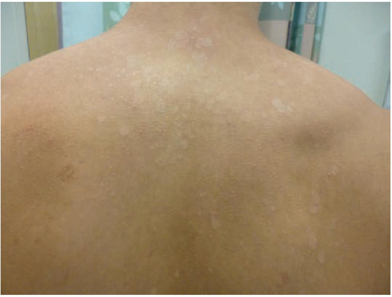
(Note: versicolor means "varying colors" - your case shows the hyperpigmented (brown) variant, while this textbook image shows the hypopigmented variant. Both are the same condition.)
Why this diagnosis fits
| Feature | Pityriasis Versicolor | This Patient |
|---|---|---|
| Organism | Malassezia yeast | - |
| Location | Central upper back, trunk | ✓ Whole back |
| Morphology | Coalescing macules/patches | ✓ Confluent |
| Pattern | Reticulated, lace-like | ✓ Net-like |
| Color | Brown (hyperpigmented) or pale | ✓ Brown |
| Itch | Mild, can occur | ✓ Present |
| Age group | Young adults (peak sebaceous activity) | ✓ Appears young adult |
| Triggers | Heat, humidity, oily skin | Common in tropical climates |
Diagnosis confirmation
A KOH (potassium hydroxide) scraping under microscopy shows the classic "spaghetti and meatballs" pattern - short hyphae (spaghetti) with round yeast spores (meatballs). This is the gold standard confirmatory test - Textbook of Family Medicine, 9e.
Treatment
Topical (first-line, most cases):
- Ketoconazole 2% shampoo - apply daily for 3-7 days, leave on 3-5 min before rinsing - Tintinalli's Emergency Medicine
- Selenium sulfide 2.5% lotion/shampoo - apply daily for 10 min for 7 days (well-studied, SOR: B)
- Clotrimazole or other azole antifungal creams twice daily
Oral (extensive/refractory disease like this widespread case):
- Fluconazole 400 mg single dose - best clinical and mycologic cure rate, no relapse at 12 months
- Ketoconazole 400 mg single dose - alternative
- Itraconazole 200 mg twice daily x 1 day/month - effective prophylaxis to prevent recurrence
Important points:
- Skin discoloration (the brownish patches) may persist for months after treatment and does not mean treatment failed
- Relapses are frequent, especially in warm/humid weather
- Prophylactic monthly antifungal shampoo is recommended to prevent recurrence
Differential diagnoses to consider
- Pityriasis rosea - has a herald patch, Christmas-tree pattern along skin lines
- Nummular eczema - more coin-shaped, intensely itchy
- Seborrheic dermatitis - greasy scale, affects face/scalp primarily
- Tinea corporis - ring-shaped lesions with active borders
⚠️ Disclaimer: This is for informational purposes only. Please see a dermatologist for formal diagnosis and treatment. A KOH scrape can confirm the diagnosis in minutes at a clinic.
How to diagnose by examination rather than microscopy
Pityriasis versicolor can be diagnosed clinically by a few bedside examination techniques without needing a microscope.
Searching sources...
Finding Sources
Finding Sources
Reading File
Reading File
Finding Sources
Finding Sources
Finding Sources
Loading Image
That textbook image from Rosen's Emergency Medicine matches your patient's presentation strikingly well - same distribution, same reticulated pattern, same mottled brown appearance on the back.
Clinical Diagnosis of Pityriasis Versicolor - No Microscope Needed
There are three bedside examination methods, ranging from simplest to most useful:
1. Visual Inspection (Pattern Recognition)
The clinical appearance alone is often sufficient in clear-cut cases like yours. Look for:
- Location: Central upper chest, back, shoulders, proximal arms - the "sebaceous zones" where Malassezia thrives on sebum
- Morphology: Flat macules that coalesce into patches with a reticulated/net-like border at the periphery
- Color: Variable ("versicolor") - can be:
- Brown/tan - on darker skin tones or sun-exposed areas (your patient)
- Pale/hypopigmented - most classic presentation in tanned or fair skin
- Pink/salmon - mildly inflamed variant
- Borders: Irregular, map-like, not sharply defined
- Surface: Flat - no raised papules, no thick plaques, no vesicles
The textbook from Rosen's Emergency Medicine confirms this is essentially a clinical diagnosis supported by microscopy - meaning the clinical picture alone is usually enough - ROSEN's Emergency Medicine, Concepts and Clinical Practice.
This is your patient's back compared to the textbook image:
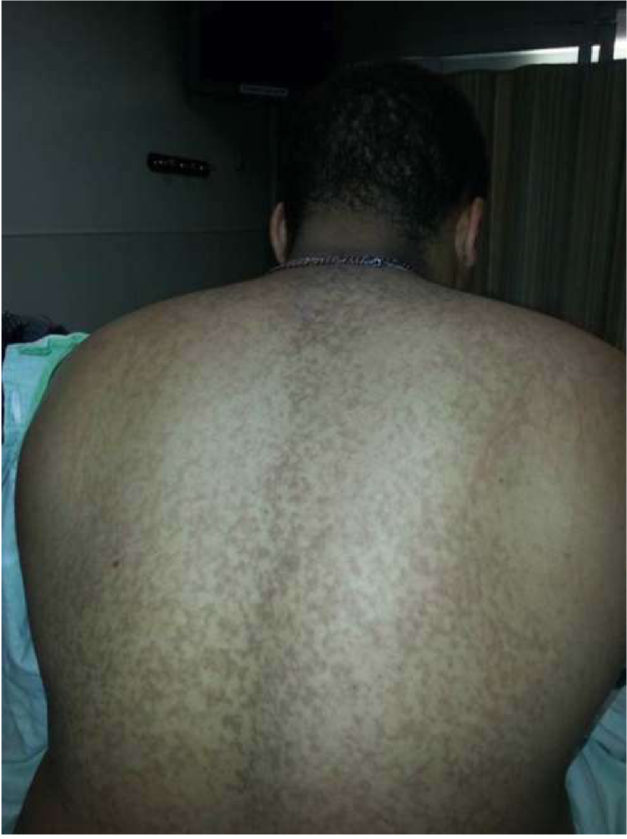
The match is very close.
2. The Scratch Sign (Besnier's Sign / Finger Nail Test)
This is the most practical bedside test - no equipment needed:
- Technique: Lightly scratch the surface of a lesion with a fingernail or the edge of a glass slide (or a tongue depressor)
- Positive result: A fine, powdery bran-like scale (furfuraceous scale) is easily lifted off the skin surface, like scratching dried paint
- Why it works: Malassezia infection makes the superficial stratum corneum fragile and loosely adherent, so minimal pressure reveals the scale that may not be visible at rest
- The scale is so fine it is sometimes described as "branny" - Dermatology 5th Edition (Tietz)
This is called the "scratch" or "nail sign" and is a classic clinical pearl for pityriasis versicolor. The scale is often invisible just looking at the skin, but becomes obvious the moment you scratch.
3. Wood's Lamp Examination (UV Light)
- Technique: Examine the skin in a darkened room under a Wood's lamp (365 nm ultraviolet light)
- Positive result: The patches show yellowish-orange or pale yellow fluorescence
- This is caused by tryptophan metabolites produced by Malassezia
- Confirmed by both Rosen's and the Tietz Textbook of Laboratory Medicine: "Wood's lamp: reddish or yellowish fluorescence in PV" - Tietz Textbook of Laboratory Medicine, 7th Edition
Important caveat: Wood's lamp fluorescence in pityriasis versicolor is not always present and is less reliable than in, say, tinea capitis. A negative Wood's lamp does not exclude the diagnosis.
Summary Table
| Method | Equipment | Finding | Reliability |
|---|---|---|---|
| Visual inspection | None | Reticulated, mottled macules on trunk | High in classic cases |
| Scratch sign (Besnier) | Fingernail only | Fine bran-like scale lifts off | High - very easy to do |
| Wood's lamp | UV lamp | Yellow-orange fluorescence | Moderate (not always positive) |
| KOH scraping (microscopy) | Microscope | "Spaghetti and meatballs" | Gold standard |
In practice: For a case like your patient's - young adult male, widespread reticulated brown patches on the entire back with itching - the combination of visual pattern + positive scratch sign gives very high diagnostic confidence without needing any lab equipment. The Wood's lamp adds further support if available.
This is a shared conversation. Sign in to Orris to start your own chat.