
Differentiated thyroid cancer
"differentiated thyroid cancer" AND management
differentiated thyroid cancer histology papillary follicular
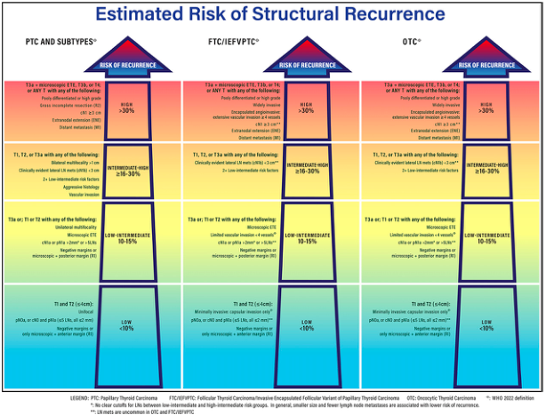
Summary : This figure presents a structured risk stratification chart for the estimated risk of structural recurrence in thyroid carcinoma, comparing three types: Papillary Thyroid Carcinoma (PTC) and subtypes, Follicular Thyroid Carcinoma/Invasive Encapsulated Follicular Variant of Papillary Thyroid Carcinoma (FTC/IEFVPTC), and Oncocytic Thyroid Carcinoma (OTC). The chart categorizes risk into four levels (High, Intermediate-High, Low-Intermediate, Low) based on specific clinical and pathological criteria. flowchart: # Risk Categories : • High (>30%) • Intermediate-High (≥16–30%) • Low-Intermediate (10–15%) • Low (<10%) # Cancer Types Compared : • PTC and Subtypes • FTC/IEFVPTC • OTC # Criteria for Each Risk Category : ## High (>30%) : • T3a + microscopic ETE, T3b, or T4; or ANY of the following: – Poorly differentiated or high grade – Gross incomplete resection (R2) – Encapsulated extension (EN) – Distant metastasis (M1) – Widely invasive (FTC/IEFVPTC, OTC) – Extensive vascular invasion (FTC/IEFVPTC, OTC) – Extrathyroidal extension (EN) (FTC/IEFVPTC, OTC) ## Intermediate-High (≥16–30%) : • T1, T2, or T3a with any of the following: – Clinically evident lateral LN mets (cN1b) or >5 pathologic LN mets (pN1b) – 2+ low-intermediate risk factors – Aggressive histology – Vascular invasion ## Low-Intermediate (10–15%) : • T3a or T2, T1 with any of the following: – Unilateral multifocality (>2 foci) – Limited vascular invasion (<5 vessels) (FTC/IEFVPTC, OTC) – cN1a or pN1a >5mm or >5 LNs – Microscopic + posterior margin (R1) – Microscopic ETE (OTC) ## Low (<10%) : • T1 and T2 (≤4cm): – Minimally invasive/capsular invasion only (FTC/IEFVPTC, OTC) – pN0/x or cN0 and pN1a (≤5 LNs, all ≤2mm) – Only microscopic + anterior margin (R1) # Colour Coding : • High risk: Red • Intermediate-High risk: Orange • Low-Intermediate risk: Yellow • Low risk: Green/Blue # Legend : • PTC: Papillary Thyroid Carcinoma • FTC/IEFVPTC: Follicular Thyroid Carcinoma/Invasive Encapsulated Follicular Variant of Papillary Thyroid Carcinoma • OTC: Oncocytic Thyroid Carcinoma • WHO 2022 definition • Notes on cutoffs and risk group distinctions # Layout : • Three vertical columns for each cancer type, each subdivided into four horizontal risk bands. • Each band lists specific clinical/pathological criteria for risk assignment. # Analysis : • The chart visually stratifies recurrence risk for three thyroid carcinoma types using consistent risk categories and criteria. • High risk is associated with advanced disease features (e.g., distant metastasis, extensive invasion). • Intermediate and low-intermediate risks are defined by combinations of nodal involvement, multifocality, and limited invasion. • Low risk is reserved for small, minimally invasive tumors with limited nodal involvement. • The structure allows for rapid comparison of risk factors and recurrence likelihood across carcinoma types.
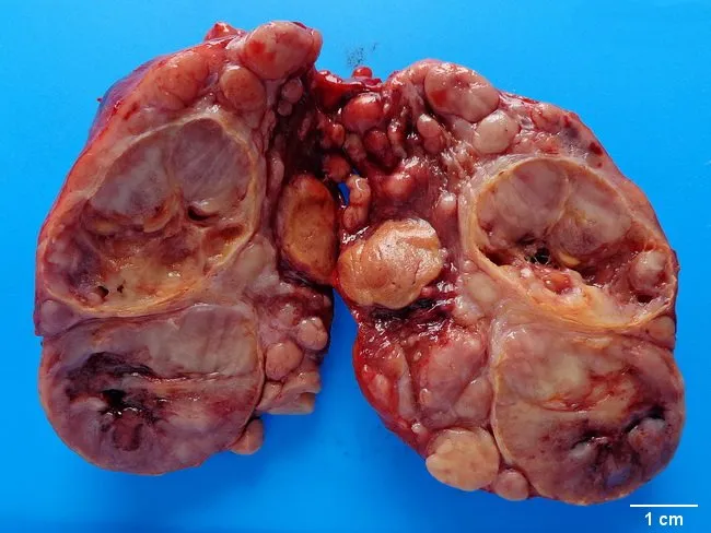
This gross pathology photograph documents a resected thyroid gland specimen, anterior view, with bilateral lobes separated for inspection. The background is a blue-friendly field and a 1 cm scale bar is present for size reference. The specimen displays multiple firm, tan to brown nodules replacing native thyroid parenchyma, with irregular contours and areas of marginal hemorrhage. The left and right lobes show expansion of the thyroid capsule, and prominent nodularity suggests follicular architecture disrupted by neoplastic growth. The caption indicates widely invasive follicular carcinoma of the thyroid, consistent with extrathyroidal extension and potential vascular invasion; gross features include invasion beyond the capsule and involvement of perithyroid soft tissue. No papillary nuclear features are assessable on gross inspection; histology would confirm follicular differentiation, capsular and vascular invasion, and tumor margins. Clinically, this specimen supports a diagnosis of aggressive differentiated thyroid carcinoma associated with higher risk of hematogenous metastasis. The imaging context is macroscopic pathology; the modality is gross photography used for documentation, education, and archival purposes. This image is relevant for surgical pathology education, thyroid cancer staging, case-based discussions, and radiologic-pathologic correlation exercises; useful for learners reviewing follicular thyroid carcinoma, aggressive variants, and the significance of extrathyroidal extension in prognosis.
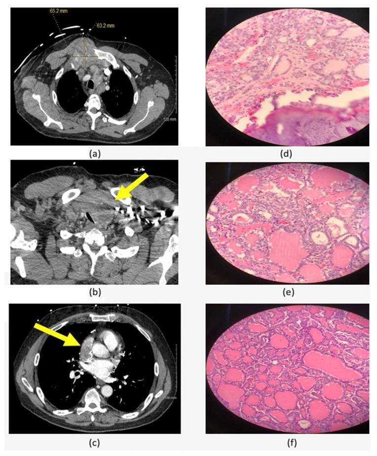
A multi-panel figure containing radiological and histopathological images of Follicular Variant of Papillary Thyroid Carcinoma (FVPTC) and its metastases. The left column displays axial CT scans: (a) shows a 6.5 cm soft tissue mass at the right manubrial costal junction with associated bone destruction; (b) illustrates a large left thyroid lobe mass causing significant tracheal compression and deviation; (c) demonstrates a 5.1 cm metastatic mass involving the superior vena cava and right atrium. The right column (d-f) presents H&E stained light microscopy images of resected tissues. Image (d) shows the chest wall metastasis, (e) shows the primary left thyroid tumor, and (f) shows the right atrial metastasis. All histological sections demonstrate characteristic features of FVPTC, including neoplastic follicular architecture with varying amounts of pink-stained colloid and cells with the nuclear features of papillary thyroid carcinoma. The composition illustrates the clinical progression and diagnostic findings of advanced differentiated thyroid cancer with rare cardiac and chest wall involvement.
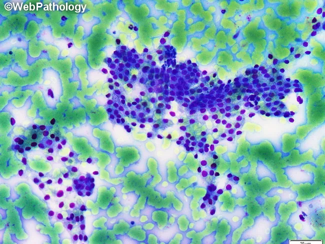
This is a light microscopy histology image of thyroid gland tissue prepared with a Romanowsky-type stain (e.g., Diff-Quik/Wright-Giemsa). The specimen shows normal-appearing follicular epithelium arranged in intact macrofollicles with abundant colloid occupying the luminal spaces. Follicular cells are evenly spaced with uniform, round to oval nuclei and scant to moderate cytoplasm, consistent with well-differentiated thyroid epithelium. The colloid background is pale blue and prominent, reflecting thyroglobulin-containing material released from follicles. The architectural pattern emphasizes large follicle units with a single layer of cuboidal to low-columnar epithelium surrounding expansive colloid pools. No prominent nuclear features of papillary carcinoma are evident, and there is no marked cellular atypia or mitotic activity in the visible fields. This image illustrates classic histology of benign thyroid tissue or colloid-rich goiter, useful for teaching follicular morphologies, thyroid folliculogenesis, and colloid dynamics. Clinically, such samples are evaluated in the context of thyroid nodules and goiter workups, with correlation to serum thyroid function tests and, when applicable, ultrasound or FNA findings. Potential educational uses include cytology-histology correlation, differential diagnosis of thyroid disease, and artifact recognition in Romanowsky-stained preparations. Images are suitable for medical education, pathology review, and database indexing. Correlative data improve student comprehension and diagnostic recall greatly.
Differentiated Thyroid Cancer (DTC)
Overview
- ~54,000 new cases/year in the United States; ~2,000 annual deaths
- Strong female predominance (3:1 female-to-male ratio)
- 10-year survival >90% for stage I/II; markedly reduced in stage IV with distant metastases
Types of DTC
1. Papillary Thyroid Carcinoma (PTC)
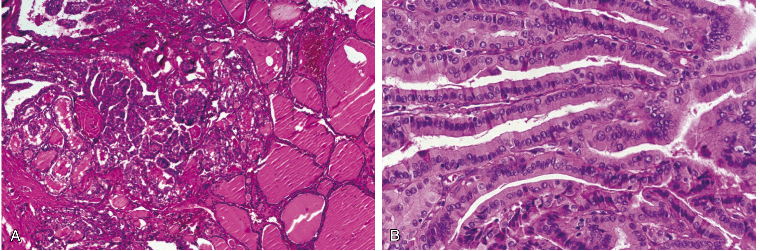
- Complex branching papillae with pseudoinclusions
- Nuclear grooves, nuclear clearing ("Orphan Annie eye" nuclei with powdery chromatin)
- Prominent nucleoli and psammoma bodies
- Multifocality is common; may invade locally and through the thyroid capsule
| Variant | Behavior |
|---|---|
| Classic | Indolent, excellent prognosis |
| Follicular variant (IEFVPTC) | Similar prognosis to classic PTC |
| NIFTP (noninvasive follicular thyroid neoplasm with papillary-like nuclear features) | No longer considered malignant; previously called encapsulated follicular variant without invasion |
| Tall cell, columnar cell, hobnail, diffuse sclerosing | Aggressive; require intensive therapy and close follow-up |
2. Follicular Thyroid Carcinoma (FTC)
- Minimally invasive (capsular invasion only): very low risk of metastasis; lobectomy suffices
- Angioinvasive FTC: more aggressive; poorer prognosis than PTC due to higher proportion presenting with stage IV disease
3. Oncocytic Carcinoma (OCA)
- New term in the 2022 WHO classification (replacing "Hürthle cell carcinoma")
- Defined as invasive malignant follicular cell neoplasm with ≥75% oncocytic cells
- Accounts for ~5% of DTCs; peak incidence in the sixth to seventh decades
- Metastases via both lymphatic and hematogenous routes; distant metastases in up to 20% at diagnosis
- 5-year overall survival ~85%; only 24% with distant metastases
- Less RAI-avid than other DTCs, complicating treatment of recurrent disease
- RAI may still improve survival in OCAs 2-4 cm in size
Key Histology Images
Diagnosis and Workup
- FNA (fine-needle aspiration): First-line for cytologic diagnosis of PTC (shows classic nuclear features). Cannot diagnose FTC (cannot detect capsular/vascular invasion).
- Ultrasound: All patients should have preoperative cervical ultrasound to evaluate central and lateral lymph node compartments for suspicious adenopathy (FNA any suspicious nodes).
- Molecular testing: BRAF V600E mutation (present in ~60% of PTCs) indicates higher recurrence risk; helps guide management when FNA is indeterminate.
- Thyroglobulin (Tg): Used for post-treatment surveillance.
- NIFTP: The encapsulated follicular variant without capsular or angioinvasion is now reclassified as NIFTP (a low-risk neoplasm, not carcinoma).
Staging (AJCC 8th Edition)
- Patients <55 years: Only stages I and II possible (no stage III/IV regardless of local invasion or nodal disease unless distant metastases present)
- Patients ≥55 years: Full staging I-IV applies
- All patients with distant metastases (M1): Stage IV regardless of age
Risk Stratification
| Risk Category | Recurrence Risk | Features |
|---|---|---|
| Low | <10% | T1-T2, intrathyroidal, pN0/pN1a (≤5 nodes, all ≤2 mm), no vascular invasion |
| Low-Intermediate | 10-15% | T2/T3a + unilateral multifocality, limited vascular invasion, cN1a or pN1a >5mm |
| Intermediate-High | 16-30% | Clinically evident lateral LN mets, >5 pathologic LN mets, aggressive histology, vascular invasion |
| High | >30% | T3b/T4, distant metastasis, poorly differentiated features, gross incomplete resection |
Treatment
Surgery
- Active surveillance: An option for small (≤1.5 cm) intrathyroidal micropapillary cancers without cervical LN mets, extrathyroidal extension, or proximity to the recurrent laryngeal nerve
- Lobectomy: Appropriate for low-risk intrathyroidal tumors >1 cm and <4 cm (T1b-T2) without metastases; protects contralateral RLN and parathyroids; avoids lifelong levothyroxine dependence in some patients
- Near-total/total thyroidectomy: Indicated for tumors >4 cm, metastases, extrathyroidal invasion, bilateral disease, or when RAI therapy is planned postoperatively; also required if aggressive pathology found after lobectomy (completion thyroidectomy)
- Therapeutic compartment-oriented neck dissection: Performed when metastatic nodes are confirmed preoperatively or intraoperatively
- Elective central neck dissection (level VI): Considered in patients at highest risk for occult central neck mets (e.g., extrathyroidal extension); not recommended routinely in low-risk patients
- Elective lateral neck dissection: Abandoned due to high morbidity and lack of survival benefit
TSH Suppression Therapy
| Risk Category | Target TSH |
|---|---|
| Low risk | 0.5-2.0 mIU/L (low-normal range) |
| Intermediate risk | 0.1-0.5 mIU/L |
| High risk | <0.1 mIU/L |
| Known metastatic disease | <0.1 mIU/L |
Radioactive Iodine (RAI / ¹³¹I) Therapy
- Remnant ablation - eliminates residual normal thyroid, enabling Tg monitoring
- Adjuvant therapy - treats microscopic residual disease
- Treatment of metastases - effective for iodine-avid distant metastases (lung/bone)
- High-risk patients: Near-universal use (gross extrathyroidal extension, incomplete resection, distant metastases)
- Intermediate-risk patients: Selectively used (aggressive histology, vascular invasion, lymph node disease)
- Low-risk patients: Generally not indicated; ATA guidelines have moved toward less aggressive use of RAI
Surveillance and Follow-Up
- Serum Tg: Primary tumor marker; should be undetectable after total thyroidectomy + RAI ablation. Rising Tg is the first sign of recurrence.
- Anti-Tg antibodies: Can interfere with Tg assays; must be monitored.
- Cervical ultrasound: Periodic assessment for locoregional recurrence.
- Whole-body RAI scan: Used in high-risk patients to detect iodine-avid residual/metastatic disease.
- Stimulated Tg: Under rhTSH stimulation - used in high-risk patients to increase sensitivity for detecting residual disease.
Advanced/Refractory Disease
- Kinase inhibitors:
- Sorafenib and Lenvatinib - approved multikinase inhibitors for progressive radioiodine-refractory DTC
- Lenvatinib showed significant PFS benefit in the SELECT trial
- BRAF/MEK inhibitors: For BRAF V600E-mutant aggressive DTC (e.g., dabrafenib + trametinib)
- External beam radiotherapy: For locally unresectable disease
- Redifferentiation therapy: Experimental use of MEK or BRAF inhibitors to restore RAI uptake in refractory tumors
Management During Special Circumstances
- Pregnancy: DTC diagnosed during pregnancy generally has a favorable prognosis. Surgery in the 2nd trimester is preferred if required; for small, stable tumors, active surveillance until postpartum is acceptable. RAI is absolutely contraindicated in pregnancy.
- Microcarcinoma (≤1 cm PTC): Active surveillance protocols (without immediate surgery) have shown favorable outcomes in selected patients in Japanese studies.
Prognosis Summary
| Feature | Favorable | Unfavorable |
|---|---|---|
| Age | <55 years | ≥55 years |
| Tumor size | ≤4 cm | >4 cm |
| Invasion | Intrathyroidal | Extra-thyroidal / vascular |
| Nodes | N0 or minimal micrometastases | Multiple large nodes, cN1b |
| Metastases | None | Distant (bone/lung/CNS) |
| Histology | Classic PTC, minimal FTC | Tall cell, hobnail, OCA, widely invasive FTC |
- Harrison's Principles of Internal Medicine 22E (2025), Chapter 397, pp. 3089-3091
- Sabiston Textbook of Surgery, Chapter 73, pp. 1508-1515
- Bailey and Love's Short Practice of Surgery 28th Edition, Chapter on Thyroid Surgery, pp. 6015-6082
- Schwartz's Principles of Surgery 11th Edition, Postoperative Management of DTC