
femoral artery
Reading File
The femoral artery is the main arterial supply to the lower limb, arising as the continuation of the external iliac artery below the inguinal ligament. It runs through the femoral triangle and adductor canal before becoming the popliteal artery.
Searching sources...
Finding Sources
Finding Sources
Good results from multiple authoritative sources. Let me read the key sections from Gray's Anatomy, the Imaging Anatomy atlas, and the Color Atlas in parallel.
Reading File
Reading File
Reading File
Excellent content. Now let me also read the Gray's Anatomy section on the femoral triangle in detail, and fetch the key anatomy diagram.
Reading File
Loading Image
Loading Image
Now let me get the Gray's Anatomy femoral artery diagram and also the medial/lateral circumflex branches:
Loading Image
Reading File
I have comprehensive content from multiple authoritative sources. Here is the full, structured answer.
Femoral Artery
Origin and Proximal Course
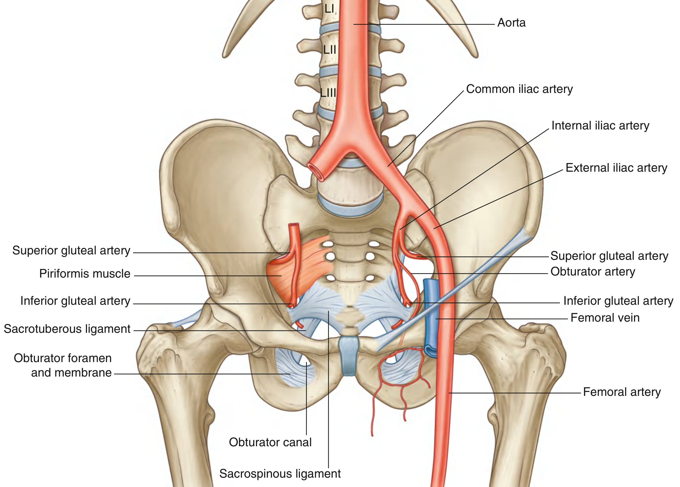
The femoral artery is the direct continuation of the external iliac artery. It begins where the external iliac artery passes posterior to the inguinal ligament to enter the femoral triangle on the anterior aspect of the upper thigh. It is palpable just inferior to the inguinal ligament, midway between the anterior superior iliac spine (ASIS) and the pubic symphysis - the mid-inguinal point.
The common femoral artery is typically 5-7 cm long and 6-9 mm in diameter. It travels within the femoral sheath, where it lies lateral to the femoral vein. The femoral nerve lies lateral to the artery but outside the femoral sheath.
- Gray's Anatomy for Students, p. 703
- Imaging Anatomy, Vol. 3, p. 359
Course Through the Thigh
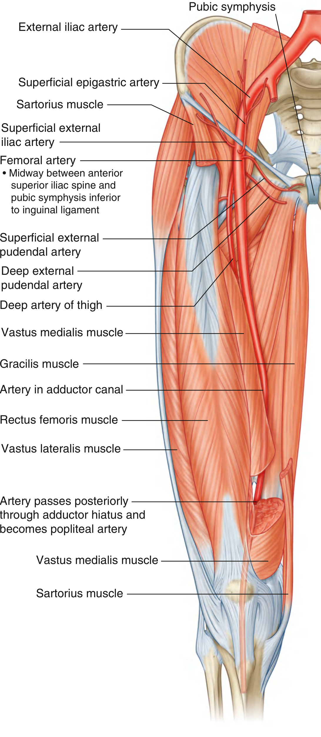
From the femoral triangle, the femoral artery passes vertically downward and then enters the adductor canal (Hunter's canal) - a musculoaponeurotic tunnel deep to the sartorius muscle. It exits the canal by passing through the adductor hiatus in the adductor magnus muscle, at which point it becomes the popliteal artery behind the knee.
Branches
Small superficial branches (given off in the femoral triangle)
These arise just below the inguinal ligament:
- Superficial epigastric artery - ascends to the anterior abdominal wall toward the umbilicus
- Superficial circumflex iliac artery - runs laterally toward the ASIS
- Superficial external pudendal artery - passes medially to the perineum
- Deep external pudendal artery - also passes medially to the perineum
Deep artery of the thigh (Profunda femoris)
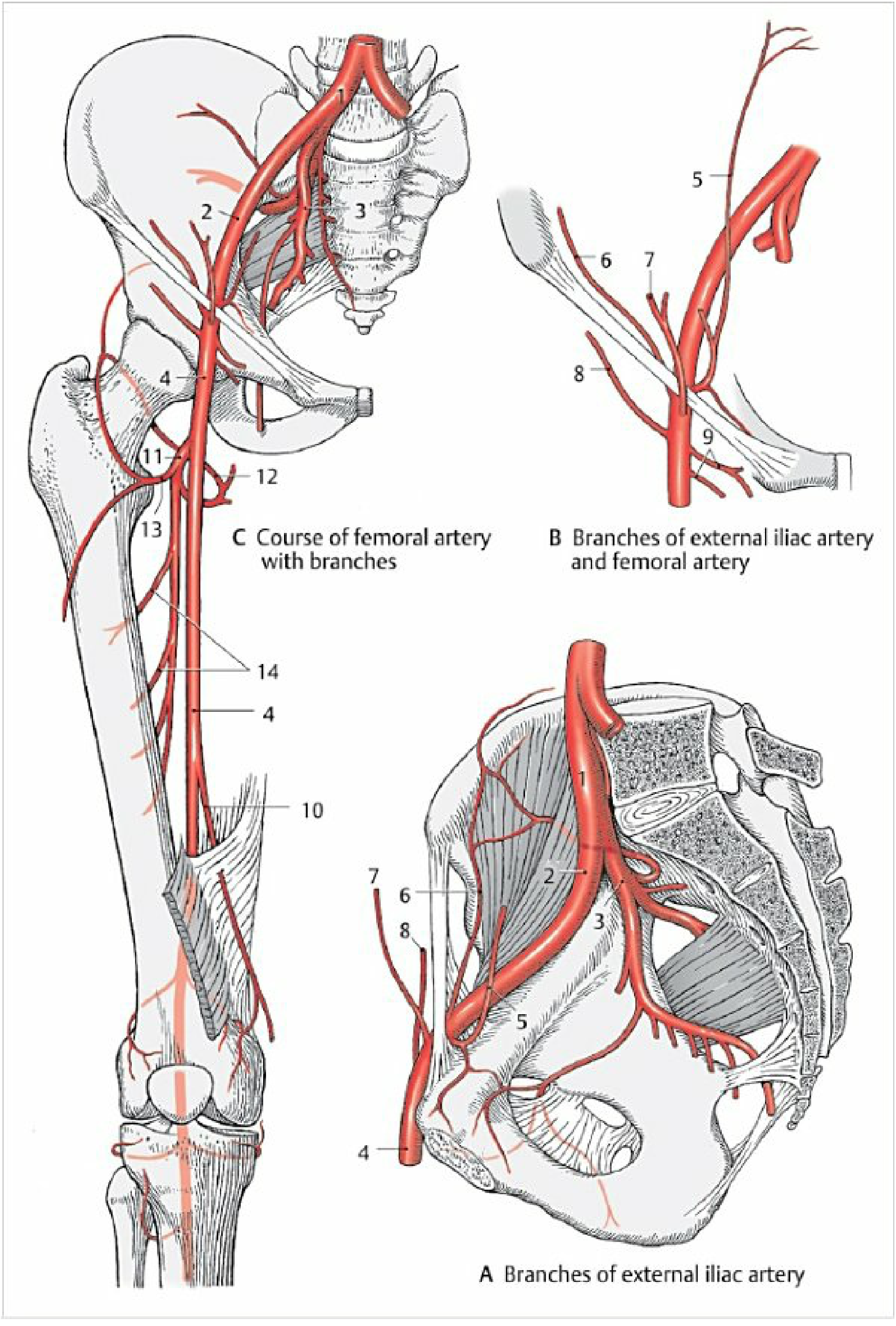
The largest and most important branch. It arises from the lateral side of the femoral artery in the femoral triangle, approximately 3-6 cm below the inguinal ligament. It passes posteriorly between pectineus and adductor longus, then between adductor longus and adductor brevis, eventually piercing adductor magnus to anastomose with popliteal branches.
Its main branches:
| Branch | Course | Supply |
|---|---|---|
| Lateral circumflex femoral a. | Passes deep to sartorius and rectus femoris; divides into ascending, descending, transverse branches | Vastus lateralis, neck/head of femur, cruciate anastomosis |
| Medial circumflex femoral a. | Passes posteromedially between pectineus/iliopsoas, then between obturator externus/adductor brevis | Hip joint, head/neck of femur |
| 1st, 2nd, 3rd perforating arteries | Pierce adductor magnus near linea aspera | Posterior compartment of thigh |
Descending genicular artery
Given off just before the femoral artery passes through the adductor hiatus. Divides into:
-
Saphenous branch - descends to the leg
-
Articular branch - contributes to the genicular anastomosis around the knee
-
Color Atlas of Human Anatomy, Vol. 2, p. 117-118
Relations in the Femoral Triangle
The femoral triangle (Scarpa's triangle) is bounded by:
- Superiorly: inguinal ligament
- Medially: adductor longus
- Laterally: sartorius
From lateral to medial within the triangle, the order is:
Nerve - Artery - Vein (NAVe)
- Femoral nerve (lateral, outside sheath)
- Femoral artery
- Femoral vein
- Femoral canal (most medial, inside sheath)
Collateral Circulation
Branches of the femoral, inferior gluteal, superior gluteal, and obturator arteries interconnect to form an anastomotic network in the upper thigh and gluteal region. The cruciate anastomosis around the hip (formed by the lateral and medial circumflex femoral arteries, the inferior gluteal artery, and the first perforating artery) is particularly important when proximal femoral artery occlusion occurs.
- Gray's Anatomy for Students, p. 669
Clinical Relevance
-
Pulse palpation: The femoral pulse is the most accessible pulse in the lower limb, felt at the mid-inguinal point. It is a key assessment point in trauma and cardiovascular examination.
-
Arterial access: Because it lies superficially just below the inguinal ligament, the femoral artery is the standard access point for cardiac catheterization, coronary angiography, and peripheral vascular interventions. A catheter introduced here can be advanced into the great vessels and left heart.
-
Compression for hemorrhage: In emergencies, the femoral artery can be compressed against the pelvic rim to control life-threatening lower limb hemorrhage.
-
Femoral artery aneurysm: The most common site for isolated femoral artery aneurysm is the common femoral artery. These are often associated with popliteal and aortoiliac aneurysms and can present with pulsatile groin masses, thrombosis, or distal embolization.
-
Superficial femoral artery (SFA) disease: The SFA (now more precisely termed the femoral artery proper, distal to the profunda origin) is the most common site for peripheral arterial disease causing claudication. Its course through Hunter's canal makes it prone to repetitive mechanical compression.
- Roberts and Hedges' Clinical Procedures in Emergency Medicine, p. 5 (femoral access)
- Color Atlas of Human Anatomy, Vol. 2, p. 118
Nomenclature Note
Historically the term "superficial femoral artery" was used for the main femoral trunk distal to the profunda origin. This term has fallen out of favor because it misleadingly suggests the vessel is not a "deep" structure - it is, in fact, entirely intramuscular through the adductor canal. The preferred anatomical terminology is simply "femoral artery" for the main trunk, with "common femoral artery" used for the proximal segment before the profunda bifurcation in clinical/radiological settings.
- Imaging Anatomy, Vol. 3, p. 360 (variant anatomy discussion)
This is a shared conversation. Sign in to Orris to start your own chat.