
Classification criteria, Etiopathogenesis, clinical features, various presentations, pathology, diagnosis and Management of IgG4 Related Disease. Describe the Histopathological, changes, Explain IgG4 related disease responder index. detailed from rheumatology textbooks and latest journals
Classification criteria, Etiopathogenesis, clinical features, various presentations, pathology, diagnosis and Management of IgG4 Related Disease. Describe the Histopathological, changes, Explain IgG4 related disease responder index. detailed from rheumatology textbooks and latest journals
"IgG4-related disease"[MeSH Terms] AND classification
IgG4 related disease 2024 2025 classification criteria responder index management guidelines
"IgG4-related disease" AND "responder index"
PMID: 32939060
PMID: 22611406
"IgG4-related disease" AND (treatment OR management OR rituximab)
PMID: 39541094
PMID: 40195520
IgG4-Related Disease (IgG4-RD): A Comprehensive Review
1. Overview and Definition
2. Classification Criteria
2019 ACR/EULAR Classification Criteria for IgG4-RD
Step 1 - Entry Criteria (Yes/No)
Step 2 - Exclusion Criteria (any one present = excluded)
| Domain | Items |
|---|---|
| Clinical | Fever; no objective response to glucocorticoids |
| Serologic | Leukopenia/thrombocytopenia unexplained; ANCA (anti-PR3 or anti-MPO); anti-SSA/Ro or SSB/La; anti-dsDNA/RNP/Sm; cryoglobulinemia |
| Radiologic | Suspicious malignancy/infection not investigated; rapid radiologic progression; long bone abnormalities (Erdheim-Chester); splenomegaly |
| Pathologic | Malignant cellular infiltrates; inflammatory myofibroblastic tumor markers; prominent neutrophilic inflammation; necrotizing vasculitis; prominent necrosis; granulomatous inflammation; macrophage/histiocytic disorder |
| Known diagnoses | Multicentric Castleman disease; Crohn's/UC (if only pancreatobiliary); Hashimoto's thyroiditis (if only thyroid) |
Step 3 - Inclusion Weighted Scoring System
| Domain/Item | Points |
|---|---|
| Histopathology | |
| Uninformative biopsy | 0 |
| Dense lymphoplasmacytic infiltrate | +4 |
| Dense lymphoplasmacytic infiltrate + obliterative phlebitis | +6 |
| Dense lymphoplasmacytic infiltrate + storiform fibrosis ± obliterative phlebitis | +13 |
| Immunostaining (IgG4+:IgG+ ratio and IgG4+ cells/hpf) | 0-16 |
| Ratio 0-40% AND <10 cells/hpf | 0 |
| Ratio ≥41% with <10 cells/hpf OR ratio 0-40% with ≥10 cells/hpf | +7 |
| Ratio 41-70% with ≥10 cells/hpf OR ratio >70% with <10 cells/hpf | +14 |
| Ratio >70% with ≥10 cells/hpf | +16 |
| Serum IgG4 Concentration | |
| Normal | 0 |
| >Normal but <2× ULN | +4 |
| 2-5× ULN | +6 |
| ≥5× ULN | +11 |
| Bilateral Lacrimal/Parotid/Sublingual/Submandibular Glands | |
| No glands involved | 0 |
| One set involved | +6 |
| ≥2 sets involved | +14 |
| Chest/Thoracic Aorta | |
| Peribronchovascular/septal thickening | +4 |
| Paravertebral band-like soft tissue | +10 |
| Pancreas and Biliary Tree | |
| Diffuse pancreas enlargement | +8 |
| Diffuse enlargement + capsule-like rim | +11 |
| Pancreas (either) + biliary involvement | +19 |
| Kidney | |
| Hypocomplementemia | +6 |
| Renal pelvis thickening | +8 |
| Bilateral renal cortex low-density areas | +10 |
| Retroperitoneum | |
| Diffuse aortic wall thickening | +4 |
| Circumferential/anterolateral soft tissue around infrarenal aorta/iliac arteries | +8 |
Classification threshold: Score ≥20 = IgG4-RD
3. Etiopathogenesis
3.1 The IgG4 Molecule Itself
- Weak binding to C1q and Fc-γ receptors (CH2 domain differences) - poor complement activation
- Low participation in antibody-dependent cell-mediated cytotoxicity
- Unique ability to undergo "Fab arm exchange" - forming bispecific "half-antibodies" incapable of cross-linking antigens and forming immune complexes
- IgG4 deposition in kidneys suggests possible immune complex injury in some organs, but this is not the dominant mechanism
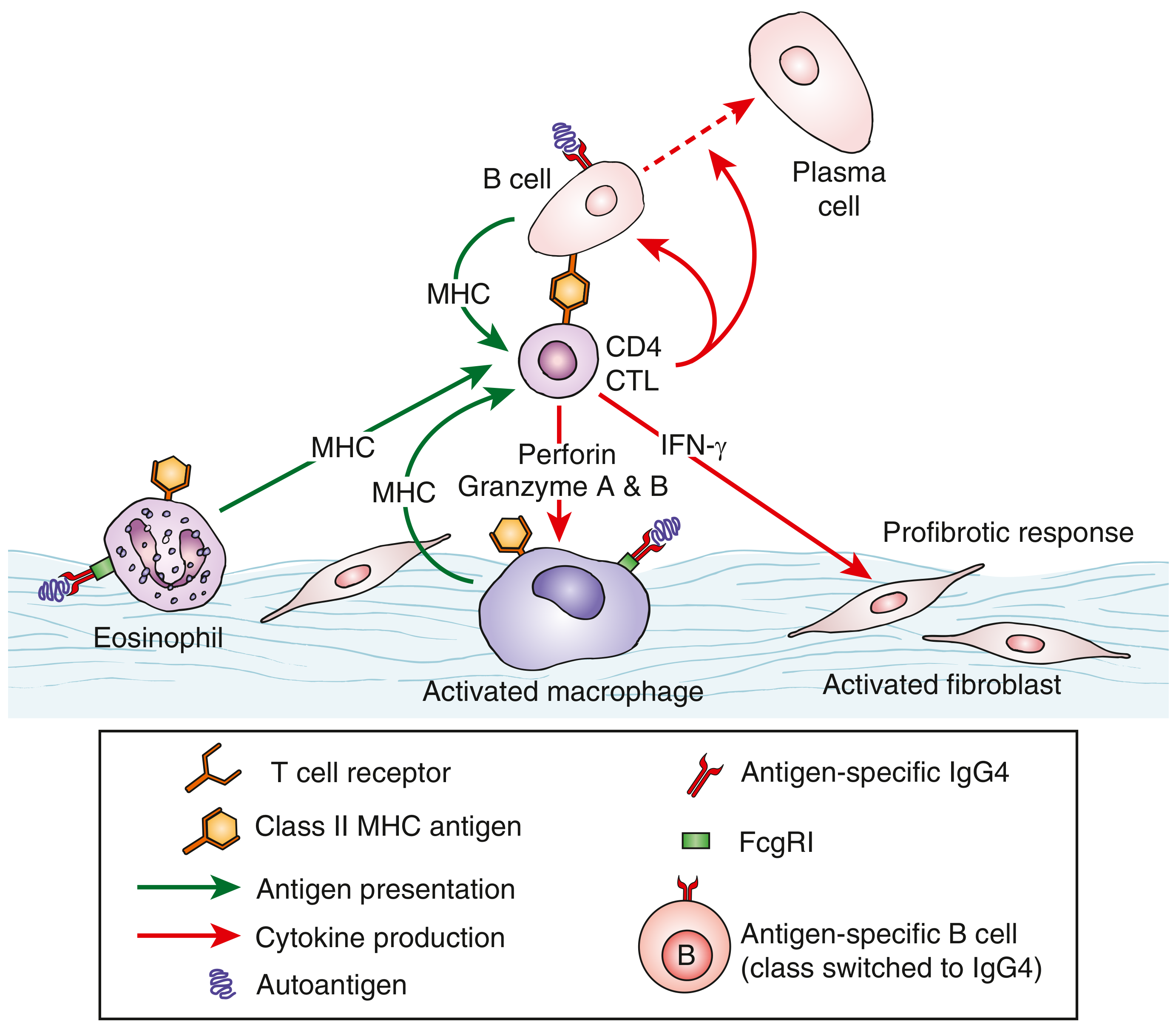
3.2 CD4+ Cytotoxic T Lymphocytes (CTLs) - Central Effectors
- Secrete perforin, granzyme A and B - directly injure target tissues
- Release IFN-γ, IL-1β, TGF-β1 - activate macrophages and fibroblasts
- Drive the profibrotic response by activating myofibroblasts
- Are found concentrated at sites of disease in tissue biopsies
- Decline after B cell depletion (confirming B cell-CTL interaction)
3.3 B Cells and Plasmablasts
- Markedly expanded plasmablast populations circulate in active disease (useful biomarker)
- B cells act as antigen-presenting cells to CD4+ CTLs via MHC class II
- A specific subset of T follicular helper (Tfh) cells drives IgG4 class switching in germinal centers
- Multiple self-antigens have been identified that drive B cell expansion (e.g., laminin-511, prohibitin, galectin-3, annexin A11, HSP70 in specific organ manifestations)
- Depletion of B cells (rituximab, inebilizumab) produces rapid, robust clinical responses
3.4 Macrophages and Fibroblasts
- Activated macrophages (M2 polarization) and activated myofibroblasts amplify TGF-β1-driven fibrosis
- B cell depletion reduces infiltrating activated myofibroblasts, thereby decreasing fibrosis
3.5 Th2 Skewing and Atopy
- Peripheral eosinophilia and elevated serum IgE (sometimes >10× ULN) are common
- These are partially independent of pre-existing atopy - IgG4-RD itself contributes to eosinophilia/IgE elevation
- Circulating Th2 memory cells found only in patients with pre-existing atopy histories
3.6 Genetic Factors
- HLA associations (HLA-DRB1 alleles) have been reported, particularly in Japanese populations with autoimmune pancreatitis
- No single causative autoantigen or gene has been definitively identified
4. Clinical Features and Organ Presentations
4.1 General Features
- Insidious onset, often without systemic symptoms (fever, weight loss are uncommon - their presence should prompt exclusion)
- Organs swell progressively, mimicking malignancy or infection
- Most patients are middle-aged to elderly men
- Multiple-organ involvement in 60-70% at diagnosis
4.2 Proliferative vs. Fibrotic Phenotypes
| Feature | Proliferative Subtype | Fibrotic Subtype |
|---|---|---|
| Origin | Glandular/epithelial tissues | Extraglandular; body regions (retroperitoneum, mediastinum) |
| Atopy | High frequency | Low frequency |
| Organ involvement | Commonly multiorgan | Commonly single-organ/body region |
| Typical organs | Lymph nodes, lacrimal, salivary, pancreas, bile ducts, kidney, lung, pituitary | Retroperitoneum, aorta, mesentery, mediastinum, pachymeninges, Riedel's thyroid |
| Serum IgG4 | High | Normal or slightly elevated |
| IgE level | High | Normal |
| Eosinophilia | Common | Unusual |
| Hypocomplementemia | Common | Unusual |
| IgG4+ cells/hpf | Dense, >50-100 | Can be sparse |
| Obliterative phlebitis | Occasional | Common |
| Treatment response | Excellent | Less robust but good if early |
4.3 Organ-Specific Presentations
Pancreas - Autoimmune Pancreatitis (AIP) Type 1
- Most recognized manifestation; presents with obstructive jaundice (painless), new-onset diabetes, weight loss
- "Sausage-shaped" pancreas on imaging; capsule-like hypodense rim
- Must be distinguished from pancreatic adenocarcinoma (the most dangerous mimicker)
Salivary and Lacrimal Glands - Mikulicz Disease / IgG4-Related Dacryoadenitis and Sialadenitis
- Bilateral, painless, firm swelling of submandibular glands (predominantly); parotid and lacrimal glands also affected
- Distinguished from Sjögren syndrome: SSA/Ro negative, milder xerostomia, bilateral submandibular >parotid
- Male-to-female ratio ~1:1 (unusual for IgG4-RD overall)
Orbits - IgG4-Related Ophthalmic Disease
- Dacryoadenitis: enlargement of lacrimal gland causing superolateral periorbital swelling
- Orbital pseudotumor, extraocular muscle swelling, infra/supraorbital nerve swelling
- Exophthalmos in retrobulbar mass
- Rare: IgG4-related optic neuropathy (decreased visual acuity)
- Main differential: MALT lymphoma, Graves' disease, GPA, sarcoidosis
Biliary Tree - IgG4-Related Sclerosing Cholangitis (IgG4-SC)
- Frequently co-occurs with type 1 AIP
- Causes biliary strictures; must be distinguished from PSC and cholangiocarcinoma
- Transmural fibrosis with obliterative phlebitis and dense IgG4+ plasma cell infiltration
- Criteria: >10 IgG4+ cells/hpf (biopsy), >50/hpf (resection), IgG4+/IgG+ ratio >40%
Kidney - IgG4-Related Kidney Disease
- Most common: tubulointerstitial nephritis (TIN) - bilateral wedge-shaped or round hypodense cortical lesions on CT
- Less common: membranous glomerulonephritis (may present as nephrotic syndrome)
- Hypocomplementemia (low C3/C4) is characteristic
- May cause chronic renal insufficiency if untreated
Retroperitoneum - Retroperitoneal Fibrosis (RPF)
- Encasing periaortic fibrosis, often extends to ureters causing hydronephrosis
- Can cause ureteral obstruction and renal failure
- Hard to distinguish from idiopathic RPF (many cases of "idiopathic" RPF are IgG4-RD)
Aorta and Large Vessels - IgG4-Related Periaortitis/Aortitis
- Diffuse wall thickening of abdominal aorta; aneurysms
- Coronary artery involvement: wall thickening, stenosis, aneurysms, coronary ectasia - unique among systemic vasculitides affecting adults
- Periaortitis (abdominal) frequently accompanies RPF
Lungs - IgG4-Related Lung Disease
- Multiple patterns: nodules, consolidation, ground-glass opacities, peribronchovascular/septal thickening
- Paravertebral band-like soft tissue in thorax is a characteristic CT finding
- Differential: malignancy, sarcoidosis, Castleman disease, inflammatory myofibroblastic tumor
Meninges - IgG4-Related Hypertrophic Pachymeningitis
- Dura mater thickening; can cause headache, cranial nerve palsies, spinal cord compression
- Differential: GPA, sarcoidosis, lymphoma, inflammatory myofibroblastic tumor
Pituitary / Thyroid / Other
- IgG4-related hypophysitis: pituitary mass, hormonal deficiencies
- Riedel's thyroiditis: rock-hard thyroid, cervical structures compression, typically fibrotic subtype
- Prostate, skin (erythematous papules head/neck), breast, mesentery, mediastinum also reported
Lymphadenopathy
- Often asymptomatic, accompanies other organ involvement
- Usually responds well to corticosteroids
- Must rule out lymphoma (Castleman disease is a key mimic)
4.4 IgG4-RD as Variable-Vessel Vasculitis
- Large: aorta and its branches (periaortitis, aortitis)
- Medium: coronary arteries (unique adult vasculitic coronary involvement)
- Small: post-capillary venules (obliterative phlebitis - histologic hallmark)
5. Pathology - Histopathological Changes
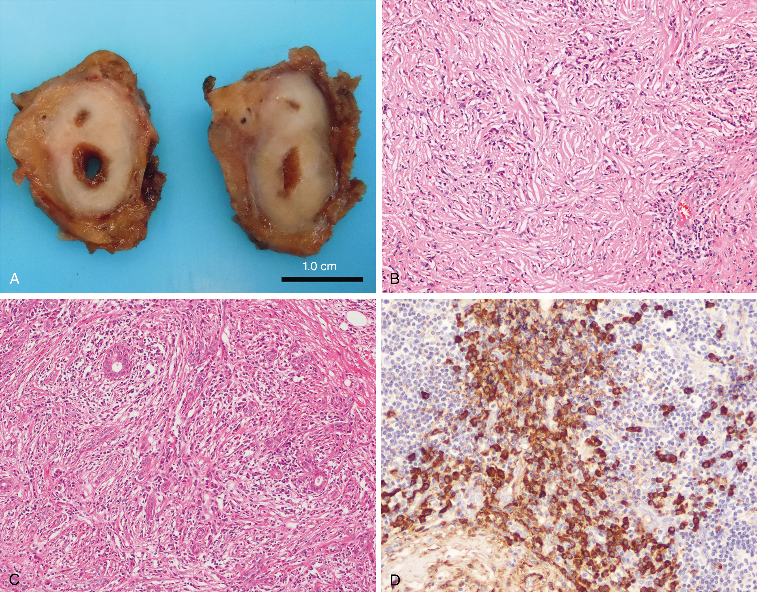
5.1 Three Cardinal Histopathological Features
5.1.1 Dense Lymphoplasmacytic Infiltrate
- Prominent lymphoplasmacytic infiltrate rich in plasma cells and lymphocytes (particularly T cells)
- The plasma cells produce predominantly IgG4 antibodies
- Germinal centers with lymphoid follicles common in the proliferative subtype
- Eosinophils present in ~50% of cases
5.1.2 Storiform Fibrosis
- The fibrosis has an irregular, whorled ("cartwheel") pattern resembling a storiform arrangement
- Fibroblasts and myofibroblasts are embedded in the infiltrate
- The fibrosis is progressive and can completely replace normal tissue architecture
- More prominent in fibrotic subtype; may be absent in early disease
5.1.3 Obliterative Phlebitis
- Inflammatory infiltrate obliterates the lumen of veins (post-capillary venules and small veins)
- Vessel wall is destroyed; lumen occluded by inflammatory cells and fibrosis
- Found in the majority of cases; more prominent in fibrotic subtype
- Obliterative arteritis also occurs in some organs (especially lung)
5.2 Immunohistochemistry (IHC)
- IgG4+ plasma cell count: Critical metric
- >10 IgG4+ plasma cells/hpf in small biopsies
- >50 IgG4+ plasma cells/hpf in large resection specimens (highly suggestive)
- Some authors use >30/hpf as threshold for glandular tissue
- IgG4+/IgG+ plasma cell ratio >40% (most important ratio; >40% = supportive, the 2019 criteria weight >70% most heavily)
- Neither the count alone nor the ratio alone is diagnostic - requires clinicopathologic correlation
5.3 Additional Microscopic Findings
- Eosinophilic infiltrate: ~50% of biopsies
- Absence of features that argue against IgG4-RD: necrotizing vasculitis, prominent necrosis, granulomas, neutrophilic infiltration, macrophage/histiocytic proliferations
- Ductal organs show pipe-stem appearance with circumferential wall thickening
- Parenchymal organs (pancreas, kidney) show diffuse enlargement
5.4 Fibrotic Lesions - A Special Consideration
- As few as 10 IgG4+ cells/hpf may be regarded as supportive if clinical context is right
- The IgG4+/IgG+ ratio >40% retains importance even with low absolute counts
- Storiform fibrosis pattern becomes the dominant diagnostic clue
6. Diagnosis
6.1 Serum IgG4 Concentration
- Elevated serum IgG4 is supportive but neither sensitive nor specific
- Sensitivity of elevated serum IgG4 in biopsy-proven IgG4-RD: only ~50-60% when cases identified histologically; ~90% when cases identified by lab query
- Specificity issue: 27% of patients with serum IgG4 ≥5× ULN do NOT have IgG4-RD
- Prozone effect: In patients with dramatically elevated IgG4 (e.g., 4-5 g/dL), nephelometry may give spuriously low readings - additional dilutions required
- Serum IgG4 is most useful as a disease activity marker and for monitoring treatment response (falls with glucocorticoid treatment)
- Other IgG subclasses (IgG1, IgG2, IgG3) are often co-elevated but IgG4 elevation is usually disproportionate
6.2 Other Laboratory Findings
- Plasmablasts (CD19+CD38+CD20-CD27+): elevated circulating plasmablasts are a sensitive biomarker of active disease, more reliable than serum IgG4 in some studies
- Eosinophilia: common, especially in proliferative subtype
- Hypocomplementemia (low C3, C4): particularly with renal involvement, may suggest immune complex deposition
- Elevated IgE: may exceed 10× ULN
- ESR/CRP: often normal or mildly elevated (not reliable markers)
- ANA, ANCA, RF: negative (positive results should prompt alternative diagnosis)
6.3 Imaging
- CT/MRI: identifies mass lesions, diffuse organ enlargement, characteristic patterns (sausage pancreas, bilateral renal cortex lesions, periaortic soft tissue, RPF)
- 18F-FDG PET/CT: maps multiorgan involvement, distinguishes active inflammatory lesions from established fibrosis, guides biopsy, monitors treatment response. Active IgG4-RD shows increased FDG uptake
- MRCP: demonstrates biliary strictures in IgG4-SC
- Endoscopic ultrasound: used for pancreatic/biliary biopsy
6.4 Histopathology/Biopsy
- Biopsy with IHC remains the gold standard for definitive confirmation
- Adequate specimen size is critical (small endoscopic biopsies may be non-diagnostic)
- Core needle biopsy often sufficient for accessible lesions
- Lacrimal gland, minor salivary gland, and submandibular gland biopsies are often the most accessible with high diagnostic yield
6.5 Integrated Diagnostic Approach
- Clinical presentation + appropriate organ involvement
- Exclude malignancy, infection, and alternative autoimmune diagnoses
- Serum IgG4 + other labs
- Cross-sectional and functional imaging
- Tissue biopsy with IHC (in most cases)
- Apply ACR/EULAR classification criteria (score ≥20)
- Response to glucocorticoid trial can support diagnosis (lack of response argues strongly against)
7. The IgG4-RD Responder Index (IgG4-RD RI)
7.1 Background and Development
- IgG4-RD is a multiorgan disease followed by multiple specialties
- Divergent approaches to assessment complicated interpretation of studies
- No validated outcome measure existed for disease activity and treatment response
7.2 Structure of the IgG4-RD RI
-
Lists all organ systems that can be involved in IgG4-RD (24 organ domains including: orbital/periorbital, salivary glands, lymph nodes, thyroid, lungs, aorta, pericardium, pancreas, biliary tree, kidneys, retroperitoneum, pachymeninges, skin, etc.)
-
For each organ system, the physician rates disease activity on a three-level scale:
- 2 = Active disease, new or worsening (within the past 30 days)
- 1 = Improved but still active (compared to prior assessment)
- 0 = No activity at that site
-
Physician Global Assessment (PGA): An overall 0-10 visual analog scale is included as a cross-check
-
Damage items: The index distinguishes between "disease activity" and "disease damage" - damage (irreversible changes due to scarring/fibrosis, scored separately) does not fluctuate with treatment
7.3 Scoring and Interpretation
- Total IgG4-RD RI score = sum of scores across all active organ domains
- Higher score = more active, more widespread disease
- Complete remission is defined as an IgG4-RD RI score of 0 (no active disease in any organ)
- Flare is typically defined as recurrence of disease activity (increase in score) after a period of remission
- In clinical trials (e.g., the MITIGATE rituximab and inebilizumab trials), the IgG4-RD RI served as the primary or key secondary efficacy outcome
- In the ACR Convergence 2025 data, the mean Responder Index in an active disease cohort was 6.09 (CI 4.63-7.55)
- Serum IgG4 and IL-5 levels correlated most significantly with the Responder Index score
7.4 International Validation
7.5 IgG4-RD Damage Index
8. Management
8.1 Indications for Treatment
- Active, symptomatic IgG4-RD in any organ
- Asymptomatic but progressive disease in vital organs (pancreas, kidney, biliary tree, aorta, meninges)
- Disease threatening organ function even if clinically silent
8.2 Remission Induction - Glucocorticoids
- Starting dose: prednisone 0.6 mg/kg/day (typically 30-40 mg/day)
- Maintained for 2-4 weeks, then tapered
- Goal: complete cessation within 10-16 weeks (some Asian centers maintain 5-10 mg/day for years)
- Response is typically swift (within days to weeks), especially in proliferative subtype
- Failure to respond = reconsider diagnosis
- Limitation: responses are often temporary; relapse is frequent, especially in multiorgan disease with high baseline IgG4
- Typical patient demographics (elderly, often diabetic, hypertensive, obese, osteoporotic)
- Pancreatic IgG4-RD itself causes glucose intolerance
8.3 Conventional DMARDs (Steroid-Sparing)
- Mycophenolate mofetil
- Azathioprine
- Methotrexate
- 6-mercaptopurine
8.4 Biologic Agents
Anti-CD20 (Rituximab) - Anti-B Cell
- Rituximab (two 1-gram IV doses 2 weeks apart) produced significant clinical benefit in the landmark prospective open-label trial (Carruthers et al., 2015, Ann Rheum Dis [PMID: 25667206])
- 29 of 30 patients showed efficacy; 77% achieved remission off prednisone at 6 months
- Relapses common by 12 months - retreatment required
- Serum IgG4 monitoring guides retreatment timing
- Mechanism: B cell depletion interrupts antigen presentation to CD4+ CTLs; reduces myofibroblast activation
Anti-CD19 (Inebilizumab) - The MITIGATE Trial - NEJM 2025
- Design: Double-blind, randomized, placebo-controlled, 52-week trial
- n = 135 patients; inebilizumab (300 mg IV on days 1, 15, week 26) vs. placebo; both groups received identical glucocorticoid tapers
- Primary endpoint: Time to first treated, adjudicated flare
| Outcome | Inebilizumab | Placebo | Statistic |
|---|---|---|---|
| Patients with ≥1 flare | 10% (7/68) | 60% (40/67) | HR 0.13 (95% CI 0.06-0.28), P<0.001 |
| Annualized flare rate | -- | -- | Rate ratio 0.14 (P<0.001) |
| Flare-free, treatment-free complete remission | Higher | Lower | OR 4.68, P<0.001 |
| Flare-free, glucocorticoid-free complete remission | Higher | Lower | OR 4.96, P<0.001 |
- Inebilizumab depletes CD19+ B cells (broader B cell depletion than rituximab, which targets CD20; CD19 is expressed on plasmablasts and plasma cell precursors not targeted by CD20)
- This trial confirms CD19-targeted B cell depletion as a major treatment advance in IgG4-RD
Anti-CTLA4 (Abatacept)
- Open-label trial of 10 patients showed modest evidence of efficacy
- Most appropriate for mild disease; not optimal without glucocorticoids or for multiorgan disease
Emerging Therapies
- Obexelimab (anti-CD19/Fc-γRIIb): Phase II trial - positive responses in 12/15 patients without glucocorticoids; Phase III ongoing
- Elotuzumab (anti-SLAMF7): SLAMF7 expressed on both CD4+ CTLs and B cells - proof-of-concept trial initiated
- Cytokine targeting: IL-13, IL-21, IL-5, IFN-γ pathways under investigation
8.5 Treatment Algorithm Summary
Active IgG4-RD
        |
Glucocorticoids (Prednisone 0.6 mg/kg/day, taper over 10-16 weeks)
        |
    [Achieved remission?]
   YES              NO
    |                |
Maintenance:    Reconsider diagnosis
- Watch/wait    If confirmed: B cell depletion
- Steroid-      (Rituximab or Inebilizumab)
  sparing DMARD
        |
  [Relapse?]
      |
B cell depletion (Rituximab/Inebilizumab)
± retreatment guided by serum IgG4 + clinical assessment (IgG4-RD RI)
9. Differential Diagnosis by Organ
| Organ | Key Mimics |
|---|---|
| Orbits | Lymphoma (MALT), Graves' disease, GPA, Sarcoidosis |
| Salivary glands | Sjögren syndrome, Lymphoma, Sarcoidosis |
| ENT/Sinuses | Allergic disease, EGPA (Churg-Strauss), GPA |
| Meninges | Idiopathic hypertrophic pachymeningitis, IMT, GPA, GCA |
| Pituitary | Neoplasms, Histiocytosis, Hypophysitis (primary/secondary) |
| Lymph nodes | Castleman disease, Lymphoma |
| Thyroid | Thyroid lymphoma, Papillary carcinoma |
| Lungs | Malignancy, IMT, Sarcoidosis, Castleman, Erdheim-Chester |
| Aorta | GCA, Takayasu's, Sarcoidosis, Erdheim-Chester |
| Retroperitoneum | Lymphoma, Sarcoma, Erdheim-Chester |
| Kidney | Lymphoma, RCC, Drug-induced TIN, GN, SLE, Sjögren |
| Pancreas | Pancreatic adenocarcinoma, Type 2 AIP |
| Biliary tree | Cholangiocarcinoma, PSC, Pancreatic cancer |
10. Nomenclature of IgG4-RD by Organ
| Organ | Old/Alternative Names | Current IgG4-RD Terminology |
|---|---|---|
| Pancreas | Autoimmune pancreatitis (AIP type 1) | IgG4-related pancreatitis |
| Salivary + Lacrimal glands | Mikulicz disease | IgG4-related dacryoadenitis and sialadenitis |
| Biliary tree | Sclerosing cholangitis | IgG4-related sclerosing cholangitis |
| Retroperitoneum | Idiopathic retroperitoneal fibrosis | IgG4-related retroperitoneal fibrosis |
| Thyroid | Riedel's thyroiditis | IgG4-related thyroiditis |
| Kidney | Idiopathic tubulointerstitial nephritis | IgG4-related tubulointerstitial nephritis |
| Aorta/periaorta | Inflammatory aneurysm, periaortitis | IgG4-related periaortitis/aortitis |
| Meninges | Idiopathic hypertrophic pachymeningitis | IgG4-related pachymeningitis |
Key References
- Firestein & Kelley's Textbook of Rheumatology, 2-Volume Set - Chapter 126: IgG4-Related Disease (comprehensive sections on histology, pathogenesis, vasculitis, classification, and treatment)
- Robbins & Cotran Pathologic Basis of Disease (10th ed., 2023) - Chapter 6: IgG4-RD histopathology and immunology
- Rheumatology, 2-Volume Set (Elsevier, 2022) - Chapters 178, 179: organ manifestations
- Carruthers MN, Stone JH, Deshpande V, Khosroshahi A. Development of an IgG4-RD Responder Index. Int J Rheumatol 2012 [PMID: 22611406]
- Perugino CA, Stone JH. IgG4-related disease: an update on pathophysiology and implications for clinical care. Nat Rev Rheumatol 2020 [PMID: 32939060]
- Stone JH, Khosroshahi A, Zhang W, Della-Torre E, Okazaki K, Tanaka Y, et al. Inebilizumab for Treatment of IgG4-Related Disease (MITIGATE Trial). N Engl J Med 2025 Mar 27 [PMID: 39541094] - Phase III RCT; HR 0.13 for flare risk with inebilizumab
- Peyronel F, Della-Torre E, Maritati F, et al. IgG4-related disease and other fibro-inflammatory conditions. Nat Rev Rheumatol 2025 May [PMID: 40195520]
- Lanzillotta M, Culver E, Sharma A, et al. Fibrotic phenotype of IgG4-related disease. Lancet Rheumatol 2024 Jul [PMID: 38574746]
Recent Evidence Update (2024-2026): The MITIGATE Phase III RCT (NEJM 2025) represents the highest quality evidence yet in IgG4-RD management, demonstrating that inebilizumab (anti-CD19) reduces flare risk by 87% (HR 0.13, P<0.001) and nearly quintuples the odds of glucocorticoid-free complete remission compared to placebo. This establishes CD19-targeted B cell depletion as a potential standard of care for relapsing/refractory IgG4-RD. The 2025 Nature Reviews Rheumatology review also clarifies the differential diagnosis framework against histiocytoses, lymphoproliferative disorders, and systemic vasculitides.