
In the condition of acute stroke for investigation why suggest the non contrast CT scan why not Contrast CT scan and why not MRI
why non contrast CT preferred over contrast CT and MRI in acute stroke imaging
non contrast CT scan acute stroke hemorrhage ischemic brain imaging
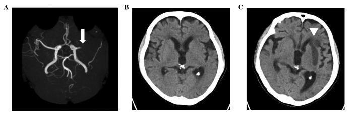
A composite clinical imaging series illustrating the progression of an acute ischemic stroke. Panel A is a Brain Magnetic Resonance Angiography (MRA) showing a high-signal representation of the Circle of Willis; a white arrow indicates an abrupt cutoff and signal loss in the left middle cerebral artery (MCA), consistent with an acute occlusion. Panel B is a non-contrast axial computed tomography (CT) scan of the brain performed on the day of the stroke, showing normal parenchymal attenuation and no acute hemorrhage or visible early ischemic changes. Panel C shows a follow-up axial non-contrast CT scan of the same patient taken six days later; a white arrowhead highlights a well-demarcated area of hypodensity (low attenuation) in the left frontal lobe and basal ganglia, indicating a mature infarction in the vascular territory of the previously identified MCA occlusion. The series highlights the diagnostic utility of vascular imaging in early stroke detection and the temporal evolution of cerebral tissue infarction on CT imaging.
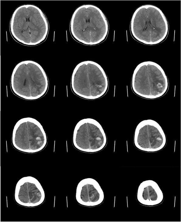
A series of twelve non-contrast axial CT scan slices of the brain demonstrating a large acute cerebral infarction with hemorrhagic transformation. The images reveal a significant area of hypodensity involving the left subcortical region and the corona radiata, consistent with cytotoxic edema and tissue death from an ischemic stroke. Within this hypodense zone, multiple focal areas of hyperdensity are visible, indicating acute intraparenchymal hemorrhage. According to the European Cooperative Acute Stroke Study (ECASS) classification, this appearance is characteristic of parenchymal hematoma type 2 (PH2), where the hemorrhage occupies more than 30% of the infarcted area and exerts a mild mass effect. Key anatomical landmarks include the lateral ventricles and cortical sulci, with the latter showing some effacement on the left side due to associated swelling. This diagnostic imaging serves as a classic educational example of secondary hemorrhagic transformation following a primary ischemic event.
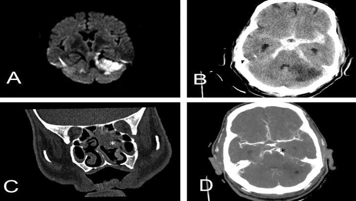
This composite of diagnostic imaging illustrates various neurovascular and otorhinolaryngological pathologies across multiple modalities. Panel A: Axial diffusion-weighted magnetic resonance imaging (DWI MRI) demonstrating hyperintensity in the left cerebellar hemisphere, consistent with restricted diffusion in acute ischemic infarction. Panel B: Non-contrast axial head CT showing diffuse hyperdensity within the basal cisterns and sulci, characteristic of extensive subarachnoid hemorrhage (SAH), alongside a hypodense region in the cerebellum representing secondary ischemic stroke. Panel C: Coronal CT scan of the paranasal sinuses revealing opacification of the nasal cavity and ethmoid cells (marked with a white star), indicating an inflammatory or infectious process such as sinusitis. Panel D: Axial brain CT angiography (CTA) showing vascular morphology, specifically highlighting two consecutive fusiform aneurysms localized in the superior cerebellar artery (marked with a black star). The set provides a comprehensive visual comparison of acute stroke, intracranial hemorrhage, infectious sequelae, and vascular malformations for medical education in radiology and neurology.
Why Non-Contrast CT (NCCT) is the First-Line Investigation in Acute Stroke
The Core Clinical Problem
Is this hemorrhagic or ischemic?
Why NCCT Specifically?
1. Hemorrhage is Immediately Bright (Hyperdense) on NCCT
"Intracranial hemorrhage is often identified on noncontrast computed tomography (CT) imaging of the head during the acute evaluation of stroke. Because CT is more widely available and may be logistically easier to perform than MRI, CT imaging is generally the preferred method for acute stroke evaluation."
- Harrison's Principles of Internal Medicine 22E
2. It is Fast - Speed is Everything in Stroke
- A proper MRI protocol takes 20-45 minutes
- Patient preparation (removing metal, IV access for contrast) adds more delay
3. Widely Available 24/7
4. No Contraindications in Emergency
5. NCCT Findings Change With Time - Clinically Useful
| Phase | Timing | NCCT Finding |
|---|---|---|
| Hyperacute | < 12 hours | Main role: exclude hemorrhage; may show hyperdense vessel sign (dense MCA) |
| Acute | 12-24 hours | Subtle loss of gray-white differentiation (cytotoxic edema) |
| Subacute | 24 hrs - 5 days | Increasingly visible hypodensity in infarcted zone |
| Old | Weeks later | Well-demarcated hypodense area (complete infarction) |
Why NOT Contrast CT?
| Problem | Explanation |
|---|---|
| Masks hemorrhage | Contrast enhancement makes blood vessels and some tissues bright white - this can obscure or mimic hemorrhage, destroying the most important diagnostic signal |
| Nephrotoxicity | Contrast agents can cause contrast-induced nephropathy, especially in elderly stroke patients who may already have CKD |
| Allergy risk | Contrast reactions (from urticaria to anaphylaxis) add another emergency on top of the existing one |
| Time delay | IV access, allergy history check, creatinine check - all add precious minutes |
| Not needed for hemorrhage detection | NCCT without contrast is already highly sensitive for acute blood (near 100% sensitivity for hemorrhagic stroke) |
Why NOT MRI as First Line?
| Problem with MRI | Detail |
|---|---|
| Time | MRI takes 20-45 min vs. CT's 3-5 min. In stroke, every minute counts |
| Workflow delay | Patient screening, metal removal, positioning all add 10-20 min before scanning begins |
| Contraindications | Pacemakers, metallic implants, cochlear implants, some older heart valves, spinal cord stimulators - all are MRI contraindications. ~11% of stroke patients cannot undergo MRI |
| Agitation/movement | Acute stroke patients are often confused, restless, or uncooperative - MRI is highly motion-sensitive and produces non-diagnostic images if the patient moves |
| Cost and availability | MRI is more expensive and less available in emergency settings at all hours |
| Equally good for hemorrhage | NCCT and MRI (with susceptibility-weighted imaging/SWI) are equally sensitive for detecting acute hemorrhage - so MRI provides no advantage for the most urgent question |
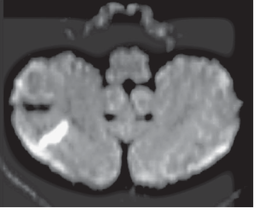
"Diffusion-weighted imaging on MRI is the most sensitive imaging sequence for the detection of acute ischemic infarction and can show infarcted tissue within minutes of stroke onset, considerably earlier than CT... MRI is particularly advantageous in demonstrating small ischemic lesions deep in the hemispheres and brainstem (a region somewhat obscured by adjacent bone on CT)."
Summary Table
| Feature | NCCT | Contrast CT | MRI (DWI) |
|---|---|---|---|
| Speed | Fast (3-5 min) | Moderate | Slow (20-45 min) |
| Hemorrhage detection | Excellent (near 100%) | Poor - contrast masks blood | Excellent (with SWI) |
| Ischemia detection | Poor in first 6 hrs | Same as NCCT | Excellent (within minutes) |
| Availability | Universal, 24/7 | Universal | Limited in emergencies |
| Contraindications | None | Allergy, renal failure | Pacemakers, metal implants |
| Patient cooperation | Easy | Easy | Difficult if agitated |
| Cost | Low | Low-moderate | High |
| First-line for stroke? | YES | No | In select centers with rapid access |
The Clinical Flow
Acute stroke symptoms
        ↓
Immediate NCCT head
        ↓
┌─────────────────────────────────┐
│ Hemorrhage (bright/hyperdense)  │ → No thrombolytics / anticoagulants
│                                 │ → BP control, neurosurgery referral
└─────────────────────────────────┘
        ↓
┌─────────────────────────────────┐
│ No hemorrhage (CT negative)     │ → Ischemic stroke likely
│                                 │ → Check thrombolysis eligibility
│                                 │ → Consider CTA to look for LVO
│                                 │ → Consider MRI/DWI if needed
└─────────────────────────────────┘