

Differentials
apical four chamber view color Doppler severe mitral regurgitation jet
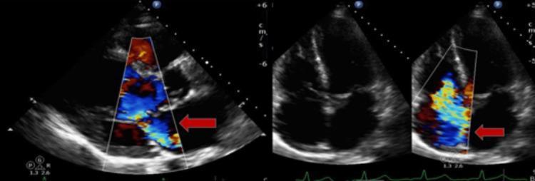
Transthoracic echocardiogram (TTE) images demonstrating valvular regurgitation using color Doppler. The image on the left shows a parasternal long-axis view (PLAX) focusing on the mitral valve. A moderate mitral regurgitation (MR) jet is identified by a red arrow, appearing as a turbulent, multicolored (blue and yellow) conical flow originating from the mitral leaflets and extending into the left atrium during systole. The images on the right side show an apical four-chamber view without and with color Doppler. The color Doppler frame on the far right highlights tricuspid regurgitation (TR), indicated by a red arrow. The TR jet exhibits significant mosaicism, suggesting moderate to severe severity as the turbulent flow occupies a substantial portion of the right atrial area. These diagnostic images are used in cardiology to assess cardiac hemodynamics, valve coaptation, and the severity of valvular heart disease.
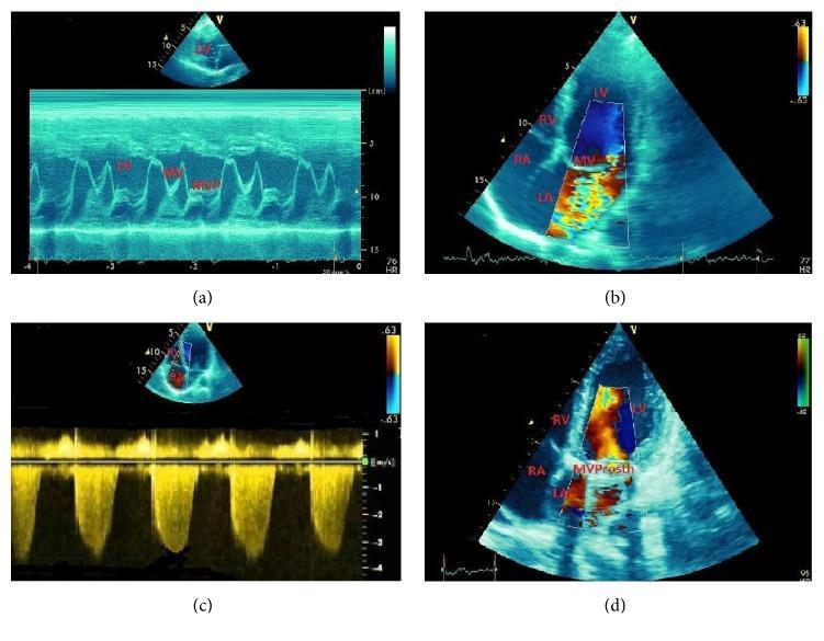
This composite of four echocardiographic images illustrates the diagnostic features and surgical outcome of mitral valve prolapse (MVP). Image (a) presents an M-mode echocardiogram showing characteristic late-systolic posterior displacement of the mitral valve (MV) leaflets, diagnostic of MVP. Image (b) shows a 2D color Doppler in an apical four-chamber view, demonstrating a large, turbulent mosaic jet of mitral regurgitation (MR) originating from the mitral valve and filling a significant portion of the left atrium (LA). Image (c) displays continuous-wave Doppler measurement used to estimate systolic pulmonary artery pressure (SPAP), which is elevated at 58 mmHg, indicating secondary pulmonary hypertension. Image (d) illustrates a postoperative apical four-chamber view following mitral valve replacement with a mechanical prosthesis (labeled 'MVProsth'). The color Doppler shows a significant reduction in regurgitation, with only minor physiologic jets visible. These images collectively demonstrate the progression from valvular pathology to hemodynamic consequences and eventual surgical correction in a patient with congenital MVP and severe MR.

This composite diagnostic image features four panels of a transthoracic echocardiogram (TTE) demonstrating severe mitral regurgitation and associated cardiac remodeling. Panel A shows a parasternal long-axis view revealing left-sided chamber enlargement. Panel B displays a color Doppler apical 4-chamber view, highlighting a large, eccentric regurgitant jet (predominantly red and yellow) that fills a significant portion of the dilated left atrium, consistent with grade 4 mitral regurgitation. Panel C presents continuous-wave spectral Doppler across the mitral valve, showing a dense, holosystolic, high-velocity signal, indicating a high-volume regurgitant flow. Panel D provides an apical view with volumetric measurements, confirming severe left atrial dilation. The images collectively illustrate functional mitral regurgitation in the setting of heart failure, characterized by valvular insufficiency and subsequent bi-atrial enlargement. The study is clinically significant for diagnosing Ortner's syndrome (cardiovocal syndrome) caused by the compression of the recurrent laryngeal nerve by the markedly enlarged left atrium.
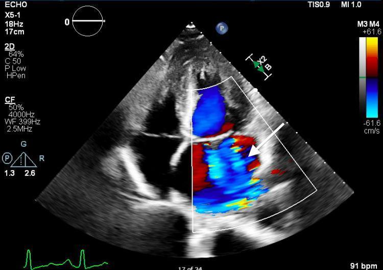
This diagnostic image is a transthoracic echocardiogram (TTE) in an apical four-chamber view with color flow Doppler. The image demonstrates severe (torrential) mitral regurgitation. A large, prominent mosaic-colored jet, indicated by a white arrow, originates at the mitral valve and extends deeply into the left atrium during systole. The mosaic pattern (a mix of red and blue hues) signifies turbulent, high-velocity flow. The color Doppler box covers the left ventricle (bottom) and the left atrium (top), showing flow directed away from the transducer into the atrium. This visual finding is a classic representation of significant valvular pathology, specifically mitral regurgitation due to valve incompetence, used in cardiology education to teach the assessment of regurgitant jet area and severity.
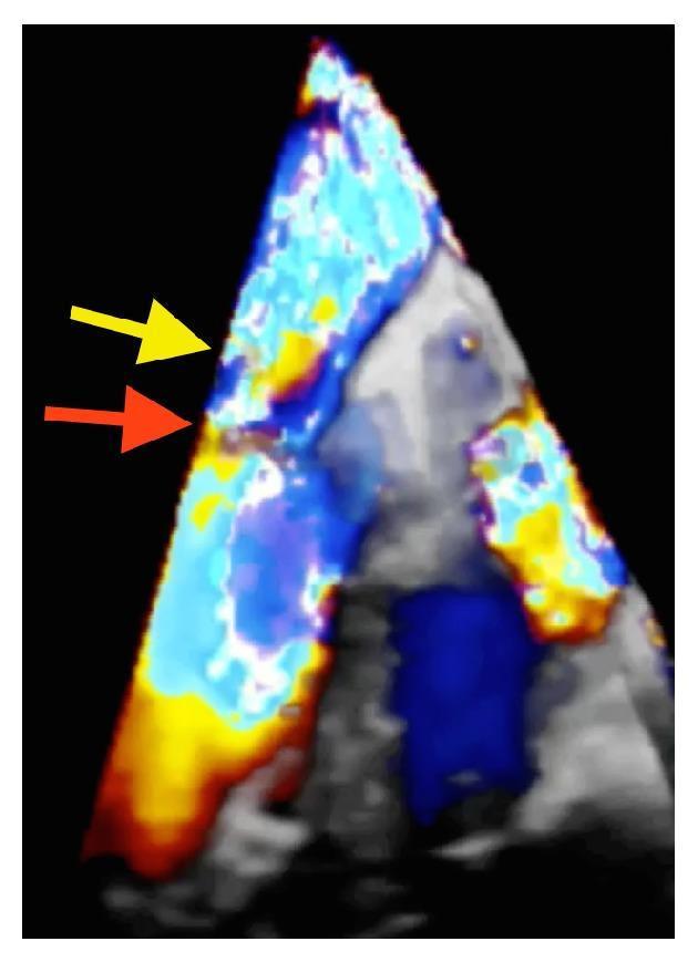
This diagnostic image is a Color 3D Transesophageal Echocardiogram (TEE) in an apical four-chamber view, demonstrating severe mitral regurgitation. The visualization combines grayscale volumetric structural data with color Doppler flow mapping to highlight hemodynamics within the left atrium. Two distinct regurgitant jets are identified: a prominent lateral jet (indicated by a red arrow) originating from a perforation in the P2 scallop of the posterior mitral leaflet, and a secondary central jet (marked by a yellow arrow). The color signals exhibit significant variegation, including shades of red, blue, and yellow, which denote high-velocity turbulent flow and aliasing typical of severe valvular insufficiency. The spatial relationship between the grayscale anatomical structures and the dynamic color signals illustrates the mechanical failure of the mitral valve apparatus, often associated with infective endocarditis and subsequent leaflet perforation. This image serves as a high-level educational example of using real-time 3D Doppler to map complex multi-jet regurgitation for surgical planning.
What the image shows
Differential diagnosis for this appearance
- Large, mosaic, holosystolic jet filling >50% of LA area, often with a wide vena contracta.
- Etiologies to consider: myxomatous/prolapse, ruptured chordae/flail leaflet, ischemic/functional MR from LV dysfunction, endocarditis with leaflet perforation, rheumatic disease.
- Similarly large central or eccentric jet filling most of the RA.
- Consider functional TR (RV/annular dilation, pulmonary hypertension), pacemaker lead-related, endocarditis (especially IV drug use), carcinoid, or primary leaflet pathology.
- If both round far-field structures (likely the two atria) show comparable filling, biventricular failure or biatrial functional AV regurgitation should be considered, which would fit the pattern of two color-filled chambers side by side in this frame.
- Less likely given the chamber geometry here, but a VSD produces a similarly mosaic, high-velocity jet, usually seen crossing the septum rather than filling an atrium.
- Excessive color gain, high wall-filter settings, or a poor angle of incidence can make even mild flow look like it fills a chamber. This has to be excluded by checking the actual color gain settings and correlating with spectral (CW/PW) Doppler and 2D structural findings.
- If this frame was captured in diastole with high color gain, normal simultaneous mitral and tricuspid inflow can create an impression of both ventricles/atria being "filled with color" without any pathology — this is the benign end of the differential and should be ruled out before assuming regurgitation.
To narrow this down further, it would help to know:
- Which view is this exactly (A4C, A5C, or subcostal) and is this a systolic or diastolic frame?
- Any clinical details on the patient (age, symptoms, known murmur, prior valve disease, IV drug use, recent fever)?
- Does the full study/report have quantitative severity grading (vena contracta, PISA, regurgitant fraction) or CW Doppler spectral tracings?