Hearing
Reading File
Asking for Preferences
Finding Sources
Finding Sources
Reading File
Reading File
Reading File
Finding Sources
Loading Image
Finding Sources
Loading Image
Reading File
Reading File
Loading Image
Loading Image
Here is a thorough overview of the anatomy of the ear, organized by its three main divisions.
Anatomy of the Ear
The ear is divided into three parts: the external ear, the middle ear, and the inner ear. Each has a distinct structural role in collecting, conducting, and transducing sound.
1. External Ear
The external ear consists of the auricle (pinna) and the external auditory canal (EAC).
- Auricle: Stratified squamous epithelium over a framework of elastic cartilage. The lobule (earlobe) is the only cartilage-free portion, containing adipose tissue instead. Sebaceous glands and hair follicles are present in the subcutaneous layer.
- External auditory canal (EAC): Approximately 2.5-3 cm long in adults. The outer one-third is cartilaginous (contains hair follicles, sebaceous glands, and apocrine glands that produce cerumen). The inner two-thirds is bony (thin squamous epithelium tightly adherent to bone, no glands or hair follicles). The narrowest point is the isthmus, at the bony-cartilaginous junction.
- Cerumen: Hydrophobic, slightly acidic (pH 6.0-6.5), formed by glandular secretions and sloughed epithelium in the cartilaginous canal. The canal is self-cleaning due to outward centrifugal migration of keratin from the tympanic membrane.
- Fissures of Santorini: Transverse slits in the cartilaginous canal that can allow spread of infection or tumor to surrounding soft tissues.
- Foramen of Huschke: A defect in the anterior bony canal from incomplete ossification; can allow disease spread to the parotid gland.
(Cummings Otolaryngology, Bailey and Love's Surgery)
2. Tympanic Membrane
The tympanic membrane (TM) separates the EAC from the middle ear.
- Semitransparent, thin (~0.1 mm), ~1 cm in diameter, average surface area ~65 mm²
- Tilted anteriorly and inferiorly (like a miniature satellite dish)
- Three layers from outside to inside:
- Stratified squamous keratinized epithelium (outer)
- Dense connective tissue core with radially arranged outer fibers and circumferentially arranged inner fibers
- Simple cuboidal epithelium of the middle ear mucosa (inner)
- Two functional regions:
- Pars tensa: the large, taut central portion
- Pars flaccida: the small, lax upper portion (Shrapnell's membrane), prone to retraction pockets
- The umbo is the most inward point of the TM where the tip of the malleus manubrium attaches
- On otoscopy, a cone of light (light reflex) radiates anteroinferiorly from the umbo
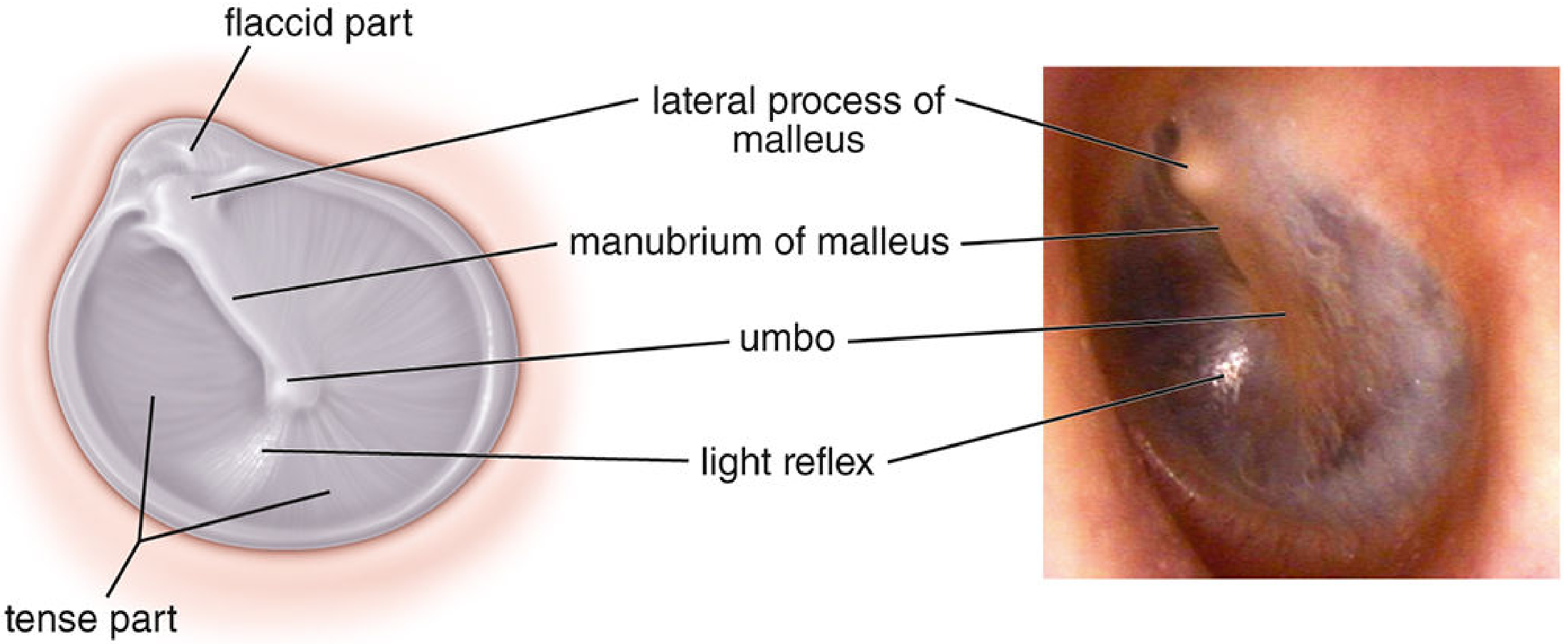
(Histology: A Text and Atlas; Bailey and Love's Surgery)
3. Middle Ear
The middle ear (tympanic cavity) contains the three ossicles - the smallest bones in the body - which form a mechanical chain transmitting vibration from the TM to the inner ear.
Ossicles (medial to lateral):
| Bone | Connection |
|---|---|
| Malleus | Handle (manubrium) attached to TM; head in the epitympanum |
| Incus | Body articulates with malleus head; long process connects to stapes |
| Stapes | Footplate sits in the oval window; connected to inner ear perilymph |
- The long process of the incus is the most vulnerable ossicle - it has a single nutrient vessel and lacks collateral circulation
- Two muscles act on the chain: tensor tympani (dampens low-frequency, attaches to malleus) and stapedius (attaches to stapes neck, protective acoustic reflex)
- The facial nerve passes through the middle ear, making it surgically vulnerable. Key landmarks include the cochleariform process, oval window, and pyramidal eminence
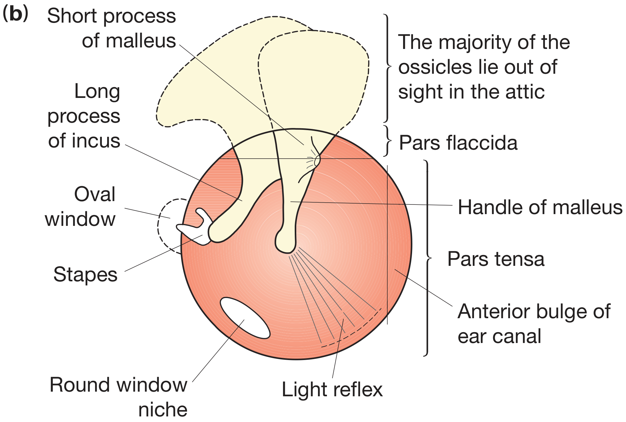
Middle ear compartments:
- Epitympanum (attic): superior to TM, contains the malleus head and incus body
- Mesotympanum: opposite the TM
- Hypotympanum: inferior to TM
- Eustachian tube: connects middle ear to nasopharynx for pressure equalization
(Cummings Otolaryngology)
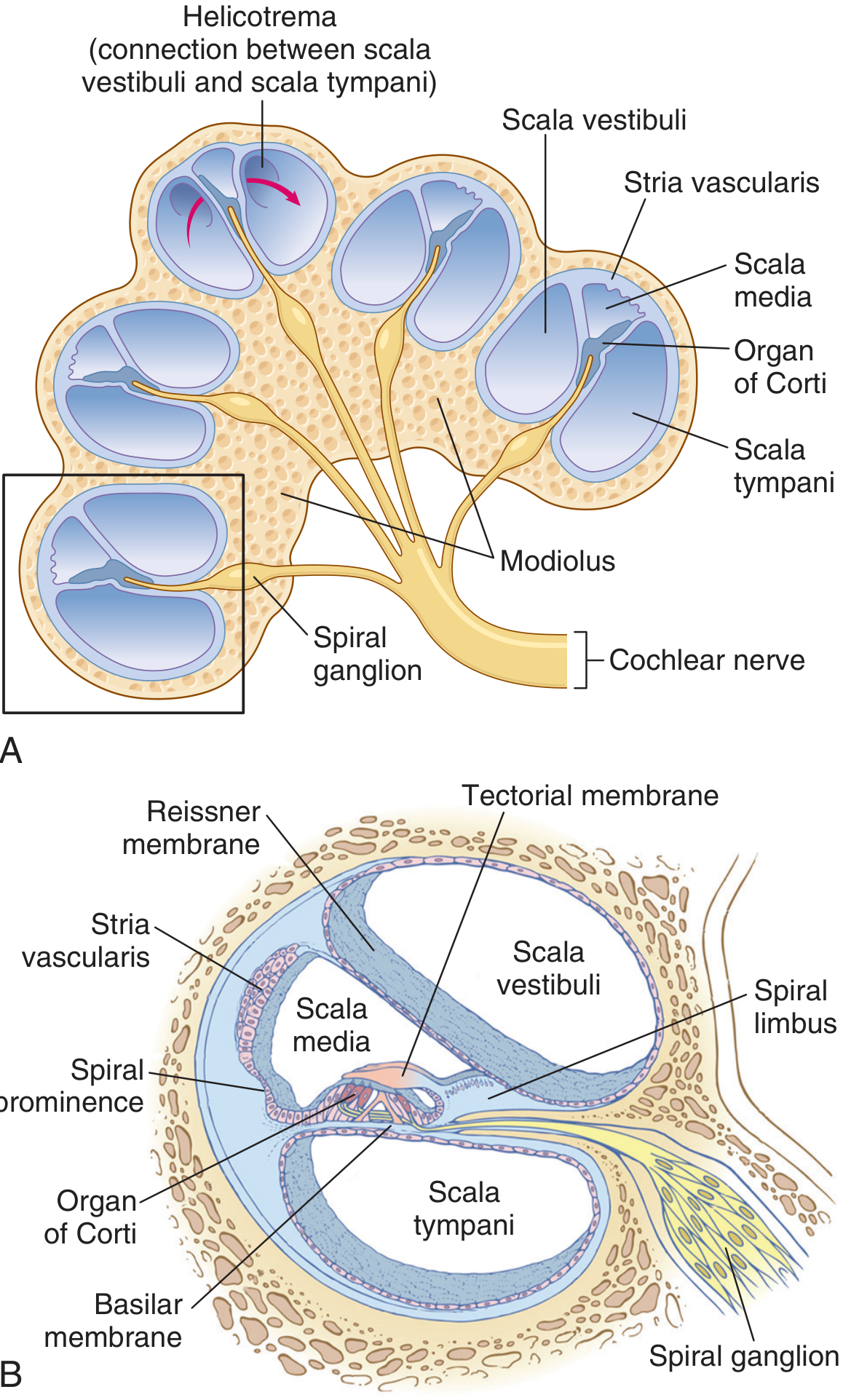
4. Inner Ear
The inner ear is housed in the dense otic capsule bone within the petrous part of the temporal bone, and contains two systems: the cochlea (hearing) and the vestibular labyrinth (balance).
Cochlea
The cochlea makes 2.75 turns around a central bony axis called the modiolus. It contains three fluid-filled compartments:
| Compartment | Fluid | Boundaries |
|---|---|---|
| Scala vestibuli | Perilymph | Upper; starts at oval window |
| Scala media (cochlear duct) | Endolymph | Middle |
| Scala tympani | Perilymph | Lower; ends at round window |
- Reissner's membrane separates the scala vestibuli from the scala media (functionally thin, maintains endolymph composition)
- Basilar membrane separates the scala media from the scala tympani
- Organ of Corti sits on the basilar membrane - contains the mechanosensory hair cells
- Stria vascularis: vascular epithelium on the outer wall of the scala media; produces endolymph (high K⁺, like intracellular fluid)
- Tectorial membrane: overlies the hair cells; deflects stereocilia during basilar membrane movement
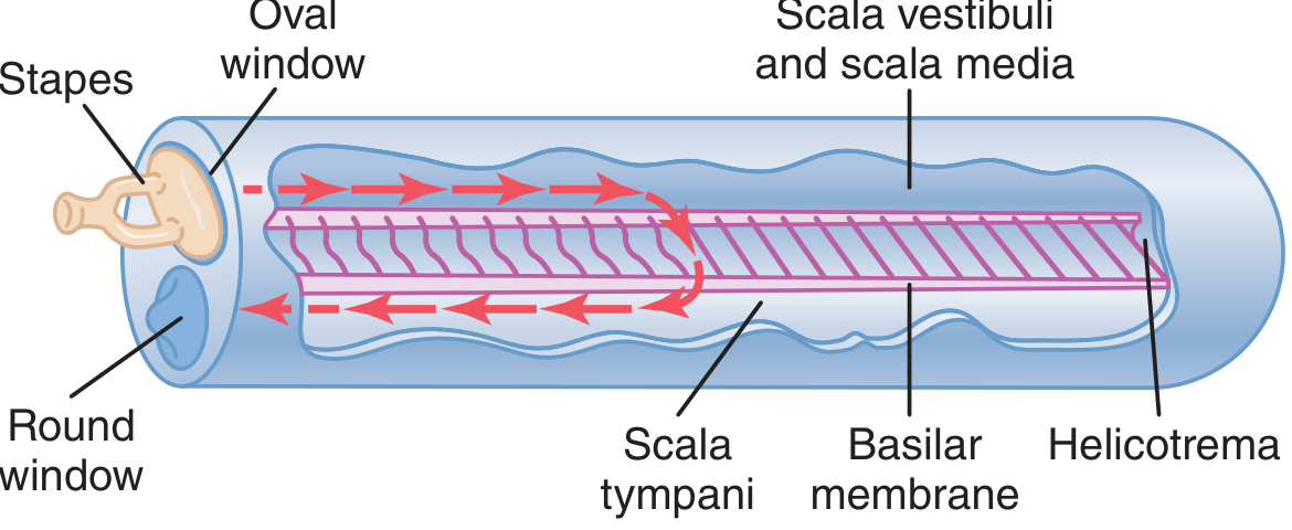
- The scala vestibuli and scala tympani communicate at the apex via the helicotrema
- Tonotopy: high frequencies activate the stiff, narrow base; low frequencies activate the wide, flexible apex
Basilar membrane dimensions:
- Base (near oval window): ~0.04 mm fiber length, stiff - responds to high frequencies
- Apex (helicotrema): ~0.5 mm fiber length, flexible - responds to low frequencies
Vestibular Labyrinth
- Saccule and utricle: detect linear acceleration and head position relative to gravity (macular organs with otoliths)
- Three semicircular canals (superior, posterior, horizontal): detect rotational/angular acceleration; each has an ampulla containing a crista with cupula
Sound transmission pathway (summary):
Sound waves → pinna/EAC → TM vibrates → malleus → incus → stapes footplate → oval window → perilymph in scala vestibuli → basilar membrane displacement → hair cell stereocilia deflect against tectorial membrane → mechanoelectrical transduction → cochlear nerve (CN VIII) → brainstem auditory nuclei
(Guyton and Hall Medical Physiology; Cummings Otolaryngology; Bailey and Love's Surgery)
Temporal Bone (Housing)
The entire ear apparatus is enclosed within the temporal bone, which has four embryological components:
- Squamous part: lateral wall of middle fossa
- Mastoid part: posterior, air-cell system
- Petrous part: dense bone housing the inner ear
- Tympanic part: forms most of the bony EAC
The outer and middle ears derive from the first and second branchial arches and grooves; the inner ear derives from the otic placode (separate embryological origin).
This is a shared conversation. Sign in to Orris to start your own chat.