
What are kidney stones from harrison
kidney stone nephrolithiasis types calcium oxalate uric acid
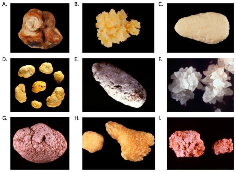
A multi-panel clinical photograph displaying a comparison chart of nine distinct kidney stone (nephrolithiasis) subtypes, labeled A through I. The specimens demonstrate significant morphological variation based on chemical composition. (A) Calcium oxalate monohydrate (Whewellite): brown, multi-lobed stone with concentric layers. (B) Calcium oxalate dihydrate (Weddellite): yellowish, sharp bipyramidal crystals. (C) Calcium phosphate (carbapatite): smooth, oval, light tan specimen. (D) Uric acid: small, rounded, yellowish-orange stones. (E) Ammonium urate: elongated, greyish stone with surface staining. (F) Struvite (triple phosphate): white, branched crystalline clusters characteristic of staghorn configurations. (G) 2,8-Dihydroxyadenine: reddish-brown, rounded stone with a bumpy, granular texture. (H) Cystine: yellowish stones with a characteristic hexagonal crystalline wax-like appearance. (I) Drug-induced (N-acetylsulfadiazine): irregular, reddish-pink stones with a rough, granular surface. This educational resource is designed for urological pathology and diagnostic training, emphasizing the use of visual stone analysis (morphoconstitutional study) to suggest underlying metabolic or pharmacological etiologies.
![This composite educational image demonstrates the post-processing analysis of a dual-energy computed tomography (DECT) scan for nephrolithiasis characterization. The top panel contains three axial non-contrast CT sections of the kidney, showing regions of interest (ROI) labeled 3, 4, and 5 identifying small renal calculi. Below, a stone composition scatter plot correlates 'Low energy [HU]' on the x-axis with 'High energy [HU]' on the y-axis. The graph features two reference slope lines: a red line for Uric Acid and a blue line for Calcium Oxide (Ca Oxide). Data points for specific stones are plotted against these lines; proximity to the red slope suggests uric acid composition, while proximity to the blue slope indicates calcium-based stones. A accompanying table provides quantitative metrics for each ROI, including Mean Hounsfield Units (HU), Standard Deviation (SD), and Volume (ml) for both low and high-energy spectra. This tool is clinically used to non-invasively differentiate stone types, such as uric acid from calcium oxalate, which informs surgical planning and medical management strategies.](https://cdn.orris.care/cdss_images/pmc_clinical_VQA_4684a7e4dbad17c89fb1f7302e99788c159da092f5bd0779f2722073438bf80b.jpg)
This composite educational image demonstrates the post-processing analysis of a dual-energy computed tomography (DECT) scan for nephrolithiasis characterization. The top panel contains three axial non-contrast CT sections of the kidney, showing regions of interest (ROI) labeled 3, 4, and 5 identifying small renal calculi. Below, a stone composition scatter plot correlates 'Low energy [HU]' on the x-axis with 'High energy [HU]' on the y-axis. The graph features two reference slope lines: a red line for Uric Acid and a blue line for Calcium Oxide (Ca Oxide). Data points for specific stones are plotted against these lines; proximity to the red slope suggests uric acid composition, while proximity to the blue slope indicates calcium-based stones. A accompanying table provides quantitative metrics for each ROI, including Mean Hounsfield Units (HU), Standard Deviation (SD), and Volume (ml) for both low and high-energy spectra. This tool is clinically used to non-invasively differentiate stone types, such as uric acid from calcium oxalate, which informs surgical planning and medical management strategies.
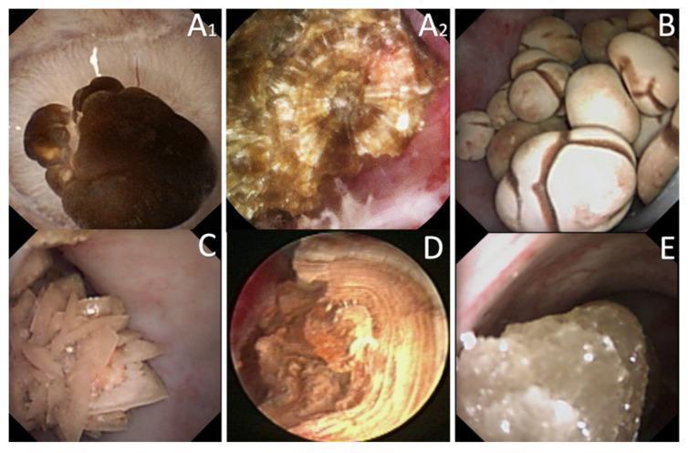
This composite of endoscopic clinical photographs demonstrates the Endoscopic Stone Recognition (ESR) of various nephrolithiasis types. Panel A1 shows a calcium oxalate monohydrate (COM) stone with a characteristic dark-brown, mammillary surface anchored to a renal papilla via Randall's plaque. A2 displays a COM stone transection revealing a radiating crystalline structure originating from a central nucleus. Panel B illustrates COM subtype Id stones with a pale, brownish-yellow budding surface. Panel C depicts calcium oxalate dihydrate (COD) stones, characterized by yellow, spiculated surfaces consisting of aggregated crystals with sharp angles. Panel D shows a uric acid (UA) stone with a smooth, homogeneous orange exterior and a transected view showing concentric laminar organization around a nucleus. Panel E presents a cystine stone, identifiable by its rough, bumpy light brown-yellow surface with a distinctive waxy luster. These images serve as a diagnostic guide for intraoperative identification of stone morphology to inform metabolic etiology and recurrence risk.
Nephrolithiasis (Kidney Stones) - Harrison's Principles of Internal Medicine, 22nd Edition
Overview
- Harrison's Principles of Internal Medicine 22E, p.2464
Stone Types
| Type | Frequency |
|---|---|
| Calcium oxalate | ~75% |
| Calcium phosphate | ~15% |
| Uric acid | ~8% |
| Struvite | ~1% |
| Cystine | <1% |
- Harrison's, p.2468
Epidemiology
- Prevalence is increasing, likely due to Westernization (dietary changes, rising BMI).
- Up to 20% of men and 10% of women will develop at least one stone in their lifetime (NHANES 2007-2016).
- Prevalence is ~50% lower in Black individuals vs. whites.
- Peak annual incidence in white men: ~3.5 cases/1000 at age 40.
- Substantial economic impact as those affected are often of working age.
- Harrison's, p.2473
Associated Medical Conditions
- GI malabsorption (Crohn's disease, gastric bypass surgery)
- Primary hyperparathyroidism
- Obesity and type 2 diabetes mellitus
- Distal renal tubular acidosis
- Harrison's, p.2478
Risk Factors
Dietary Risk Factors
- Low fluid intake / low urine volume - the single most important modifiable risk factor
- High sodium intake - increases urine calcium
- High animal protein intake - increases urine uric acid and calcium, decreases citrate
- High oxalate intake - increases urine oxalate
- Low calcium intake - paradoxically increases risk (dietary calcium binds intestinal oxalate)
- High fructose intake - increases urine oxalate, calcium, and uric acid
Non-dietary Risk Factors
- Highest incidence in middle-aged white men
- Obesity, high BMI
- Hot climate / geographic "stone belt" regions
- Anatomic abnormalities (e.g., medullary sponge kidney, horseshoe kidney)
- Genetic factors - monogenic disorders cause nephrolithiasis; GWAS studies reveal polygenic contributors
Urinary Risk Factors (the proximate determinants)
- Hypercalciuria - most common urinary abnormality
- Hyperoxaluria
- Hypocitraturia - citrate is the main natural inhibitor of crystallization
- Hyperuricosuria
- Low urine volume
- High or low urine pH (depending on stone type)
- Harrison's, p.2494-2534
Clinical Presentation
- Renal colic: Sudden onset severe flank pain radiating to the groin; classically colicky but often constant; nausea and vomiting common
- Hematuria (gross or microscopic) in ~90% of cases
- Urinary urgency/frequency when stone is near the ureterovesical junction
- Stones <5 mm usually pass spontaneously; >10 mm unlikely to pass without intervention
Diagnosis
- Non-contrast CT (CT-KUB): Gold standard - detects virtually all stone types, determines size and location
- Plain X-ray (KUB): Calcium-containing stones are radiopaque; uric acid stones are radiolucent
- Renal ultrasound: Useful to detect hydronephrosis; avoids radiation (preferred in pregnancy)
- Urinalysis: Hematuria; crystal type may suggest stone composition; pH helps (low pH - uric acid; high pH - struvite)
- 24-hour urine collection: Essential for metabolic evaluation in recurrent stone formers - measures calcium, oxalate, uric acid, citrate, sodium, creatinine, pH, volume
Prevention by Stone Type
Calcium Oxalate
- Increase fluid intake (goal urine volume >2 L/day)
- Reduce sodium and animal protein
- Maintain adequate dietary calcium (~1200 mg/day with meals)
- Thiazide diuretics (e.g., hydrochlorothiazide, indapamide, chlorthalidone) - reduce urine calcium
- Potassium citrate - increases urine citrate, reduces crystallization
- DASH diet or Mediterranean diet - strong observational data for reduced stone risk
Calcium Phosphate
- Same as above for calcium oxalate
- Monitor urine pH (supplemental alkali can worsen by raising pH - since these patients already have high pH)
- Reduce dietary phosphate
- Harrison's, p.2665-2667
Uric Acid
- Urine pH is the key factor - uric acid stones form at persistently low pH
- Alkali supplementation (potassium citrate/bicarbonate) - goal urine pH 6.5 throughout day and night
- Reduce purine-containing foods (animal flesh)
- Increase fruits and vegetables
- If above measures fail: xanthine oxidase inhibitor (allopurinol or febuxostat) reduces urine uric acid by 40-50%
- Note: serum uric acid does NOT reliably predict urine uric acid excretion
- Harrison's, p.2669-2671
Cystine
- High fluid intake is essential (highest priority)
- Tiopronin (preferred) or D-penicillamine - bind cystine covalently to increase solubility
- Potassium citrate/bicarbonate to alkalinize urine (goal pH 7.5)
- Avoid sodium salts (sodium increases cystine excretion)
- Harrison's, p.2673
Struvite (Infection Stones)
- Form only when the upper urinary tract is infected with urease-producing bacteria (e.g., Proteus mirabilis, Klebsiella)
- Urease splits urea into ammonium, raising urine pH to 7.2-8.0 - an alkaline environment that precipitates magnesium ammonium phosphate
- Also called "infection stones" or "triple-phosphate stones"
- Can form staghorn calculi that fill the renal pelvis and calyces
- Require complete surgical removal + eradication of the infection; stone fragments harbor bacteria and lead to recurrence
- Harrison's, p.2675
Summary Table
| Stone Type | Key Risk Factors | Prevention |
|---|---|---|
| Calcium oxalate | Low urine volume, hypercalciuria, hyperoxaluria, hypocitraturia | Fluids, thiazides, K-citrate, low sodium/protein |
| Calcium phosphate | High urine pH, hypercalciuria, distal RTA, hyperparathyroidism | Thiazides, monitor pH carefully with alkali |
| Uric acid | Low urine pH, high purine intake, gout | Alkali (K-citrate), low purine diet, allopurinol |
| Struvite | Urease-producing UTI | Complete stone removal + antibiotics |
| Cystine | Cystinuria (genetic) | Tiopronin, K-citrate, high fluid intake |