
Sympathetic and parasympathetic ganglions
I'll look this up in the medical library for you right away.
Searching sources...
Reading File
Finding Sources
Finding Sources
Good results. Let me pull detailed content from the most relevant sources - the anatomy atlas (THIEME) and Gray's Anatomy.
Reading File
Finding Sources
Excellent - I found a detailed section on the autonomic nervous system. Let me read more of this content and also fetch the key diagram.
Reading File
Loading Image
Finding Sources
Great content found. Let me now pull the detailed text from the key physiology sources on ganglion locations, structure, and differences.
Reading File
Reading File
Reading File
Excellent content. Let me get parasympathetic ganglion details now and fetch one more key image.
Reading File
Loading Image
I now have comprehensive information from multiple authoritative textbooks plus detailed diagrams. Let me compile the full answer.
Sympathetic and Parasympathetic Ganglia
Both divisions of the autonomic nervous system (ANS) use a two-neuron chain between the CNS and the target organ: a preganglionic neuron (in the CNS) synapses onto a postganglionic neuron (in a peripheral ganglion), which then innervates the effector. The location of that ganglion is the key structural difference between the two divisions.
Overview Diagram
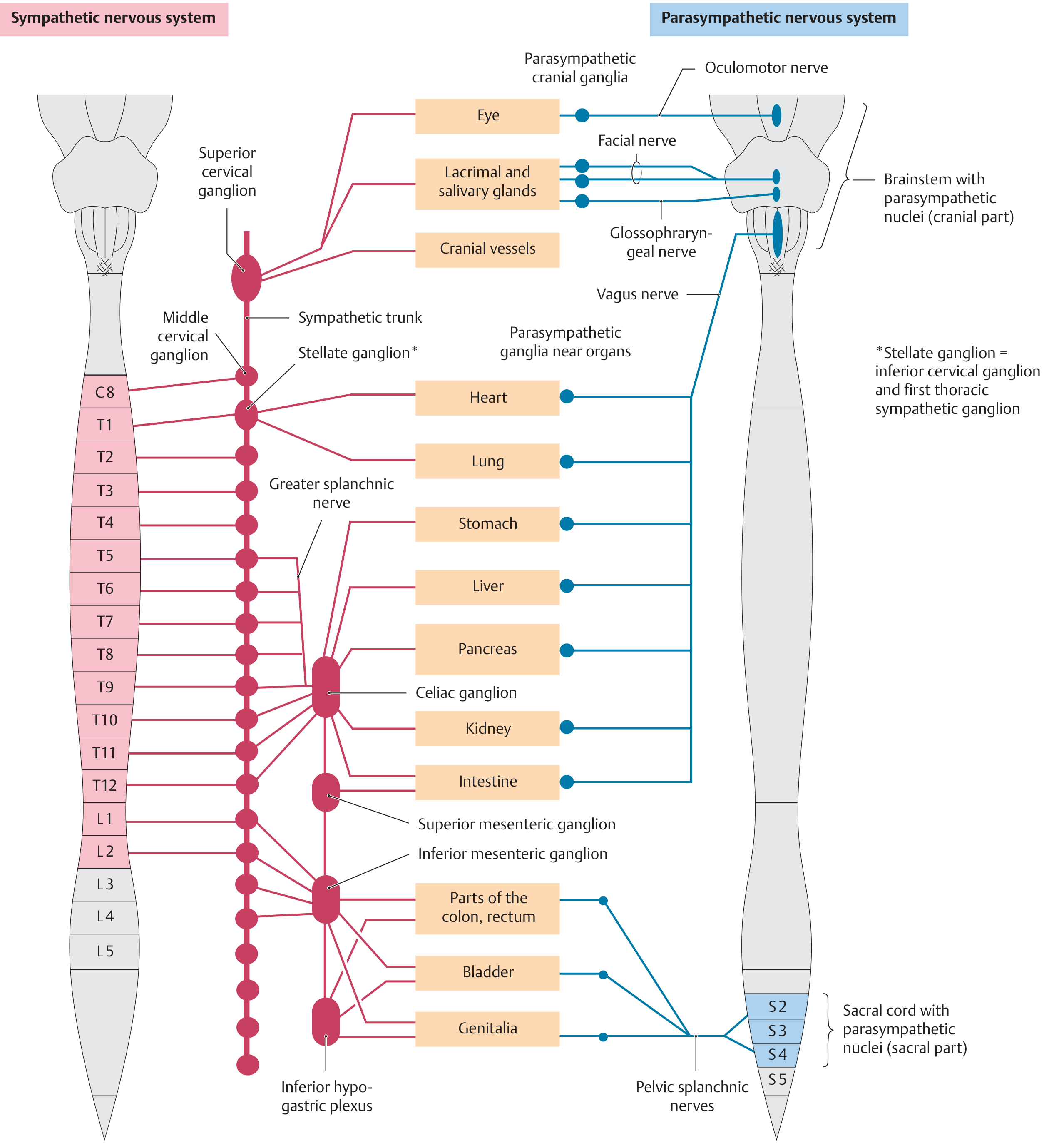
Sympathetic Ganglia
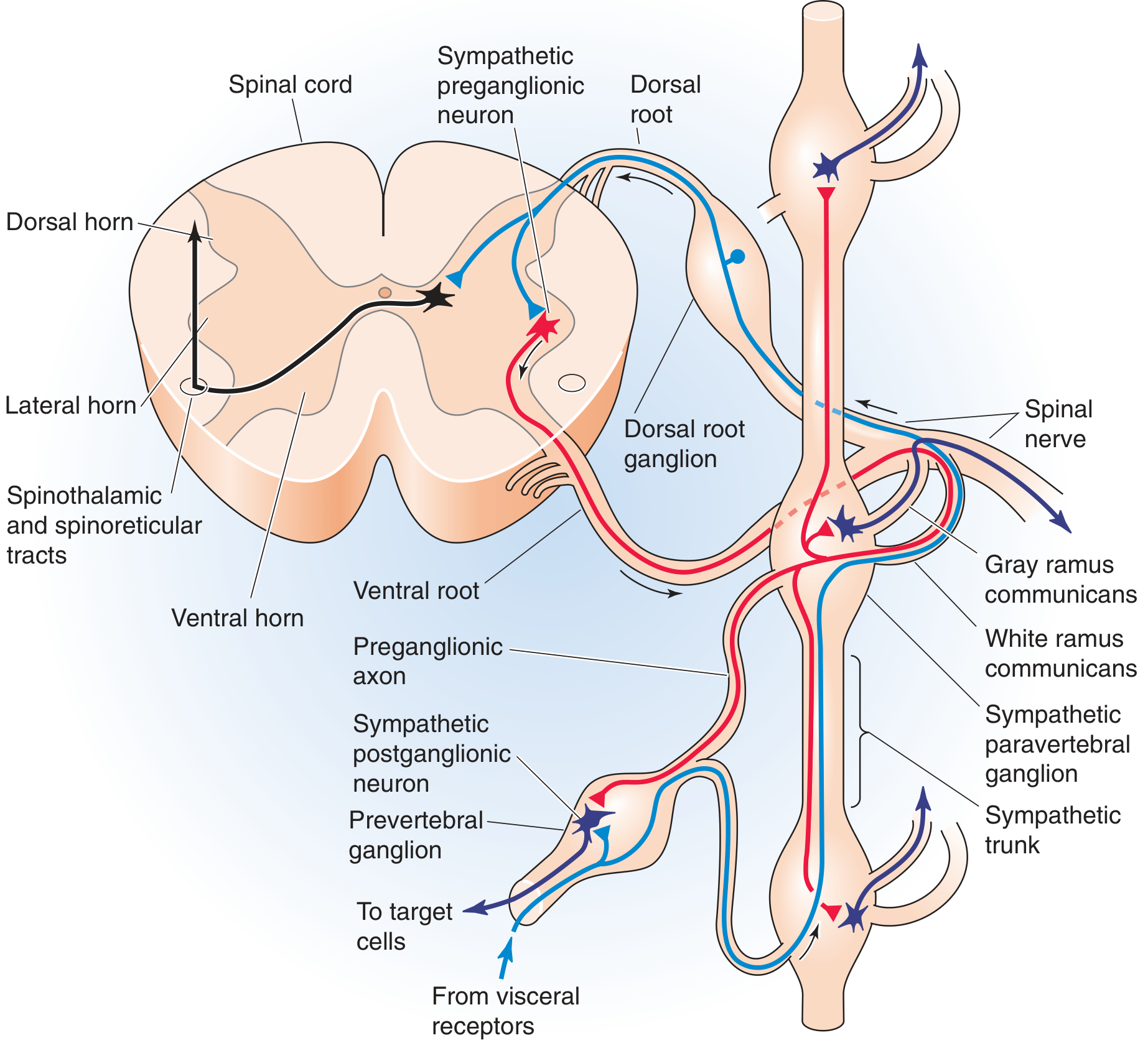
Origin of preganglionic neurons: Lateral horns of the thoracolumbar spinal cord (T1-L3). Preganglionic axons exit via ventral roots and white rami communicantes.
Sympathetic ganglia are located close to the spinal cord and far from target organs, meaning postganglionic fibers are long. There are two types:
1. Paravertebral Ganglia (Sympathetic Chain / Sympathetic Trunk)
- Form two bilateral chains running alongside the vertebral column from the cervical region to the sacrum
- Contain postganglionic neuronal cell bodies
- Connected to spinal nerves via white rami communicantes (incoming, myelinated preganglionic) and gray rami communicantes (outgoing, unmyelinated postganglionic)
- White rami exist only at T1-L3; gray rami exist at all spinal levels (C2/C3 to coccyx)
- Named cervical ganglia include:
- Superior cervical ganglion - projects to head, neck, heart
- Middle cervical ganglion
- Stellate ganglion (inferior cervical + 1st thoracic, fused) - projects to heart and arm
- Thoracic ganglia (T1-T12)
- Lumbar and sacral ganglia
A preganglionic fiber entering the chain has three possible fates:
- Synapse in that same segmental ganglion
- Travel up or down the chain to synapse in a higher or lower ganglion
- Pass through without synapsing to reach a prevertebral ganglion
2. Prevertebral Ganglia (Collateral Ganglia)
- Lie anterior to the aorta along its major branches (unpaired, midline)
- Receive preganglionic fibers that bypass the paravertebral chain (via splanchnic nerves)
- Supply visceral organs of the abdomen and pelvis
- Named for adjacent arteries:
| Ganglion | Location | Supplies |
|---|---|---|
| Celiac ganglion | Along celiac artery | Stomach, liver, pancreas, kidney |
| Superior mesenteric ganglion | Along superior mesenteric artery | Small intestine, ascending/transverse colon |
| Aorticorenal ganglion | Along renal artery | Kidney, adrenal |
| Inferior mesenteric ganglion | Along inferior mesenteric artery | Descending colon, rectum, bladder, genitalia |
Key features of sympathetic ganglia:
- Each preganglionic fiber synapses on up to ~200 postganglionic neurons - this divergence explains the broad, diffuse ("mass discharge") nature of sympathetic responses
- Neurotransmitter at preganglionic synapse: acetylcholine (nicotinic receptors)
- Neurotransmitter at target organ: norepinephrine (adrenergic receptors)
Parasympathetic Ganglia
Origin of preganglionic neurons: Brainstem (craniosacral outflow) and sacral spinal cord (S2-S4). Preganglionic fibers travel with cranial nerves and pelvic splanchnic nerves.
Parasympathetic ganglia are located close to or within the target organs, so preganglionic fibers are long and postganglionic fibers are very short. This produces focal, discrete effects (unlike the diffuse sympathetic response).
Cranial Parasympathetic Ganglia (4 named ganglia)
| Ganglion | Cranial Nerve | Preganglionic Origin | Target |
|---|---|---|---|
| Ciliary ganglion | CN III (oculomotor) | Edinger-Westphal nucleus (midbrain) | Constrictor pupillae, ciliary muscle (lens) |
| Pterygopalatine ganglion (sphenopalatine) | CN VII (facial) via greater petrosal nerve | Superior salivatory nucleus (rostral medulla) | Lacrimal gland, nasal/palatal mucosa |
| Submandibular ganglion | CN VII via chorda tympani | Superior salivatory nucleus | Submandibular and sublingual salivary glands |
| Otic ganglion | CN IX (glossopharyngeal) via lesser petrosal nerve | Inferior salivatory nucleus + nucleus ambiguus | Parotid gland |
Note: The ciliary ganglion is also traversed (not synapsed) by postganglionic sympathetic fibers from the superior cervical ganglion, which pass through to dilate the pupil.
Vagal (CN X) Ganglia
The vagus nerve carries the majority of parasympathetic output. Its preganglionic fibers originate in the nucleus ambiguus and dorsal motor nucleus of the vagus in the medulla. They run long courses to synapse in intramural ganglia (ganglionic plexuses embedded within the walls of target organs):
- Heart (cardiac plexus)
- Lungs (pulmonary plexus)
- Entire GI tract from pharynx to the distal colon (splenic flexure)
- Liver, pancreas, kidneys
Sacral Parasympathetic Ganglia
- Preganglionic neurons arise from S2-S4 and travel via pelvic splanchnic nerves
- Synapse in ganglia near or in the walls of pelvic organs:
- Descending colon, rectum, sigmoid
- Bladder (detrusor)
- Genitalia
Key features of parasympathetic ganglia:
- Ganglia are located in or near target organs - responses are localized and discrete
- Each preganglionic fiber synapses on far fewer postganglionic neurons than in sympathetic (less divergence)
- Neurotransmitter at preganglionic synapse: acetylcholine (nicotinic)
- Neurotransmitter at target organ: acetylcholine (muscarinic receptors)
Key Comparison: Sympathetic vs. Parasympathetic Ganglia
| Feature | Sympathetic | Parasympathetic |
|---|---|---|
| CNS origin | Lateral horn, T1-L3 (thoracolumbar) | Brainstem + S2-S4 (craniosacral) |
| Ganglion location | Near spinal cord (para- or pre-vertebral) | Near or within target organs (intramural) |
| Preganglionic fiber length | Short | Long |
| Postganglionic fiber length | Long | Short |
| Divergence | High (~200 post per pre) | Low (focal) |
| Response pattern | Diffuse, mass discharge | Discrete, localized |
| Pre-synaptic transmitter | Acetylcholine (nicotinic) | Acetylcholine (nicotinic) |
| Post-synaptic transmitter | Norepinephrine (adrenergic) | Acetylcholine (muscarinic) |
| Overall function | "Fight or flight" | "Rest and digest" |
Sources: General Anatomy and Musculoskeletal System (THIEME Atlas), p. 114-115 | Medical Physiology (Boron & Boulpaep), p. 502-506 | Costanzo Physiology 7th Ed., p. 56-57
This is a shared conversation. Sign in to Orris to start your own chat.