
23 year old female having fungal infection at abdominal area and back of leg, management?
tinea corporis fungal skin infection abdomen ringworm
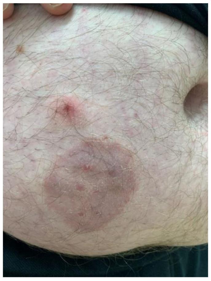
This clinical photograph displays a dermatological presentation of tinea corporis (zoonotic dermatophytosis) on the abdomen of an adult male. The primary feature is a well-circumscribed, annular, erythematous plaque approximately 4-5 cm in diameter. The lesion exhibits a classic 'ringworm' morphology with a slightly raised, scaly inflammatory border and a partially cleared central area containing subtle papular elements. Superior to the primary plaque is a secondary, smaller, raised erythematous papule with a central punctum or crust. The surrounding skin shows mild diffuse erythema and scattered smaller inflammatory macules. Hair follicles are visible throughout the affected and unaffected regions. This image serves as an educational example of fungal infection (likely Trichophyton mentagrophytes) transmitted from a pet (guinea pig) to a human host, emphasizing the visual diagnostic features of annular inflammatory skin lesions.
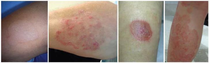
This composite of clinical photographs illustrates various clinical presentations of tinea corporis, a common dermatophyte fungal infection. The images depict the characteristic morphology of 'ringworm' across different skin areas. Key diagnostic features visible include well-demarcated, erythematous, annular (ring-shaped) patches and plaques. Several lesions demonstrate classic central clearing, where the center of the ring appears less inflamed than the periphery. The advancing borders are notably active, exhibiting varying degrees of elevation and fine scaling. The presentation varies from discrete, solitary circular lesions to larger, confluent, and irregularly shaped polycyclic patterns. The color intensity ranges from faint pink to vivid red, representing different stages of inflammatory response. These visual characteristics are essential for distinguishing tinea corporis from other annular dermatoses such as nummular eczema, granuloma annulare, or subacute cutaneous lupus erythematosus. The collection serves as an educational tool for identifying the hallmark features of cutaneous fungal infections in a clinical setting.
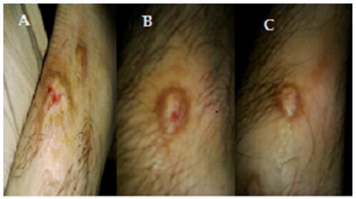
This sequence of three clinical photographs (A, B, and C) documents the progression and resolution of a Tinea corporis infection on a patient's forearm under treatment. Image A (Before treatment) shows a classic fungal skin lesion with intense central erythema, localized inflammation, and evidence of epidermal breakdown or erosion. Image B (After 1 week) demonstrates the characteristic 'ringworm' morphology, with the development of a raised, erythematous peripheral border (annular configuration) while the central redness begins to subside. Image C (After 2 weeks) shows significant healing, characterized by a marked reduction in inflammation, a more uniform skin tone, and the formation of a dry scab or resolving plaque at the primary site. The surrounding lightly pigmented skin and hair follicles remain largely unaffected throughout the timeline, illustrating the localized nature of the dermatophyte infection and the therapeutic response to the topical treatment.
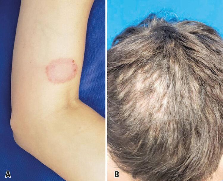
This composite clinical photograph displays two dermatological manifestations of fungal infection (dermatophytosis). Image A shows a classic presentation of tinea corporis on the left upper limb. The lesion is a well-circumscribed, erythematous, annular (ring-shaped) patch with a raised, inflammatory border and central clearing, characteristic of a fungal skin infection. Image B demonstrates a more severe involvement of the scalp (tinea capitis) in a pediatric patient. It reveals diffuse thinning of the hair and significant areas of non-scarring alopecia, particularly in the frontoparietal region. The scalp shows signs of underlying inflammation and scaling. These images illustrate the zoonotic transmission of Trichophyton mentagrophytes, showing both the common 'ringworm' presentation in an adult and a more extensive scalp involvement leading to hair loss in a child. This material is useful for teaching clinical recognition of dermatophytosis and its varied presentations across different anatomical sites and age groups.
tinea corporis dermatophytosis treatment topical antifungal
Fungal Skin Infection - Management (23-Year-Old Female)
Clinical Features to Confirm Diagnosis
- Annular (ring-shaped) lesions with central clearing and a raised, scaly, erythematous advancing border
- Pruritis is common
- KOH (potassium hydroxide) preparation is the most important confirmatory test - scrape the leading edge of the lesion and look for hyphae under microscopy (sensitivity 77-88%)
Management
1. Topical Antifungals (First-line for localized disease)
| Drug Class | Examples | Notes |
|---|---|---|
| Allylamines (preferred) | Terbinafine 1% cream/gel | Once daily for 7 days is highly effective; fungicidal - higher and more rapid cure rate; OTC available |
| Allylamines | Naftifine, Butenafine | Shorter treatment duration vs. azoles |
| Azoles | Clotrimazole, Miconazole, Ketoconazole, Econazole | Fungistatic; effective but require longer treatment; less expensive |
| Ciclopirox | Ciclopirox olamine | Also has some antibacterial coverage |
2. Oral (Systemic) Antifungals
- Infection is widespread (both abdominal area AND back of leg suggests this patient may benefit)
- Poor response to topical therapy
- Recurrent or extensive involvement
| Drug | Dose | Duration |
|---|---|---|
| Terbinafine (preferred) | 250 mg once daily | 2 weeks |
| Itraconazole | 100-200 mg daily (or 2 × 100 mg daily) | 1-2 weeks |
| Fluconazole | 150 mg once weekly | 2-4 weeks |
| Griseofulvin | Weight-based daily dosing | 4-6 weeks (less preferred) |
Note: Oral itraconazole is equal in outcomes to oral terbinafine. Ketoconazole is not preferred orally due to risk of severe hepatotoxicity and QT prolongation. - Goodman & Gilman's Pharmacological Basis of Therapeutics
Non-Pharmacological Measures
- Keep affected areas dry - moisture promotes fungal growth
- Wear loose, breathable clothing (cotton preferred); avoid tight-fitting or occlusive garments
- Avoid sharing towels, clothing, or bedding
- Treat any household contacts or pets if they are a potential source
- Do not use corticosteroid-containing antifungal combinations (e.g., clotrimazole + betamethasone) - these mask the infection and can cause "tinea incognito" with worsening of disease
- Maintain good personal hygiene; pat skin dry after bathing
Differential Diagnoses to Exclude
| Condition | Distinguishing features |
|---|---|
| Nummular eczema | No central clearing; responds to steroids |
| Granuloma annulare | No scale; not pruritic; does not respond to antifungals |
| Psoriasis (inverse) | Silvery scale; nail pitting; KOH negative |
| Pityriasis rosea | Herald patch; "Christmas tree" distribution |
| Contact dermatitis | History of irritant/allergen exposure |
Follow-up
- Review in 2-4 weeks to assess response
- If no improvement with topical therapy after 2 weeks, switch to or add oral antifungal
- Recurrence suggests reinfection source (pets, partner) or immunosuppression - consider fasting blood glucose to rule out diabetes mellitus in recurrent/extensive cases
- Consider fungal culture if KOH is negative but clinical suspicion persists
- Textbook of Family Medicine 9e, Chapter 33 (Tinea Corporis & Tinea Cruris sections)
- Goodman & Gilman's Pharmacological Basis of Therapeutics, Table 75-8 (Cutaneous antifungal therapy recommendations)