
What is a fascia? Also describe topographic anatomy of pectoral region
Reading File
Now let me search the medical library for both topics simultaneously.
Finding Sources
Finding Sources
Reading File
Reading File
Reading File
Reading File
Reading File
I need to go back and get the full muscles of the pectoral region content (the section was cut off earlier):
Reading File
Now let me also fetch the pectoral region diagram image:
Loading Image
I now have all the information needed for a thorough response. Here is the complete answer:
1. Fascia
Definition
Fascia is connective tissue containing varying amounts of fat that:
- Separates, supports, and interconnects organs and structures
- Enables movement of one structure relative to another
- Allows transit of vessels and nerves from one area to another
(Gray's Anatomy for Students, p. 40)
Categories of Fascia
A. Superficial (Subcutaneous) Fascia
- Lies just deep to and attached to the dermis of the skin
- Made of loose connective tissue usually containing a large amount of fat
- Thickness varies considerably between body regions and individuals
- Functions:
- Allows skin movement over deeper structures
- Acts as a conduit for vessels and nerves coursing to and from the skin
- Serves as an energy (fat) reservoir
B. Deep Fascia
- Usually consists of dense, organized connective tissue
- The outer layer attaches to the deep surface of superficial fascia and forms a thin fibrous covering over deeper body regions
- Inward extensions form:
- Intermuscular septa - compartmentalize muscle groups with similar functions and innervations
- Investing fascia - surrounds individual muscles and groups of vessels/nerves
- Retinacula - thickenings near joints that hold tendons in place and prevent bowstringing during joint movement
Special Named Deep Fascial Layers
| Name | Location | Notes |
|---|---|---|
| Transversalis fascia | Between parietal peritoneum and abdominal wall muscles | Separates peritoneum from deep muscle fascia |
| Endothoracic fascia | Thorax | Analogous layer within the thoracic cavity |
| Clavipectoral fascia | Pectoral region | Encloses subclavius and pectoralis minor |
Clinical Importance of Fascia
Fascias are clinically significant because they limit the spread of infection and malignant disease. When infection or tumor crosses a fascial plane, surgical clearance becomes far more extensive.
A classic example: tuberculosis of an intervertebral body can spread laterally into the psoas muscle; pus fills the muscle but is contained by the psoas fascia, which extends inferiorly into the groin, causing the pus to point below the inguinal ligament (psoas abscess).
2. Topographic Anatomy of the Pectoral Region
The pectoral region is external to the anterior thoracic wall and helps anchor the upper limb to the trunk.
Compartments
The pectoral region has two compartments:
| Compartment | Contents |
|---|---|
| Superficial | Skin, superficial fascia, and breasts |
| Deep | Muscles and associated structures |
Nerves, vessels, and lymphatics in the superficial compartment emerge from the thoracic wall, the axilla, and the neck.
Superficial Compartment: The Breast
- The mammary glands are modified sweat glands in the superficial fascia, anterior to the pectoral muscles and the anterior thoracic wall
- Consist of 15-20 lactiferous ducts converging at the nipple, surrounded by the areola
- Suspensory ligaments of the breast (Cooper's ligaments) - condensations of connective tissue stroma that attach to the dermis and support the breast; carcinoma creates tension on these, causing skin pitting
- The breast lies on deep fascia covering pectoralis major, separated from it by the retromammary space (loose connective tissue that allows mobility)
- The base of the breast extends:
- Vertically: rib II to rib VI
- Horizontally: sternum to midaxillary line
Vascular Supply
- Laterally: axillary artery branches (superior thoracic, thoraco-acromial, lateral thoracic, subscapular arteries)
- Medially: branches from the internal thoracic artery
- Perforating branches of the 2nd-4th intercostal arteries
Venous Drainage
Drains via axillary, internal thoracic, and intercostal veins.
Innervation
Anterior and lateral cutaneous branches of intercostal nerves II-VI. The nipple is specifically innervated by the 4th intercostal nerve.
Lymphatic Drainage
Lymph drains to axillary, supraclavicular, and parasternal nodes, and may spread to abdominal nodes or even the opposite breast - making containment of metastatic breast cancer difficult.
Deep Compartment: Muscles of the Pectoral Region
Each pectoral region contains three muscles: pectoralis major, pectoralis minor, and subclavius. All originate from the anterior thoracic wall and insert into bones of the upper limb.
| Muscle | Origin | Insertion | Innervation | Function |
|---|---|---|---|---|
| Pectoralis major | Medial half of clavicle, anterior sternum, 1st-7th costal cartilages, external oblique aponeurosis | Lateral lip of intertubercular sulcus of humerus | Medial and lateral pectoral nerves | Adduction, medial rotation, and flexion of the humerus |
| Subclavius | Rib I at junction with costal cartilage | Groove on inferior surface of middle third of clavicle | Nerve to subclavius | Pulls clavicle medially, stabilizes sternoclavicular joint; depresses tip of shoulder |
| Pectoralis minor | Anterior surfaces of ribs III-V and overlying deep fascia | Coracoid process of scapula | Medial pectoral nerves | Depresses tip of shoulder; protracts scapula |
Clavipectoral Fascia
A continuous layer of deep fascia - the clavipectoral fascia - encloses the subclavius and pectoralis minor, attaching to the clavicle above and to the floor of the axilla below. Nerves, vessels, and lymphatics pass through this fascia between the subclavius and pectoralis minor, or pass under the inferior margin of pectoralis major and minor to communicate between the pectoral region and the axilla.
The muscles of the pectoral region collectively form the anterior wall of the axilla.
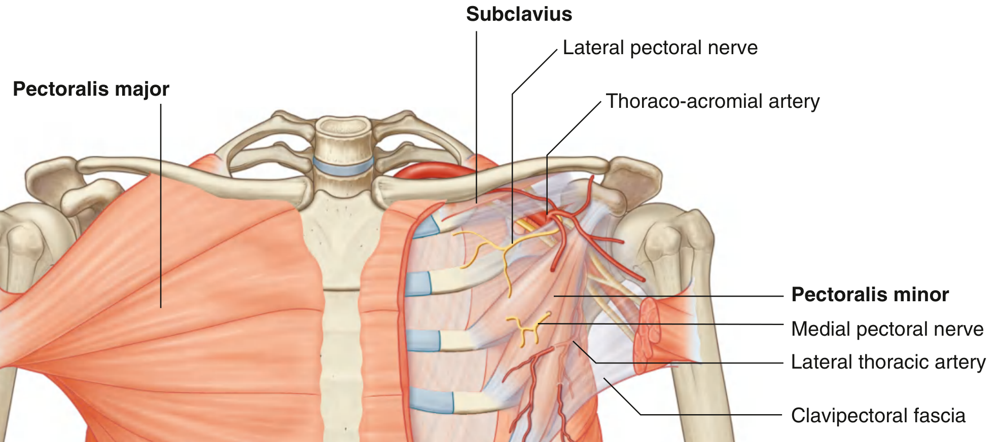
Fig. 3.18 - Muscles and Fascia of the Pectoral Region (Gray's Anatomy for Students)
Source: Gray's Anatomy for Students, Chapter 3 (Thorax), pp. 40, 177-178
This is a shared conversation. Sign in to Orris to start your own chat.