Side swipe injury etiology mechanism of injury associated injury signs and symptoms and investigations and evaluation treatment explain in detail with appropriate illustrative diagrams and images
Reading File
Finding Sources
Finding Sources
Finding Sources
Searching the Web
sideswipe injury elbow mechanism associated injuries treatment orthopedics
Reading File
Finding Sources
Finding Sources
Searching PubMed
"sideswipe injury" elbow
Reading Article
Reading Article
Reading File
Finding Sources
Reading File
Finding Sources
Searching the Web
sideswipe injury elbow "traffic elbow" fracture dislocation classification injuries vascular nerve
Reading Web Page
sideswipe injury mechanism associated injuries treatment signs symptoms
Finding Sources
Reading File
Reading File
Reading File
Loading Image
Loading Image
Loading Image
Generating Image
Now I have all the necessary content. Let me generate the comprehensive detailed answer.
Sideswipe Injury (Traffic Elbow / Baby Car Injury)
Definition
A sideswipe injury is a high-energy traumatic injury to the elbow and/or upper limb sustained when a passenger or driver rests their elbow on, or protrudes their arm through, a vehicle window - and the exposed limb is struck by a passing vehicle or a fixed object. It is also called "traffic elbow" or "baby car injury" in the literature. Because no standardized classification covers all its presentations, it is broadly termed a complex elbow injury.
Etiology
Sideswipe injuries occur in the following settings:
| Setting | Mechanism |
|---|---|
| Car/bus passenger | Elbow resting on or protruding through an open window, struck by a passing vehicle |
| Driver | Arm resting on the window sill during a side-impact collision |
| Train passenger | Window seat with elbow outside, struck by a fixed object or passing vehicle at a rail crossing (reported by Pandey et al., 2021, PMID 34858714) |
| Tricycle passenger | Limb protrusion struck by a fixed or moving object (can involve the knee rather than the elbow) |
| Mountain/narrow road collisions | Vehicle squeezing through a narrow gap, arm struck against a rock face or another vehicle |
Vehicle-to-vehicle collisions are the most common type of side-impact collision, but fixed-object collisions account for approximately 37% of serious-to-fatal side-impact injuries. Alcohol intoxication is a significant contributory factor in many reported series. The injuries tend to cluster in the late afternoon hours (1700-1900 hrs) when road traffic is dense.
Mechanism of Injury
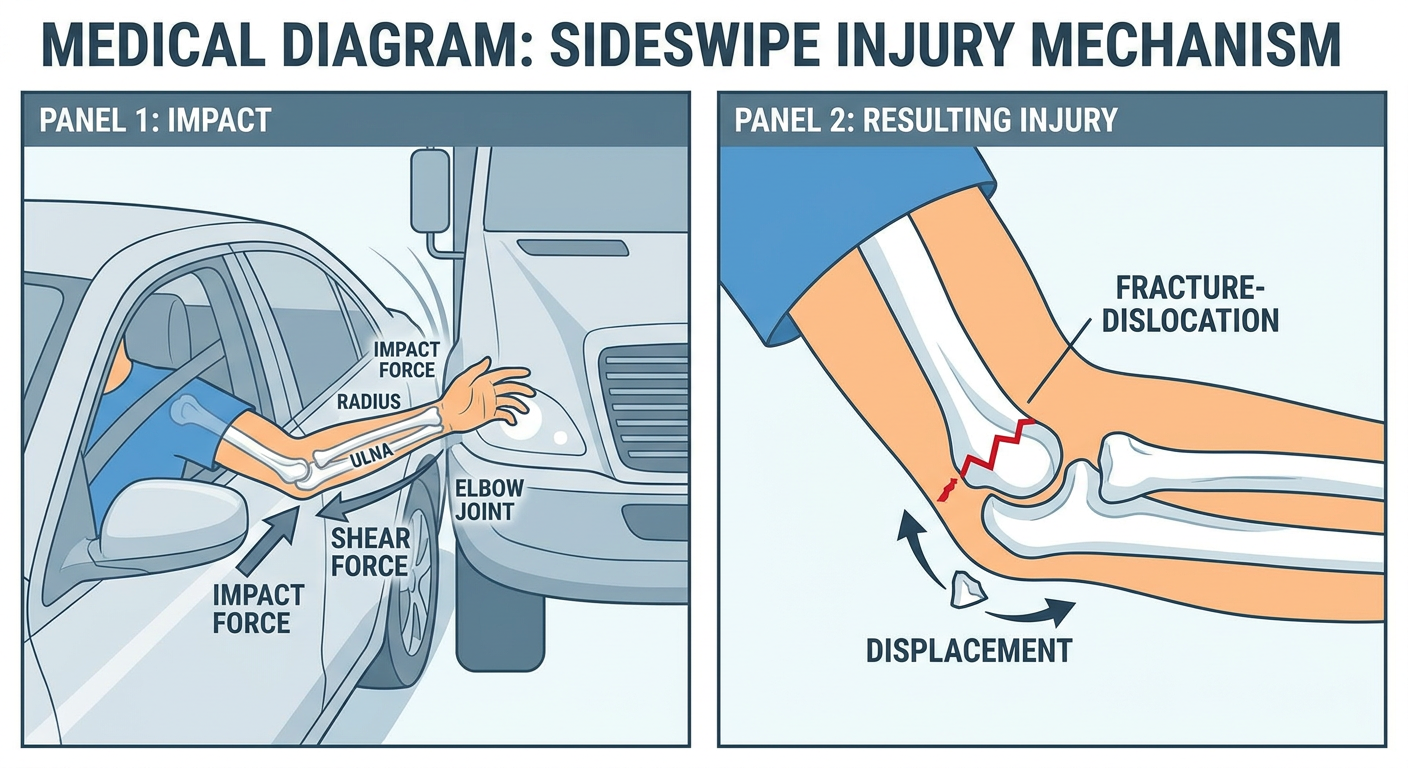
The key biomechanical principle involves a violent lateral blow to the elbow and proximal forearm while the arm is in a semi-extended or extended position resting outside the vehicle. The forces involved are:
- Direct impact force - a lateral or posterolateral crushing force directed at the elbow region from the striking object
- Shear force - tangential scraping/grinding forces that produce extensive soft tissue stripping and degloving
- Compressive-torsional force - the combination of compression along the forearm axis with a rotational component, accounting for Monteggia-pattern and forearm fractures
- Avulsion force - the momentum of the vehicle can pull tissues off the bone (periosteal stripping, tendon avulsion)
These simultaneous multi-directional forces explain why a single sideswipe event can produce:
- Open wounds with bone exposure
- Comminuted multi-bone fractures
- Joint dislocation
- Neurovascular injury
- Soft tissue degloving/loss
As described in Pye's Surgical Handicraft (22nd edition): "This is usually a severe injury, often compound and with considerable comminution. An example of this is the 'side-swipe' injury caused by a violent blow to the elbow hanging out of a car window."
Elbow Anatomy Relevant to Sideswipe Injury
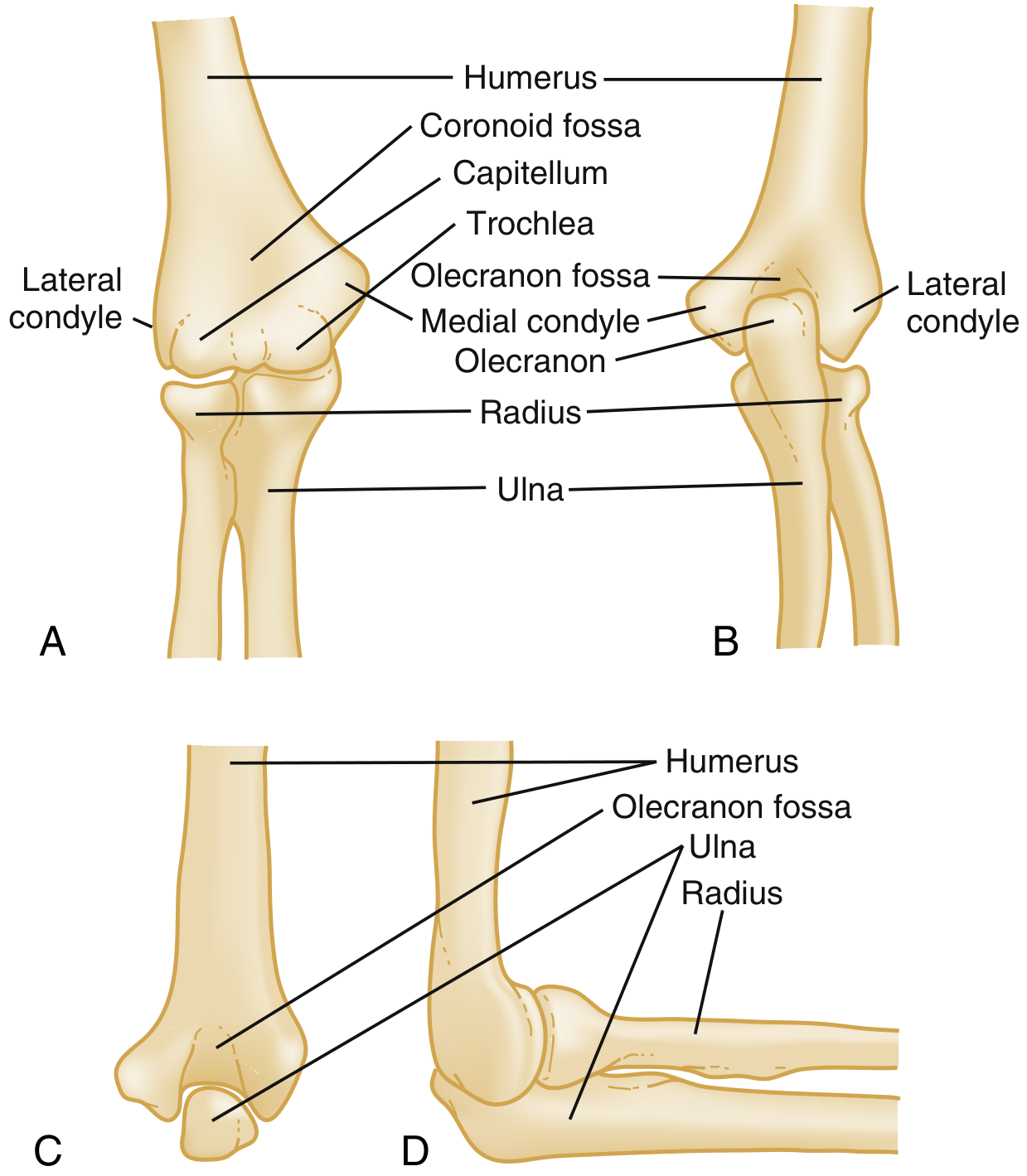
(Bony anatomy of the elbow: humerus, capitellum, trochlea, olecranon fossa, medial and lateral condyles, radius, and ulna - Rosen's Emergency Medicine)
The following structures at the elbow are at risk in sideswipe injury:
- Distal humerus (condyles, epicondyles, supracondylar region)
- Olecranon and coronoid process of the ulna
- Radial head and neck
- Brachial artery - passes anterior to the elbow joint and is vulnerable to laceration or transection
- Median nerve - runs medial to the brachial artery anteriorly
- Radial nerve / posterior interosseous nerve - passes around the radial neck laterally
- Ulnar nerve - passes posterior to the medial epicondyle in the cubital tunnel
- Brachial plexus - can be injured in very high-energy mechanisms
Associated Injuries
Sideswipe injuries characteristically produce a complex pattern rather than a single isolated injury. The most frequent combinations are:
Osseous (Bone) Injuries
| Bone Structure | Injury |
|---|---|
| Distal humerus | Comminuted distal humeral fracture (most common bony injury) |
| Proximal ulna | Olecranon fracture, coronoid fracture, Monteggia fracture-dislocation |
| Radial head/neck | Radial head fracture (part of "terrible triad") |
| Humeral shaft | Shaft fracture (especially at junction of middle and distal thirds) |
| Both forearm bones | Radius + ulna shaft fractures ("both bones forearm") |
The most frequent fracture pattern reported across series is shaft humerus combined with distal humerus and/or proximal ulna fractures.
Monteggia Fracture-Dislocation
Pandey et al. (2021) reported an open proximal forearm Monteggia fracture-dislocation with bone and soft tissue loss as the consistent pattern in their railway track sideswipe series.
Soft Tissue Injuries
- Degloving injuries of the forearm and elbow (skin stripped off while underlying structures remain)
- Open wounds with exposed bone - Gustilo-Anderson Grade I to Grade III
- Laceration/rupture of the triceps tendon
- Rupture of collateral ligaments (lateral collateral ligament, ulnar collateral ligament)
- Elbow joint capsule disruption
- Compartment syndrome of the forearm
Neurovascular Injuries
| Structure | Injury | Clinical Effect |
|---|---|---|
| Brachial artery | Laceration, transection, intimal tear | Absent radial/ulnar pulse, ischemia, compartment syndrome |
| Radial nerve / PIN | Contusion, laceration, avulsion | Wrist drop, inability to extend fingers/thumb, patchy dorsal hand sensory loss |
| Ulnar nerve | Contusion, laceration | Loss of intrinsic muscle function, small finger sensory loss |
| Median nerve | Contusion, laceration | Thenar wasting, "OK sign" weakness, lateral thumb sensation loss |
| Brachial plexus | Traction avulsion (high energy) | Complete upper limb paralysis (rare) |
Vascular injury to the brachial artery can occur in up to 10-20% of patients with severe periarticular elbow fractures (Campbell's Operative Orthopaedics, 15th ed.). Crucially, due to the rich collateral blood supply around the elbow, the hand may remain warm and pink despite complete brachial artery disruption - so absent pulse alone cannot rule out vascular injury.
Associated Lower Limb Injuries
When two simultaneous impacts occur (e.g., the body hits the door frame and the arm is struck by an external object), associated lower limb injuries have been reported.
Signs and Symptoms
Local Examination (Elbow and Upper Limb)
Inspection:
- Open wound with active bleeding
- Exposed bone fragments (open fracture)
- Visible degloving with skin loss
- Deformity of the elbow (may show posterolateral displacement as in fracture-dislocation)
- Swelling and hematoma
- Inability to use the upper limb
Palpation:
- Severe localized pain
- Crepitus over fracture sites
- Loss of normal bony landmarks (in elbow dislocation, the equilateral triangle formed by the two epicondyles and olecranon tip is disrupted)
- Tenderness over the radial head, medial/lateral epicondyles, olecranon
Functional Assessment:
- Inability to flex/extend the elbow
- Inability to pronate/supinate the forearm
- Wrist drop (radial nerve injury)
- Inability to extend MCP joints/thumb (posterior interosseous nerve)
- Patchy sensory changes over the hand
Neurovascular Status (Critical Assessment)
| Nerve | Motor Test | Sensory Test |
|---|---|---|
| Radial nerve | Wrist extension, finger/thumb MCP extension | Dorsum of hand (first web space) |
| Median nerve | "OK sign" (thumb-index pinch), thumb opposition | Lateral palm, thumb, index, middle fingers |
| Ulnar nerve | Finger abduction (small finger), intrinsic grip | Medial palm, ring and small fingers |
Vascular:
- Radial and ulnar pulse palpation (but note: absence may not be apparent if collaterals are maintaining perfusion)
- Capillary refill time
- Hand color and temperature
- Sensation (as paresthesia may indicate ischemia)
Systemic Assessment
Patients should be managed under Advanced Trauma Life Support (ATLS) protocol on arrival, as sideswipe injuries can be associated with:
- Hemorrhagic shock (open wounds with major vessel involvement)
- Associated thoracic, abdominal injuries (if vehicle occupant in multi-trauma)
Investigations and Evaluation
Imaging
Plain Radiography (First-Line)
- AP and lateral views of the elbow (mandatory)
- AP lateral view of the entire forearm including wrist (to exclude Monteggia, Galeazzi variants)
- Lateral view of the humerus if shaft fracture suspected
- Findings: fracture lines, dislocation, intra-articular fragments, degree of comminution
On lateral radiograph, look for:
- Posterior fat pad sign - indicates hemarthrosis even with subtle fractures
- Anterior fat pad ("sail sign") - also indicates joint effusion
- Disruption of the radiocapitellar line
CT Scan (Gold Standard for Surgical Planning)
- CT with 3D reconstruction is essential for:
- Defining fracture pattern precisely
- Identifying coronoid fractures (often missed on plain films)
- Assessing degree of comminution
- Guiding surgical approach and fixation strategy
- Classifying with the Wrightington or OTA/AO classification
Vascular Studies
- Duplex Doppler ultrasound - non-invasive first-line
- CT Angiography (CTA) - preferred when vascular injury is suspected clinically
- Formal angiography - reserved for cases requiring interventional planning
MRI
- Not typically used in the acute setting
- Useful for ligamentous injury assessment if stability is uncertain after initial treatment
Laboratory Investigations
- Full blood count, urea/electrolytes, group and crossmatch
- Coagulation profile
- Injury Severity Score (ISS) documentation
Treatment
Treatment of sideswipe injury is challenging because it does not fit a single standard injury pattern. A staged, multidisciplinary approach is the standard of care.
Phase 1 - Emergency Resuscitation (On Arrival)
- ATLS protocol - Airway, Breathing, Circulation
- IV antibiotics - Immediate broad-spectrum cover (e.g., cefazolin + metronidazole + aminoglycoside for open fractures)
- Tetanus prophylaxis
- Wound lavage - Copious irrigation of open wounds with normal saline
- Hemorrhage control - Pressure dressings, tourniquet if exsanguinating hemorrhage
- Temporary splintage - Above-elbow posterior splint to immobilize and reduce pain
Phase 2 - Wound Debridement and Fracture Stabilization (Urgent Surgery)
Open Fracture Management (Gustilo Principles):
- The priorities are: wound irrigation and debridement, IV antibiotics, tetanus coverage, fracture stabilization, and appropriate soft tissue management (Rockwood and Green's, 10th ed.)
- Contaminated/devascularized bone is excised - EXCEPT when dealing with articular surface fragments, where preservation is preferred to avoid posttraumatic arthritis
Fracture Fixation Options:
| Method | Indication |
|---|---|
| External fixation | Initial stabilization of unstable open fractures, spanning fixator across elbow, allows soft tissue management |
| ORIF with plate and screws | Definitive fixation after soft tissue recovery; distal humerus bicolumnar plating |
| Intramedullary nailing | Humeral shaft fractures |
| Tension band wiring | Olecranon fractures (comminuted = plate preferred) |
| Radial head ORIF or replacement | Radial head fractures depending on fragment size |
| External fixator (hinged) | Complex instability; allows early motion |
For Monteggia Fractures (common in sideswipe series):
- ORIF of the ulna fracture with anatomic restoration of ulnar bow
- Radial head will usually reduce after ulnar fixation; if not, open reduction of radial head required
Phase 3 - Vascular Repair
Algorithm:
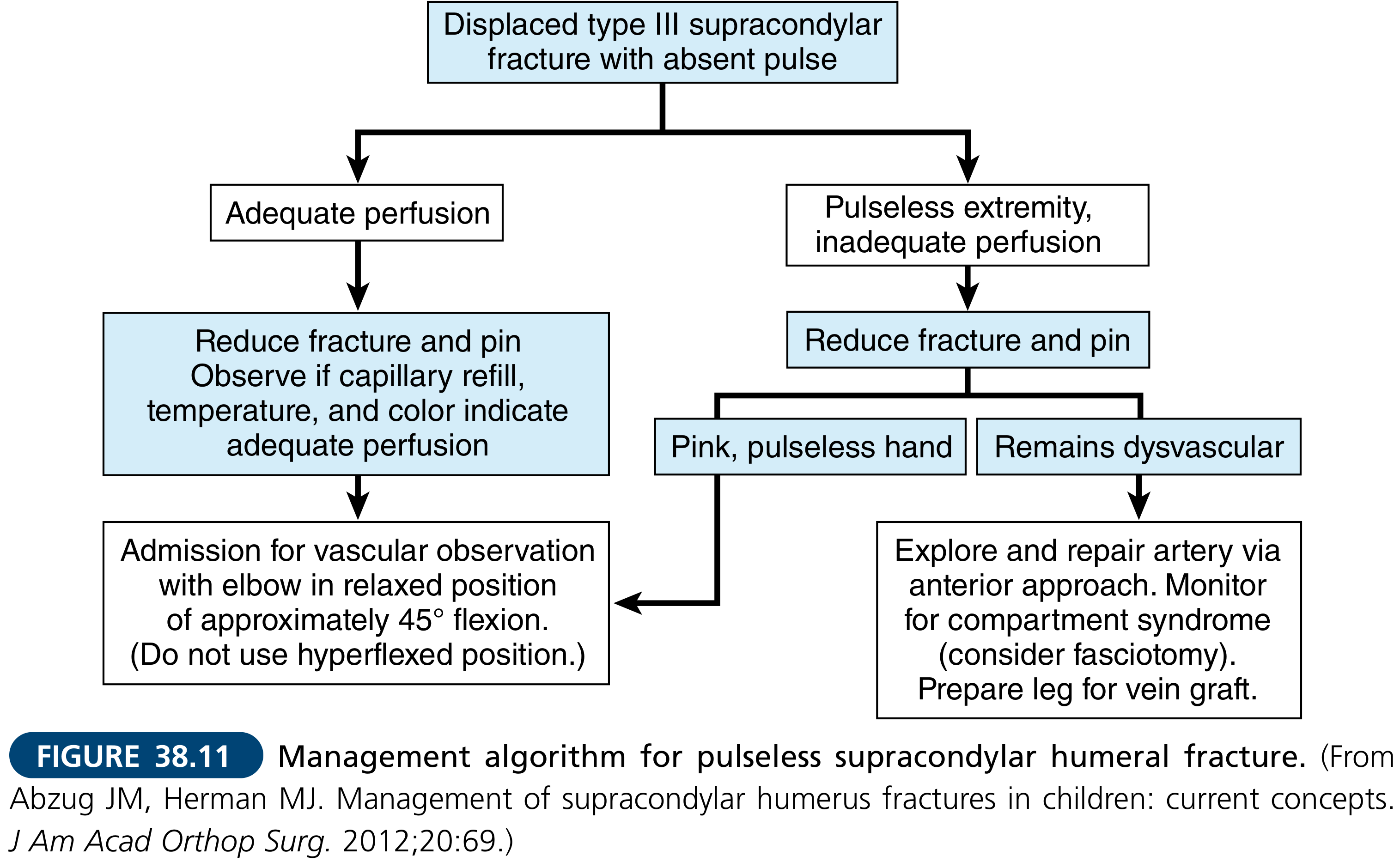
(Management algorithm for pulseless elbow fracture with vascular injury - Campbell's Operative Orthopaedics)
- Brachial artery injury requires urgent vascular surgery consultation
- If hand is pulseless and ischemic after fracture reduction: explore and repair brachial artery via anterior approach with vein graft from the leg
- Collateral circulation may maintain a pink, pulseless hand - this warrants close observation
- Compartment syndrome must be monitored; fasciotomy is performed at low threshold
Phase 4 - Nerve Exploration
- Radial nerve: exploration is performed for established motor deficit (wrist drop, PIN palsy); nerve is explored through posterior or lateral approach
- In Pandey et al.'s series, all four patients underwent radial nerve exploration as part of the staged protocol
- Ulnar and median nerve injuries: primary repair where technically feasible; nerve grafting for segmental loss
- Outcome note: Nerve injury is the most important factor for dismal functional outcomes; only ~25% of ulnar nerve injuries fully recover (Campbell's, 15th ed.)
Phase 5 - Soft Tissue Reconstruction
- Wound closure should not be performed primarily in contaminated injuries
- After debridement, wounds are left open and covered with moist dressings or negative pressure wound therapy (NPWT/VAC)
- Secondary closure at 48-72 hours if wound is clean
- Skin grafting for degloving injuries
- Flap reconstruction (local or free) for exposed bone/hardware where direct closure is not possible
Phase 6 - Rehabilitation
- Early range of motion exercises (30-120 degrees active) once stability achieved - critical to prevent stiffness
- Physiotherapy initiated within first 3 weeks
- Splinting in functional position between exercises
- Stiffness is the most common long-term outcome even in successfully managed cases (Pye's)
- Heterotopic ossification prophylaxis (indomethacin or single-dose radiation) considered for high-energy injuries
Complications
| Complication | Comment |
|---|---|
| Elbow stiffness and contracture | Most common; often permanent |
| Nonunion | Especially with bone loss; may require bone grafting |
| Infection / osteomyelitis | Risk increases with open/contaminated wounds (11% deep infection in some series) |
| Nerve palsy (permanent) | Radial nerve most common; brachial plexus injury rare but devastating |
| Vascular compromise / limb loss | Amputation required if vascular repair fails or delayed |
| Compartment syndrome | Forearm; urgent fasciotomy required |
| Heterotopic ossification | Leads to additional stiffness |
| Hardware failure | Especially in comminuted fractures with osteopenic bone |
| Post-traumatic arthritis | Especially with articular surface comminution |
Key Points Summary
- Sideswipe injury = violent lateral blow to the elbow protruding from a vehicle window - a high-energy, complex, open injury
- The classic triad is: open fracture + soft tissue loss + neurovascular injury
- Most common fracture pattern: distal humerus + proximal ulna (Monteggia type) + radial head fractures
- Brachial artery injury in 10-20% of severe cases; a warm pink pulseless hand does not exclude arterial injury
- Radial nerve palsy is the most common nerve injury and the main determinant of poor outcome
- Management is staged: debridement first, then fixation, then soft tissue reconstruction
- Prognosis is guarded; stiffness, contracture, and nerve deficits are common
Sources:
- 22nd Edition Pye's Surgical Handicraft, p. 187 - Fracture-dislocation of the elbow / sideswipe injury
- Rosen's Emergency Medicine, 9th ed. - Elbow trauma, neurovascular assessment
- Campbell's Operative Orthopaedics, 15th ed. 2026 - Vascular injury in elbow fractures
- Rockwood and Green's Fractures in Adults, 10th ed. 2025 - Open distal humerus fractures
- Pandey R et al. "An unusual mechanism of sideswipe injury in passengers traveling in a train and review of literature." Int J Burns Trauma 2021 [PMID: 34858714]
- Oyewole OA et al. "Sideswipe Injury of the Knee." Niger Med J 2020 [PMID: 33487856]
This is a shared conversation. Sign in to Orris to start your own chat.