
Whqt is a plastic anemia causes etiology ant treatment
"aplastic anemia"[MeSH Terms] AND treatment
aplastic anemia bone marrow biopsy hypocellular
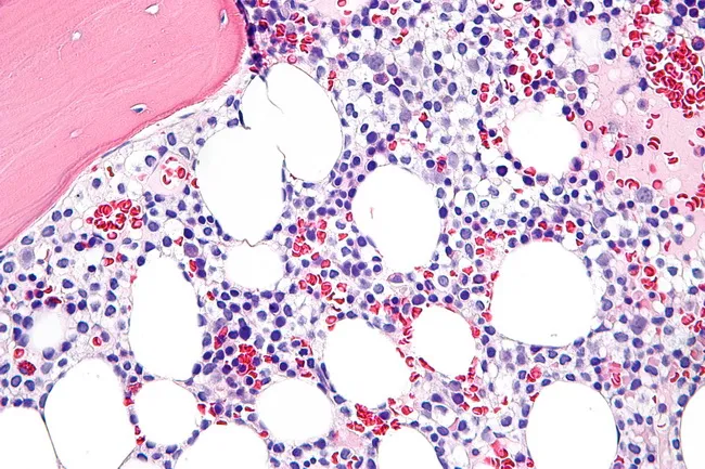
Histopathology: Bone marrow biopsy prepared for light microscopy shows diffuse infiltration by hairy cells rather than well-formed nodules. The marrow is often hypercellular in established disease but can be hypocellular in early stages, mimicking aplastic anemia. Hairy cells appear monotonous with oval nuclei and abundant clear cytoplasm that yields a characteristic fried egg morphology on H&E sections. In some cases, hair-like cytoplasmic projections (hairy indentations) are accentuated with specialized staining such as DBA.44 immunostain, supporting a hairy cell leukemia (HCL) diagnosis. The tumor cells may display spindled forms; the immunophenotype typically includes DBA.44 positivity, and cells may express CD11c, CD25, and CD103 on broader panels. Normal hematopoietic elements, particularly myeloid precursors, are reduced and can show dysplastic changes reminiscent of myelodysplastic syndrome. Plasma cells and mast cells may be mildly increased. Significant reticulin fibrosis is common, contributing to difficult marrow aspirates and making core biopsy essential to determine the extent of marrow replacement. Although marrow evaluation is informative, many cases of HCL can be diagnosed from peripheral blood smear and immunophenotyping alone; however, the biopsy provides baseline disease burden and a reference for assessing therapeutic response and fibrosis progression. This image exemplifies classic HCL marrow involvement with fried egg cells and conspicuous reticulin fibrosis.
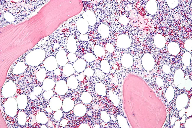
Light microscopy of a bone marrow biopsy from hairy cell leukemia (HCL) shows diffuse infiltration by small to medium lymphoid cells with abundant clear cytoplasm and oval, occasionally reniform nuclei; the cytoplasm creates a fried egg appearance. Hairy projections may be highlighted by immunostaining and by DBA.44 positivity, along with CD11c, CD25, and CD103 markers. The marrow is commonly hypercellular in established disease, whereas early involvement may be hypocellular and mimic aplastic anemia. Prominent reticulin fibrosis is typical and can hinder aspirate yield, making core biopsy essential for assessing infiltration and baseline disease burden. Normal hematopoietic elements, especially the myeloid lineage, are reduced and may display dysplastic changes resembling myelodysplastic syndrome. Plasma cells and mast cells may be mildly increased. In some cases, hairy cells assume a spindle-shaped morphology. The neoplastic cells are typically monotonous with minimal prominent nucleoli, and cytoplasm may exhibit cytoplasmic processes. Unlike several small B-cell lymphomas, discrete cellular aggregates are uncommon, with diffuse replacement predominating. Immunohistochemistry and flow cytometry confirm the diagnosis via characteristic immunophenotype. Clinically, marrow involvement informs staging, prognosis, and treatment planning, though peripheral blood smear and immunophenotypic profiling often suffice for diagnosis. This image, captured at high magnification on a hematoxylin and eosin stained section, showcases classic hairy cell features.
Aplastic Anemia - Causes, Etiology, and Treatment
Definition
- Harrison's Principles of Internal Medicine, 22nd Ed., p. 852
Epidemiology
- Incidence: 2 per million/year in Europe and Israel; 5-7 per million/year in Thailand and China
- Affects both sexes equally
- Bimodal age distribution: major peak in teens and twenties, second peak in older adults
Etiology (Causes)
Acquired Causes
| Category | Examples |
|---|---|
| Radiation | Medical radiation therapy, accidental/occupational exposure; damages DNA in actively dividing cells |
| Chemicals | Benzene (most notorious); industrial solvents |
| Drugs - dose-dependent | Chemotherapy agents, conditioning regimens for transplant (all recipients affected predictably) |
| Drugs - idiosyncratic | Chloramphenicol, NSAIDs, sulfonamides, antiepileptics, gold - no clear dose-response; rare but unpredictable |
| Viral infections | EBV (infectious mononucleosis), seronegative hepatitis (non-A, non-B, non-C), Parvovirus B19, HIV-1 |
| Immune diseases | Eosinophilic fasciitis, hypoimmunoglobulinemia, large granular lymphocytosis (LGL), thymoma/thymic carcinoma, graft-versus-host disease |
| PNH | Paroxysmal nocturnal hemoglobinuria (closely related; clonal overlap) |
| Pregnancy | Rare association |
| Idiopathic (immune) | Most cases - no identifiable trigger; immune-mediated destruction is presumed |
Constitutional/Inherited (IBMFS - Inherited Bone Marrow Failure Syndromes)
| Syndrome | Features |
|---|---|
| Fanconi anemia | Physical anomalies (radial defects, cafe-au-lait spots), childhood onset |
| Dyskeratosis congenita | Telomere biology disorder; nail dystrophy, oral leukoplakia, skin pigmentation |
| Shwachman-Diamond syndrome | Exocrine pancreatic insufficiency + marrow failure |
| GATA2 deficiency | Can present as marrow failure in normal-appearing adults |
| RUNX1 familial platelet disorder | Germline predisposition |
| Diamond-Blackfan anemia | Pure red cell aplasia variant |
- Harrison's, p. 852-853
Pathogenesis
1. Immune-Mediated (Extrinsic) - Most Common
- Stem cells are antigenically altered by drugs, infectious agents, or unknown environmental insults
- This triggers a cellular immune response: activated Th1 cells produce cytokines (IFN-γ, TNF-α) that suppress hematopoietic progenitors
- Evidence: immunosuppressive therapy directed against T cells restores hematopoiesis in 60-70% of patients
2. Intrinsic Stem Cell Abnormality
-
5-10% of patients have inherited defects in telomerase → premature senescence of HSCs
-
An additional 50% of cases have unusually short telomeres (possibly from undiscovered telomerase defects or excessive HSC replication)
-
Genetically altered stem cells may also express neoantigens, making them targets for T-cell attack
-
Robbins & Kumar Basic Pathology, p. 443
Clinical Features
- Pancytopenia causes three major symptom clusters:
- Anemia: insidious weakness, pallor, dyspnea (slowly progressive)
- Thrombocytopenia: petechiae, ecchymoses, mucosal bleeding
- Neutropenia: recurrent serious infections
- No splenomegaly - if present, another diagnosis should be sought
- Diagnosis: bone marrow biopsy showing hypocellularity (>70% fat spaces), pancytopenia on CBC
Severity Classification (International AA Study Group)
| Severity | Criteria |
|---|---|
| Severe (SAA) | BM cellularity <25%, plus ≥2 of: neutrophils <0.5×10⁹/L, platelets <20×10⁹/L, reticulocytes <20×10⁹/L |
| Very Severe (vSAA) | SAA criteria + neutrophils <0.2×10⁹/L |
| Moderate/Non-severe | Does not meet above thresholds |
Treatment
1. Hematopoietic Stem Cell Transplantation (HSCT)
- Treatment of choice for young patients (ideally <40 years) with severe/very severe AA who have a matched sibling donor
- Often curative
- Requires conditioning with immunosuppressive chemotherapy/radiation
- Minimize pre-transplant transfusions - sensitization to alloantigens causes high engraftment failure rates
2. Immunosuppressive Therapy (IST)
- For patients who are not transplant candidates (older age, no matched donor, or non-severe disease)
- Standard regimen: Anti-thymocyte globulin (ATG) + Cyclosporine A (CsA)
- Horse ATG (hATG) preferred over rabbit ATG based on clinical evidence
- Restores hematopoiesis in 60-70% of patients
- Eltrombopag (thrombopoietin receptor agonist) added to IST has significantly improved response rates - now standard in frontline treatment of SAA
- A 2024 meta-analysis (PMID 38576005) confirmed IST + eltrombopag improves complete response rates vs IST alone
- A 2026 meta-analysis (PMID 41569462) further evaluated immunosuppressive and pharmacological therapies with/without equine ATG
3. Supportive Care
- Red cell transfusions (minimize pre-transplant)
- Platelet transfusions for bleeding risk
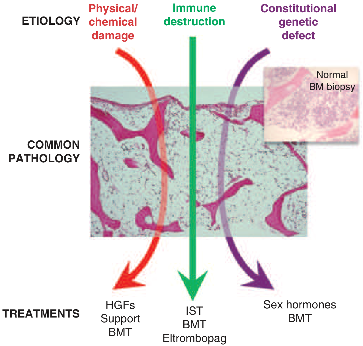
- Hematopoietic growth factors (G-CSF, erythropoietin) - primarily for physical/chemical damage etiology
- Antibiotics/antifungals for infection prophylaxis and treatment
4. Constitutional/Inherited Disease
- Sex hormones (androgens like danazol) - for telomere biology disorders and Fanconi anemia
- BMT - also applicable
Special Notes
- If an inciting drug is identified, withdrawal sometimes leads to recovery (but rare)
- Aplastic anemia has no splenomegaly - differentiates it from myelophthisic anemia and leukemia
- Monitor for clonal evolution to MDS or PNH (especially after IST)
Summary Table
| Aspect | Key Points |
|---|---|
| Definition | Pancytopenia + hypocellular bone marrow |
| Main cause | Immune-mediated T-cell destruction of HSCs (idiopathic) |
| Key drugs | Chloramphenicol, NSAIDs, sulfonamides, chemotherapy |
| Key infections | Seronegative hepatitis, EBV, parvovirus B19, HIV |
| Diagnosis | BM biopsy (>70% fat), CBC pancytopenia |
| 1st line if young + matched donor | HSCT (curative) |
| 1st line if no donor / older | hATG + Cyclosporine + Eltrombopag |
| No splenomegaly | Key distinguishing feature |