
sarcoidosis as an article i can read from whole as a neet pg aspirant and i shouldnt need anything else also include videos from osmosis
osmosis sarcoidosis video youtube
sarcoidosis granuloma histology chest X-ray clinical findings
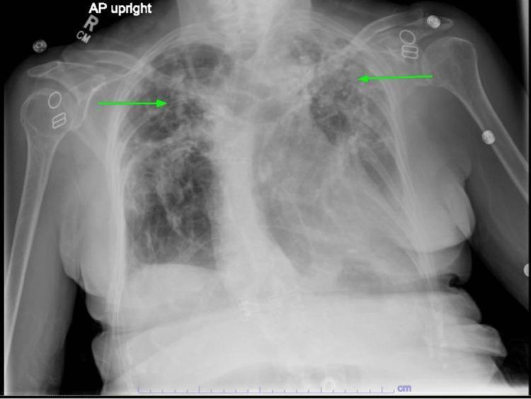
This diagnostic chest x-ray (AP upright projection) demonstrates bilateral upper lung predominant pathology, highlighted by green arrows. The imaging shows extensive areas of increased opacity and reticular markings consistent with chronic fibrotic changes, particularly pronounced in the left apical and upper lobe regions. These findings are characteristic of stage IV sarcoidosis with pulmonary fibrosis and associated architectural distortion, including apical scarring and traction bronchiectasis. The lower lung fields remain relatively lucent compared to the apices. Visible anatomical structures include the bony thorax, mediastinal silhouette, and bilateral humeri. Metallic densities are noted overlying the shoulder regions, likely representing external ECG leads or medical hardware. The radiograph serves as a clinical example of parenchymal lung disease progression and the typical upper-lobe distribution of sarcoidosis-related fibrosis.
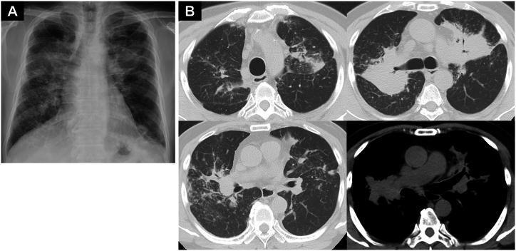
This composite diagnostic image features a posterior-anterior (PA) chest X-ray and axial chest computed tomography (CT) scans illustrating systemic sarcoidosis. (A) The chest X-ray shows bilateral, ill-defined hilar and mid-zone opacities, consistent with lymphadenopathy and pulmonary parenchymal involvement. (B) The CT scans (lung and mediastinal windows) provide detailed views of extensive mediastinal and bilateral hilar lymphadenopathy. Significant parenchymal findings include bilateral tumor-like consolidations and diffuse micronodular (granular) shadows with a perilymphatic distribution, characteristic of Stage II or III pulmonary sarcoidosis. Some lesions appear as large, dense masses mimicking malignancy, while others are small and scattered. The mediastinal window specifically highlights the conglomerate enlargement of the paratracheal and subcarinal lymph nodes. These findings, in conjunction with elevated ACE and sIL-2R levels mentioned in clinical context, demonstrate the multisystemic nature of the disease and its potential for varied radiological manifestations.
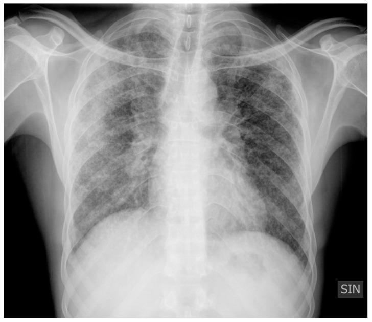
Anteroposterior (AP) chest X-ray demonstrating characteristic findings of pulmonary sarcoidosis, categorized as Scadding Stage II. The image reveals bilateral, symmetric hilar lymphadenopathy, characterized by prominent, well-defined densities at the lung roots. The lung parenchyma shows diffuse abnormalities, including multiple micronodules with a peribronchovascular distribution and extensive reticulonodular opacities scattered throughout both lung fields. Additionally, there is a localized area of pulmonary consolidation visible in the right lung. These findings represent a combination of thoracic adenopathy and active parenchymal infiltrates. The overall radiographic pattern is typical of a granulomatous lung disease, illustrating the transition from isolated nodal involvement to interstitial lung disease. This diagnostic image is intended for educational use in pulmonology and radiology to identify nodal and parenchymal patterns in sarcoidosis.
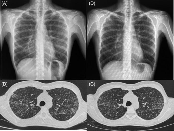
This composite clinical diagnostic image presents a longitudinal comparison of thoracic imaging findings in a patient with pulmonary sarcoidosis. Panels (A) and (D) are posteroanterior chest X-rays. Image (A) reveals diffuse, bilateral, discrete pulmonary infiltrates distributed throughout both lung fields. Image (D), taken over a year later, demonstrates the chronicity of these findings with stable bilateral interstitial opacities. Panels (B) and (C) show axial non-contrast chest CT scans at the level of the upper lobes. Image (B) highlights diffuse, bilateral micronodular pulmonary infiltrates with a distinct perilymphatic distribution, particularly along the peribronchovascular bundles and interlobar fissures. Image (C) shows the same anatomical section months later, indicating persistent micronodularity without significant radiological progression or resolution. The series illustrates the typical radiological presentation of pulmonary sarcoidosis, characterized by micronodular interstitial patterns that remain relatively stable over time, and demonstrates the utility of multi-modal imaging (X-ray and CT) in monitoring chronic granulomatous lung disease.
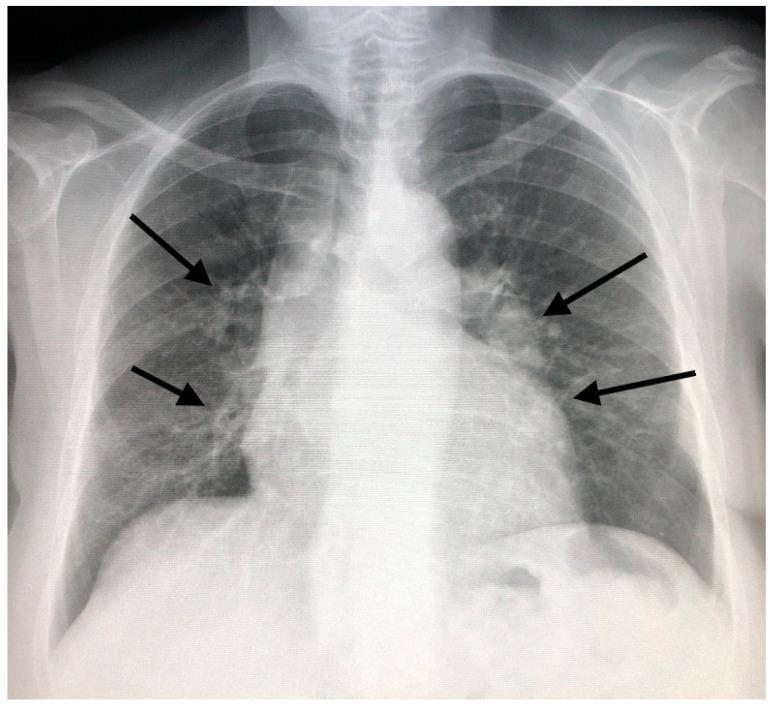
This diagnostic image is a posteroanterior (PA) chest X-ray of an adult patient. The primary findings include bilateral, diffuse pulmonary infiltrates characterized by reticulonodular opacities distributed throughout the lung fields. There is notable minimal widening of the mediastinum, particularly in the hilar and paratracheal regions, indicated by black arrows. These visual features—bilateral hilar lymphadenopathy combined with parenchymal lung disease—are consistent with Stage II sarcoidosis according to the Scadding classification system. The heart size appears within normal limits, and the costophrenic angles are clear. This image serves as a clinical example of interstitial lung disease (ILD) where the radiographic pattern suggests a granulomatous process. Educational focus is placed on recognizing the combination of adenopathy and infiltrative patterns in the diagnosis of thoracic sarcoidosis.
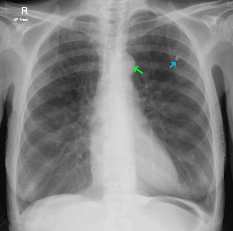
This diagnostic image is an anteroposterior (AP) erect chest X-ray demonstrating several key clinical findings. The lungs show signs of hyperinflation, characterized by increased radiolucency and a flattened appearance of the bilateral diaphragmatic domes, consistent with chronic obstructive pulmonary disease (COPD). Two specific findings are highlighted with color-coded arrows: a blue arrow points to a small, well-defined calcified granuloma located in the left upper lung zone, and a green arrow indicates an enlarged aortic knob, often associated with chronic hypertension. The cardiac silhouette appears within normal size limits, though the overall lung volume is increased. This image serves as an educational example for identifying radiographic signs of obstructive lung disease alongside common incidental thoracic findings like granulomas and vascular changes.
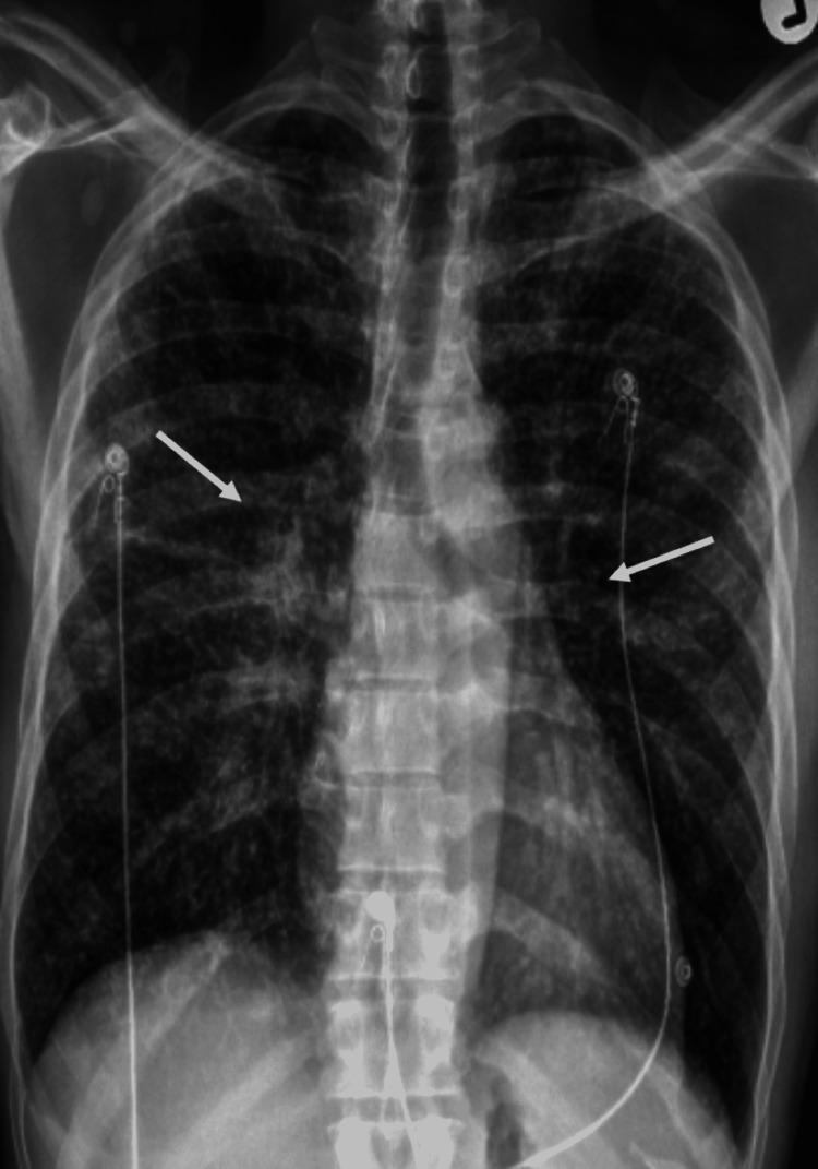
This diagnostic image is an anteroposterior (AP) chest X-ray demonstrating clinical progress in a patient with systemic sarcoidosis. The primary focus is on the bilateral hilar regions, where white arrows indicate a marked reduction in the size and density of hilar lymphadenopathy compared to previous findings. The lung parenchyma shows residual but improved fine reticulonodular opacities, characteristic of granulomatous lung disease. Several medical devices are visible, including ECG monitoring electrodes with wires overlying the thoracic cage and what appear to be central venous catheter lines entering via the subclavian regions. The osseous structures, including the spine and ribs, are visualized, and the cardiac silhouette appears stable. This radiograph serves as an educational example of radiographic response to corticosteroid therapy in a patient presenting with hypercalcemia and noncaseating granulomas.
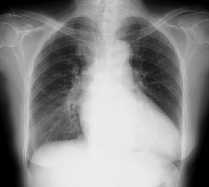
A posterior-anterior (PA) chest X-ray demonstrating a combination of thoracic findings. The image reveals prominent bilateral hilar lymphadenopathy (BHL), characterized by well-defined, bulky opacities in both hilar regions, consistent with Stage II sarcoidosis. Additionally, there is evidence of pulmonary edema, visualized as increased perihilar haziness and interstitial markings radiating from the central lung zones, which obscures the sharp margins of the pulmonary vasculature. The cardiac silhouette appears slightly enlarged, and there is a mild reduction in the clarity of the costophrenic angles. The musculoskeletal structures and overlying soft tissues are unremarkable. This radiographic presentation is significant for assessing systemic conditions such as cardiac sarcoidosis, where lymphadenopathy and acute congestive heart failure findings may coexist.
osmosis sarcoidosis youtube video site:youtube.com
sarcoidosis skin lupus pernio erythema nodosum uveitis
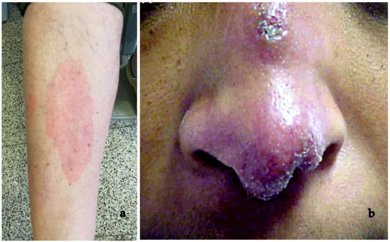
This composite clinical photograph displays two distinct dermatologic manifestations of systemic sarcoidosis. Panel (a) illustrates erythema nodosum, an acute inflammatory condition characterized by a large, ill-defined, erythematous patch on the pretibial region of the lower leg. The lesion appears as a warm, tender-looking plaque, which is a common early non-specific sign of sarcoidosis, often associated with Löfgren syndrome. Panel (b) shows lupus pernio, a pathognomonic chronic cutaneous finding. It presents as a violaceous (reddish-purple), indurated, and disfiguring plaque affecting the central face, specifically the bridge, tip, and alae of the nose. The texture is nodular with visible scaling and crusting along the nasal borders. These images contrast the transient, reactive nature of erythema nodosum against the chronic, granulomatous infiltration seen in lupus pernio, providing key diagnostic markers for the clinical evaluation of sarcoidosis in dermatology and internal medicine.
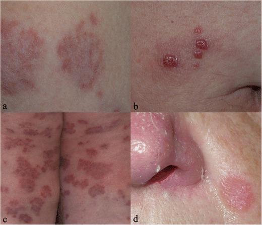
A composite of four clinical photographs (21a–d) demonstrating various cutaneous manifestations of sarcoidosis. Figure 21a illustrates erythema nodosum, characterized by ill-defined, subcutaneous erythematous nodules typically found on the lower extremities. Figure 21b displays 'specific' sarcoid lesions appearing as small, raised, reddish-brown papules with a clustered distribution on the skin. Figure 21c shows extensive involvement with multiple large, annular erythematous plaques that are relatively flat-topped and widely distributed. Figure 21d depicts lupus pernio, a classic clinical sign manifesting as chronic, indurated, violaceous or reddish-pink plaques and papules involving the middle face, specifically the alar rim and tip of the nose, resulting in localized swelling and textural changes. These images provide a comparative overview of the clinical morphology of sarcoidosis, ranging from non-specific reactive lesions to diagnostic granulomatous plaques and papules, supporting medical education in dermatology and internal medicine.
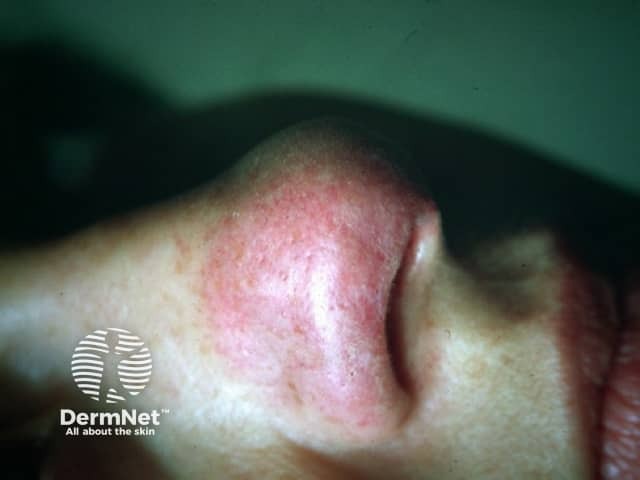
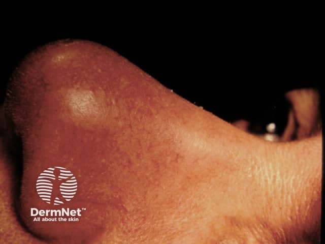
This high-resolution clinical photography shows cutaneous lupus pernio, the characteristic violaceous, indurated plaques of sarcoidosis involving the nasal dorsum and surrounding integument. Acquisition modality: bedside clinical photography, full-face anterior view, with close-up focus on the nose (nasal pyramid, alar rims) to document contour irregularities and color. The lesion appears as erythematous-violaceous plaques with firm, raised induration, subtle scaling, and blending erythema into adjacent normal skin. Dermal granulomatous inflammation in lupus pernio typically yields palpable plaques and nodules; in this image, the lesion is non-ulcerated, symmetric along the nasal region, with mild edema and vascular prominence. In the clinical context, lupus pernio strongly suggests sarcoidosis and often indicates chronic systemic involvement, particularly of the lungs and upper airways; dermatologic findings may precede or accompany pulmonary disease. Differential diagnoses include granulomatous rosacea, tuberculoid granulomatous lesions, and other granulomatous dermatoses; biopsy would show noncaseating granulomas with tight epithelioid granulomas and multinucleated giant cells. This image supports diagnostic correlation, educational case illustration, and differential diagnosis discussion for dermatology, rheumatology, pulmonology, and pathology. It has clinical relevance for prognosis and monitoring response to therapy, including corticosteroids or steroid-sparing agents, and for documenting treatment outcomes. Correlation with biopsy and systemic evaluation remains essential for definitive diagnosis.
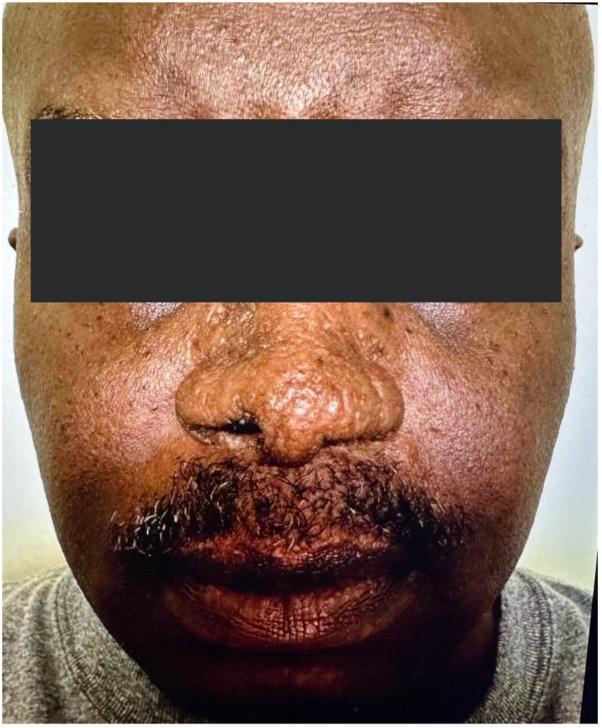
This clinical photograph shows the frontal view of a patient with cutaneous sarcoidosis, specifically lupus pernio, following systemic treatment. The image focuses on the mid-face region, including the nose, cheeks, and upper lip. While there is evidence of post-inflammatory texture changes and some subtle residual thickening, there is a marked absence of the highly characteristic indurated, violaceous, or reddish-brown shiny nodules and plaques typically seen in active lupus pernio. The nasal bridge and alae appear relatively smooth and the previous bulbous distortion has largely resolved. The skin tone on the cheeks and perioral area is more uniform, though some mild follicular prominence and chronic scarring may persist. This image serves as an educational example of the therapeutic response of cutaneous sarcoidosis to Janus kinase (JAK) inhibitor therapy, illustrating the significant reduction in granulomatous infiltration and the restoration of normal facial contours.
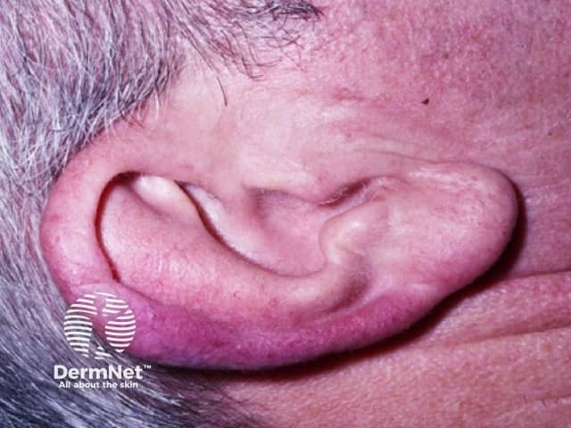
This is a high-resolution clinical photograph of a cutaneous lesion in the auricular region, captured in a lateral view of the external ear. The focal area is consistent with lupus pernio, the chronic, violaceous plaque-form of cutaneous sarcoidosis that commonly involves the nose, cheeks, ears, and lips, with the pinna often affected. The lesion appears as a well-defined, violaceous to reddish-purple plaque on the helix/pinna, with subtle surface sclerosis and mild edema; surrounding skin shows faint erythema without obvious ulceration. In cutaneous sarcoidosis, such plaques reflect dermal granulomatous infiltration; histology (if obtained) would typically show noncaseating granulomas composed of epithelioid histiocytes and Langhans-type giant cells, often with sparse lymphocytic rim. Clinically, lupus pernio is a marker of chronic sarcoidosis and may correlate with systemic involvement, including pulmonary, ocular, and cardiac disease; therefore, thorough evaluation (chest imaging, ACE levels, calcium metabolism, hepatic function) is recommended. Differential considerations include granulomatous rosacea, actinic granuloma, other granulomatous dermatitis, and infectious etiologies in the appropriate context. This image is valuable for dermatology education, sarcoidosis characterization, and monitoring therapeutic response to corticosteroids, immunomodulators, or biologic agents. This photographic depiction supports clinical diagnosis and aids multidisciplinary management decisions.
Clinical photography of a distal finger demonstrates lupus pernio, a cutaneous manifestation of sarcoidosis. This high-resolution close-up captures violaceous, indurated plaques and nodules along the dorsolateral aspect of a digit, with subtle erythema and a slightly shiny surface. The lesion is firm to palpation and demonstrates the characteristic chronic, non-ulcerating plaque morphology seen in cutaneous sarcoidosis. Anatomically, the lesion involves dermal and subcutaneous tissue of the finger pad and proximal phalanx region, consistent with acral sarcoid involvement. In the context of radiology dermatology correlation, lupus pernio often correlates with systemic sarcoidosis, including pulmonary, ocular, and cardiac involvement; thus this image has diagnostic significance beyond cosmetic appearance. Differential diagnoses include granulomatous rosacea, infectious granulomas, granulomatous dermatitis, and other inflammatory nodules; however, the violaceous hue, chronicity, and acral localization favor sarcoidosis. Clinically, recognition of lupus pernio prompts multidisciplinary assessment and potential immunosuppressive therapy. This image is useful for medical education, dermatology reference, and research in sarcoidosis phenotypes, disease activity monitoring, and treatment response evaluation. It can support case discussions on pathophysiology, imaging correlation, and prognostic implications in chronic sarcoid disease. This lesion's appearance reinforces the need for holistic evaluation to exclude systemic sarcoidosis and guide multidisciplinary management and care.
https://www.youtube.com/watch?v=9p3UVhk3TWU. Now let me read the core sarcoidosis sections from key textbooks:sarcoidosis non-caseating granuloma histopathology epithelioid cells giant cells
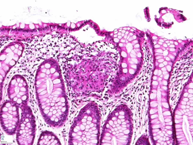
This high-quality histopathology image displays a colectomy specimen from Crohn disease, mounted on a slide and examined by light microscopy after hematoxylin and eosin (H&E) staining. The transverse section of colonic wall shows mucosa and submucosa with inflammatory changes, including chronic inflammatory infiltrate and fibrosis. In the center of the image, a well-formed non-caseating epithelioid granuloma is evident, composed of aggregates of epithelioid histiocytes with occasional multinucleated giant cells, surrounded by a rim of lymphocytes. The granulomatous focus may extend through multiple layers and can be encountered in inflamed or non-inflamed segments, and even in mesenteric lymph nodes as described in Crohn disease. The granulomas are non-necrotizing, which helps distinguish them from tuberculous granulomas. Adjacent tissue may show transmural involvement, fissuring ulcers, edematous stroma, and architectural distortion typical of Crohn disease. Importantly, granulomas are present in only about 40–60% of Crohn cases, so their absence does not exclude the diagnosis. Clinically, recognition of epithelioid granulomas supports diagnosis, informs differential with sarcoidosis and infectious granulomatous colitis, and correlates with transmural disease activity relevant for surgical planning and prognosis. This image is a valuable teaching resource for pathology, gastroenterology, and surgical pathology education.
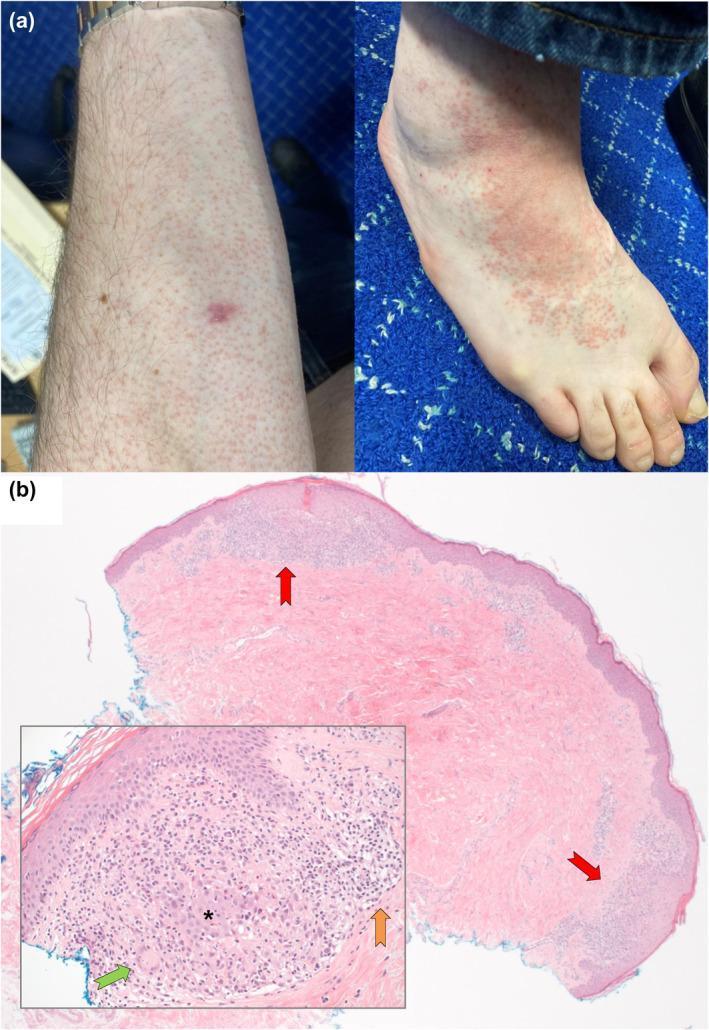
Educational panel illustrating clinical and histopathological findings of cutaneous sarcoidosis. Figure (a) contains two clinical photographs: the left shows diffuse orange-red micropapules on the forearm; the right displays similar lesions on the dorsal foot, which have coalesced into irregular, erythematous plaques. Figure (b) presents a histopathology section (H&E stain) at X25 magnification, identifying multiple well-formed, non-caseating granulomas in the superficial papillary dermis (indicated by red arrows). An inset at X250 magnification provides a high-power view of a single naked granuloma, characterized by a dense collection of epithelioid histiocytes (asterisk) and Langhans-type multinucleated giant cells (green arrow). A sparse peripheral rim of lymphocytic inflammation is also visible (orange arrow). The presentation demonstrates the typical morphological and cellular patterns used to diagnose granulomatous skin diseases.
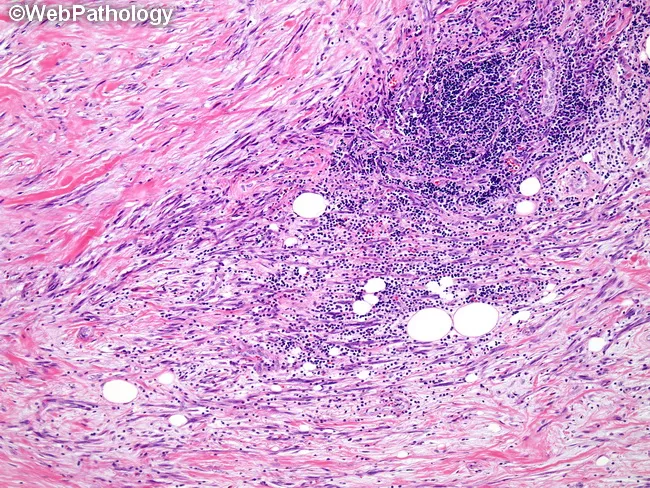
This histopathology slide represents a tissue biopsy examined under light microscopy following Hematoxylin and Eosin staining. The specimen displays dense fibrous stroma with scattered inflammatory cells and well-formed granulomatous nodules. Within the nodules, aggregates of epithelioid macrophages form tightly packed, elongated cells with eosinophilic cytoplasm. Occasional multinucleated giant cells, likely of Langhans-type, are present at the granuloma periphery. A surrounding cuff of small lymphocytes and plasma cells is evident, reflecting chronic inflammatory activity. The background shows mild edema and preserved tissue architecture in adjacent areas. There is no definitive, confluent caseous necrosis in this field, though processing artifacts can obscure subtle features. Overall, the image depicts granulomatous inflammation of uncertain etiology, requiring correlation with clinical data and ancillary studies. Diagnostic significance lies in identifying granulomas as a non-specific inflammatory pattern that prompts targeted infectious and non-infectious workups. Differential considerations include sarcoidosis (noncaseating granulomas), mycobacterial or fungal infections, foreign body reaction, and granulomatous dermatitis. This image is educational for histopathology training, pathology workflow, differential diagnosis formulation, and integration with radiologic or clinical findings to guide further testing and management decisions. Clinicians may use this pattern to decide requests for special stains, cultures, and targeted imaging and therapeutic plans thereafter.
Sarcoidosis - Complete NEET PG Article
Watch First - Osmosis Video (Highly Recommended)
1. Definition and Introduction
- Compatible clinical + radiographic + laboratory findings
- Biopsy showing non-caseating granulomas in one or more organs
- Exclusion of other causes of granulomatous disease (TB, fungal, berylliosis, etc.)
NEET PG High-Yield: Sarcoidosis = non-caseating granulomas. TB = caseating granulomas. This distinction is tested repeatedly.
2. Epidemiology
| Feature | Detail |
|---|---|
| Age of onset | Bimodal - peak 25-35 years and a second peak at 45-65 years |
| Sex | Females slightly more affected |
| Race | African-Americans have highest incidence and severity in the US; Scandinavians have the highest worldwide prevalence |
| Geography | Worldwide; highest prevalence in Scandinavia |
| Mortality | <5% overall; higher in Black patients |
- Prevalence is 10-40/100,000 in most populations
- African-Americans are 3x more likely to develop sarcoidosis vs. Caucasians, and tend to have more severe, multi-organ disease
3. Etiology and Pathogenesis
Etiology
- Genetic susceptibility - HLA-DRB1 alleles (especially HLA-DR3, DR11, DR12, DR14, DR15, DR17) influence disease susceptibility and phenotype
- Environmental/Infectious triggers - Possible antigens include mycobacterial antigens (Mycobacterium tuberculosis, M. avium), Propionibacterium acnes, organic dust, and beryllium
Pathogenesis (Step-by-Step)
- Antigen presentation: An unknown antigen (possibly mycobacterial) is presented by antigen-presenting cells (APC/macrophages) via MHC class II to naive CD4+ T-helper cells
- Th1 response: CD4+ T cells differentiate into Th1 subtype, releasing IFN-γ and TNF-α
- Macrophage activation: IFN-γ activates macrophages → epithelioid cells → multinucleated giant cells
- Granuloma formation: Activated macrophages and CD4+ T cells organize into compact granulomas
- Cytokine cascade: TNF-α, IL-12, IL-18 propagate and maintain the granulomatous response
- Resolution or fibrosis: Most granulomas resolve; some undergo fibrosis leading to organ damage
Key immunology point: CD4+ Th1 cells and TNF-α are central - this is why anti-TNF agents (infliximab) work in refractory sarcoidosis.
4. Histopathology - The Gold Standard
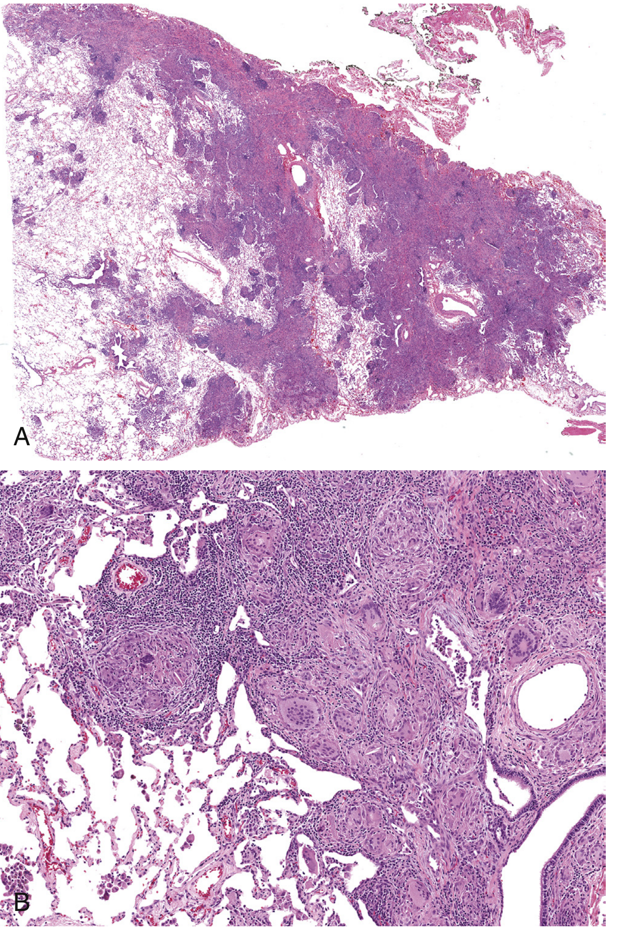
Features of Sarcoidal Granuloma:
- Non-caseating (no central necrosis) - distinguishes from TB
- Naked granulomas - tight, well-formed aggregates of epithelioid macrophages (histiocytes) with sparse lymphocytic rim
- Multinucleated giant cells (Langhans-type) are present
- Schaumann bodies (laminated calcific concentric inclusions within giant cells)
- Asteroid bodies (star-shaped crystalline inclusions in giant cells)
- Perilymphatic distribution - granulomas follow lymphatic routes along bronchovascular bundles, interlobular septa, and pleura
- Variable fibrosis surrounding granulomas
NEET PG: Schaumann bodies and asteroid bodies are characteristic but NOT specific to sarcoidosis. The defining feature is non-caseating granuloma.
5. Clinical Features - Organ by Organ
5.1 Pulmonary (90-95% of cases)
- Dry cough, dyspnea, chest pain
- Asymptomatic in up to 50% (incidental finding on CXR)
| Stage | Description | Spontaneous Remission |
|---|---|---|
| Stage 0 | Normal CXR | - |
| Stage I | Bilateral hilar lymphadenopathy (BHL) only | 55-90% |
| Stage II | BHL + pulmonary infiltrates | 40-70% |
| Stage III | Pulmonary infiltrates only (no BHL) | 10-20% |
| Stage IV | Pulmonary fibrosis (irreversible) | 0% |
NEET PG: Stage I has the best prognosis and highest spontaneous remission rate. Stage IV (fibrosis) is irreversible. Bilateral hilar lymphadenopathy in a young patient = think sarcoidosis first.
- Bilateral symmetrical hilar lymphadenopathy - "potato nodes" (discrete, non-compressing)
- Reticulonodular shadows in middle and upper lung fields
- "Eggshell calcification" in hilar nodes (rare, more common in silicosis)
- Perilymphatic micronodules (along bronchovascular bundles, fissures, subpleural)
- Ground-glass opacities
- Bilateral hilar and mediastinal lymphadenopathy
- Traction bronchiectasis and honeycombing in Stage IV
- Restrictive pattern most common (↓TLC, ↓FVC, ↓FEV1, FEV1/FVC normal or ↑)
- Obstructive pattern in endobronchial disease or fibrocystic Stage IV
- ↓Diffusing capacity (DLCO) - earliest PFT abnormality
- PFTs may be normal despite infiltrates on CXR
5.2 Lymph Nodes (75-90%)
- Bilateral hilar lymphadenopathy (BHL) - most characteristic finding
- Mediastinal, cervical, axillary, inguinal nodes may also enlarge
- Nodes are typically non-tender, rubbery, discrete
- Nodes compress but do not invade adjacent structures
5.3 Skin (25-35%)
| Lesion | Description | Significance |
|---|---|---|
| Lupus pernio | Violaceous indurated plaques on nose, cheeks, earlobes, lips | Pathognomonic of chronic sarcoidosis; indicates pulmonary involvement |
| Papules | Small, red-brown papules on face, neck, back | Most common specific skin lesion |
| Plaques | Larger, annular raised plaques | Chronic disease |
| Nodules | Subcutaneous nodules (Darier-Roussy) | Subcutaneous sarcoidosis |
| Scar sarcoidosis | Granulomas infiltrating old scars/tattoos | Highly characteristic |
| Lesion | Description | Association |
|---|---|---|
| Erythema nodosum | Tender red-purple nodules on shins | Acute sarcoidosis, good prognosis |
NEET PG High-Yield: Lupus pernio = specific, granulomatous, pathognomonic. Erythema nodosum = non-specific, reactive, good prognosis marker.
5.4 Eyes (25-50%)
- Uveitis - most common ocular manifestation
- Anterior uveitis (iritis) - most frequent; pain, redness, photophobia
- Posterior uveitis - vitreous inflammation, choroidal granulomas
- Sicca syndrome (keratoconjunctivitis sicca) - dry eyes
- Band keratopathy - calcium deposits in cornea (due to hypercalcemia)
- Lacrimal gland enlargement
- Optic neuritis (neurosarcoidosis)
NEET PG: Sarcoidosis is one of the top causes of uveitis - always think of it in a young patient with bilateral uveitis + BHL on CXR.
5.5 Cardiac (~5% clinically; up to 25% autopsy)
- Conduction abnormalities - most common cardiac manifestation
- Bundle branch block (right or left)
- Complete heart block (may need pacemaker)
- VT/VF - sudden cardiac death
- Cardiomyopathy - dilated or restrictive
- Pericarditis (uncommon)
- Cor pulmonale - due to pulmonary hypertension
NEET PG: Complete heart block in a young person + BHL on CXR = cardiac sarcoidosis until proven otherwise.
5.6 Nervous System (~5-10%) - Neurosarcoidosis
- Facial nerve palsy (CN VII) - most common cranial nerve involved
- Can be bilateral
- Part of Heerfordt's syndrome (see below)
- Other cranial neuropathies (CN II, VIII, IX, X)
- Hypothalamic-pituitary axis involvement: diabetes insipidus, hyperprolactinemia
- Peripheral neuropathy
- Meningitis (granulomatous)
- Space-occupying lesions (mass lesions)
5.7 Classic Syndromes (NEET PG Favorites!)
| Syndrome | Components | Mnemonic |
|---|---|---|
| Löfgren syndrome | Bilateral hilar lymphadenopathy + erythema nodosum + fever + polyarthritis | Acute, good prognosis, spontaneous remission in 80-90% |
| Heerfordt-Waldström syndrome | Parotitis + fever + facial nerve palsy + uveitis | "Uveoparotid fever" |
| Mikulicz syndrome | Bilateral parotid + lacrimal gland enlargement | Uveoparotid, non-tender |
| Panda sign | Bilateral parotid + lacrimal uptake on Gallium scan | Gallium-67 scan finding |
| Lambda sign | Bilateral hilar + right paratracheal uptake on Gallium scan | Combined with panda = highly specific |
Löfgren syndrome is the most favorable presentation - acute onset, typically self-limited.
5.8 Musculoskeletal
- Arthritis - large joints (knees, ankles), part of Löfgren's
- Bone involvement - lytic lesions in phalanges (Perthes-Jüngling lesions = osteolytic lesions in small bones of hands and feet)
- Myopathy (rare)
5.9 Hepatic and Splenic
- Liver involved in ~50-80% (mostly asymptomatic, granulomas on biopsy)
- Splenomegaly in ~20-30%
- Rarely causes portal hypertension or cirrhosis
5.10 Renal
- Hypercalciuria → nephrolithiasis (calcium oxalate/calcium phosphate stones)
- Hypercalcemia (~10-15%)
- Mechanism: activated macrophages in granulomas express 1-alpha-hydroxylase → excess conversion of 25-hydroxyvitamin D → 1,25-dihydroxyvitamin D (calcitriol) → ↑intestinal calcium absorption and ↑bone resorption
- Granulomatous interstitial nephritis (direct renal infiltration)
- Nephrocalcinosis
NEET PG: Hypercalcemia in sarcoidosis = unregulated extra-renal 1-alpha-hydroxylase in granuloma macrophages → excess calcitriol. Hypercalcemia worsens with vitamin D supplementation and improves with corticosteroids.
5.11 Salivary/Parotid Glands
- Parotid enlargement - may be bilateral
- Part of Heerfordt's and Mikulicz syndromes
6. Investigations
6.1 Laboratory Tests
| Test | Finding | Significance |
|---|---|---|
| Serum ACE (Angiotensin Converting Enzyme) | Elevated in 60-80% of active sarcoidosis | Produced by epithelioid cells in granulomas; used for diagnosis and disease monitoring, NOT screening |
| Serum calcium | Hypercalcemia in ~10-15% | Due to unregulated 1-alpha-hydroxylase |
| 24-hr urinary calcium | Hypercalciuria (more common than hypercalcemia) | Often precedes hypercalcemia |
| Serum LDH | May be elevated | Non-specific |
| Serum alkaline phosphatase | Elevated with liver involvement | |
| ESR, CRP | Elevated in active disease | Non-specific |
| FBC | Lymphopenia (peripheral blood), mild anemia | CD4+ cells sequestered in lungs |
| BAL (Bronchoalveolar Lavage) | ↑CD4+/CD8+ ratio (>3.5) | Normal ratio ~2; elevated in sarcoidosis |
| Serum soluble IL-2 receptor (sIL-2R) | Elevated | More sensitive than ACE; useful in monitoring |
| 1,25-dihydroxyvitamin D | Elevated | Due to granuloma macrophage 1-alpha-hydroxylase |
NEET PG: ACE levels parallel disease activity and fall with treatment. However, ACE is also elevated in other conditions (TB, histoplasmosis, leprosy) - it is NOT diagnostic alone.
6.2 Imaging
- See Scadding stages above
- BHL with "potato nodes" in stages I and II
- Perilymphatic micronodules (most common pattern)
- Bilateral hilar and mediastinal adenopathy
- Subpleural nodules
- Ground glass opacities
- Honeycombing in Stage IV

- Highly sensitive for active sarcoidosis
- Panda sign: bilateral parotid + lacrimal gland uptake
- Lambda sign: bilateral hilar + right paratracheal uptake
- Panda + Lambda together = highly specific for sarcoidosis
- More sensitive than Gallium-67
- Useful for detecting active disease sites and guiding biopsy
- Screening for cardiac sarcoidosis and pulmonary hypertension
- For neurosarcoidosis evaluation
6.3 Bronchoscopy and Biopsy
- Transbronchial lung biopsy (TBLB) - preferred initial biopsy; yield ~40-90% depending on radiographic stage
- EBUS-TBNA (Endobronchial Ultrasound-guided Transbronchial Needle Aspiration) - excellent for sampling hilar/mediastinal nodes; yield ~80-90%
- Bronchoalveolar lavage (BAL) - ↑lymphocytes, ↑CD4:CD8 ratio >3.5 (supportive, not diagnostic)
- Surgical (VATS) lung biopsy - if bronchoscopy non-diagnostic
- Skin biopsy - safest and easiest when skin lesions are present (except erythema nodosum - which is non-specific)
- Lymph node biopsy (peripheral nodes)
NEET PG: The Kveim-Siltzbach test (intradermal injection of sarcoid spleen extract → granuloma formation in 4-6 weeks) was historically used but is now obsolete due to biosafety concerns and lack of standardized reagent.
6.4 Pulmonary Function Tests
- Restrictive pattern: ↓FVC, ↓TLC, ↓DLCO, FEV1/FVC normal
- Obstructive in endobronchial disease (less common)
- DLCO is the most sensitive PFT marker
7. Differential Diagnosis
| Condition | Key Distinguishing Feature |
|---|---|
| Tuberculosis | Caseating granulomas; AFB positive; Mantoux positive |
| Berylliosis | Identical histology to sarcoidosis; occupational exposure history |
| Histoplasmosis | Fungal; complement fixation test; soil exposure |
| Lymphoma | Malignant cells on biopsy; no granulomas |
| Primary biliary cholangitis | Liver granulomas; AMA positive |
| Crohn's disease | GI symptoms; non-caseating granulomas in bowel |
| Wegener's (GPA) | Necrotizing granulomas; ANCA positive; renal involvement |
NEET PG: Berylliosis (chronic beryllium disease) is histologically indistinguishable from sarcoidosis - only exposure history differentiates them.
8. Diagnosis Summary
- Compatible clinical and radiographic picture
- Biopsy showing non-caseating granulomas in ≥1 organ
- Exclusion of other granulomatous diseases (AFB stain, fungal stains, cultures)
- Löfgren syndrome - clinical diagnosis; biopsy not needed
- Heerfordt syndrome - clinical diagnosis possible
- Asymptomatic Stage I BHL in the appropriate clinical setting
9. Treatment
9.1 Who Needs Treatment?
- Symptomatic pulmonary disease or progressive infiltrates
- Hypercalcemia or hypercalciuria
- Cardiac involvement (arrhythmia, cardiomyopathy)
- Neurosarcoidosis
- Uveitis not responding to topical therapy
- Hepatic or renal impairment
- Disfiguring skin disease (lupus pernio)
- Threatened organ damage
9.2 Treatment Ladder
| Line | Drug | Notes |
|---|---|---|
| 1st line | Oral corticosteroids (Prednisolone 0.5 mg/kg/day) | Mainstay; 6-24 months; monitor for relapse on tapering |
| 2nd line | Methotrexate | Most commonly used steroid-sparing agent; also hydroxychloroquine (for skin/joint disease) |
| 2nd line | Azathioprine | Alternative steroid-sparing agent |
| 2nd line | Hydroxychloroquine | Especially for skin, joint, and hypercalcemia; not for pulmonary disease |
| 3rd line | Infliximab (anti-TNF-α) | Refractory disease; chronic cutaneous, pulmonary, and neurosarcoidosis |
| 3rd line | Leflunomide, Thalidomide | Second-line alternatives |
| Advanced | Lung transplantation | End-stage pulmonary fibrosis (Stage IV) |
NEET PG: Corticosteroids are first-line for all symptomatic sarcoidosis. Hydroxychloroquine is preferred for skin and joint disease. Infliximab (anti-TNF) for refractory/chronic disease.
- Steroids are the treatment of choice for hypercalcemia in sarcoidosis (they suppress granuloma macrophage 1-alpha-hydroxylase activity)
- Avoid high vitamin D intake and sun exposure
- Hydration and bisphosphonates may help in severe cases
9.3 Cardiac Sarcoidosis Treatment
- High-dose corticosteroids (prednisolone 1 mg/kg/day)
- ICD (implantable cardioverter-defibrillator) for VT/VF risk
- Permanent pacemaker for complete heart block
- Heart transplantation in end-stage cardiomyopathy
10. Prognosis
| Type | Outcome |
|---|---|
| Löfgren syndrome | 80-90% remit spontaneously within 2 years; excellent prognosis |
| Stage I (BHL only) | 55-90% spontaneous remission |
| Stage II | 40-70% spontaneous remission |
| Stage III | 10-20% spontaneous remission |
| Stage IV | No spontaneous remission; irreversible fibrosis |
| Chronic/refractory sarcoidosis | Seen in Black patients, lupus pernio, hypersplenism, age >40 |
- Cardiac sarcoidosis (sudden death)
- Progressive pulmonary fibrosis (respiratory failure)
- Neurosarcoidosis
- African-American patients
11. NEET PG High-Yield Summary Table
| Feature | Details |
|---|---|
| Type of granuloma | Non-caseating (non-necrotizing) |
| Giant cell inclusions | Schaumann bodies, Asteroid bodies |
| Most common organ | Lung (90%); Bilateral hilar LN |
| Most specific skin sign | Lupus pernio (pathognomonic) |
| Best prognosis presentation | Löfgren syndrome |
| Stage with most fibrosis | Stage IV (irreversible) |
| Best stage prognosis | Stage I |
| Mechanism of hypercalcemia | ↑ 1-alpha-hydroxylase in granuloma macrophages → excess 1,25-dihydroxy-VitD |
| Serum marker | ↑ACE (60-80%), ↑sIL-2R |
| BAL finding | ↑CD4:CD8 ratio (>3.5) |
| Gallium scan signs | Panda sign + Lambda sign |
| Most common cardiac arrhythmia | Complete heart block |
| Facial nerve palsy syndrome | Heerfordt syndrome |
| Parotid + lacrimal enlargement | Mikulicz syndrome |
| Acute polyarthritis + BHL + EN | Löfgren syndrome |
| First-line treatment | Oral corticosteroids (prednisolone) |
| Skin disease treatment | Hydroxychloroquine |
| Refractory disease treatment | Infliximab (anti-TNF) |
| Obsolete test | Kveim-Siltzbach test |
| Occupational mimic | Berylliosis (histologically identical) |
12. Mnemonics for NEET PG
- Schaumann bodies/Asteroid bodies in granulomas
- Calcium raised (hypercalcemia - 1-alpha-hydroxylase)
- ACE elevated (produced by epithelioid cells)
- Restrictive lung disease on PFTs
- Erythema nodosum (acute, good prognosis)
- Fever
- Uveitis
- Uveoparotitis (parotid enlargement)
- Palsy (facial nerve - CN VII)
- Bilateral hilar lymphadenopathy
- Erythema nodosum
- Arthritis + fever
13. Additional Images