
Boundaries of prussak space
Reading File
Prussak's space (also called the superior recess of the tympanic membrane) is a small but clinically important recess in the middle ear, bounded by:
- Lateral wall: Shrapnell's membrane (pars flaccida of the tympanic membrane)
- Medial wall: Neck of the malleus and the lateral surface of the head of the malleus
- Superior wall (roof): Lateral ligament of the malleus (also described as the notch of Rivinus / bony tympanic notch)
- Anterior wall: Anterior malleolar fold
- Posterior wall: Posterior malleolar fold
- Floor: Lateral process of the malleus (the short process)
Let me confirm this from the textbooks.
Searching sources...
Finding Sources
Searching the Web
Prussak space boundaries anatomy middle ear
Now let me read the detailed textbook section from Shambaugh (which had the most direct description) and also fetch the diagram image:
Reading File
Reading File
Loading Image
Here is a complete account of the boundaries of Prussak's space, sourced from multiple authoritative ENT textbooks.
Prussak's Space (Superior Recess of the Tympanic Membrane)
Prussak's space is the lateral epitympanic recess, situated inferior to the tympanic diaphragm. It was first described by Alexander Prussak in 1867 and is the most common site of origin for pars flaccida cholesteatoma.
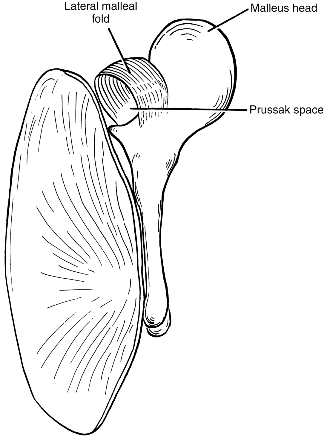
Prussak space - bounded superiorly by the lateral malleal fold and medially by the malleus head/neck (KJ Lee's Essential Otolaryngology)
Boundaries
| Wall | Structure |
|---|---|
| Lateral | Pars flaccida (Shrapnell's membrane) + lower edge of the scutum (outer attic wall) |
| Medial | Neck and head of the malleus |
| Roof (superior) | Lateral malleal fold (= lateral malleal ligament fold) - this also forms the floor of the lateral malleal space above, and separates Prussak's space from the upper unit of the attic |
| Floor (inferior) | Short (lateral) process of the malleus / neck of malleus |
| Anterior | Anterior malleal fold (anterior malleolar fold) |
| Posterior | Posterior malleal fold - opens into the posterior pouch of von Tröltsch and then into the mesotympanum |
Key Points
- Extent: It extends from the scutum (posterosuperiorly) to the umbo (inferiorly), reaching about 0.4 mm superior to the roof of the external auditory canal.
- Largest cross-section: ~2.6 mm, at the level of the roof of the EAC.
- Ventilation: Prussak's space has its own independent ventilation route, separate from the upper unit of the attic (the anterior and posterior epitympanic compartments).
- Clinical significance: Dysfunction of its ventilation - due to thickened mucus closing the posterior pocket - leads to pars flaccida retraction, adhesion to the malleus neck, and ultimately cholesteatoma formation. Keratin accumulates here, causing scutum erosion and medial displacement of the ossicular chain.
Sources
- Shambaugh Surgery of the Ear, p. 60: "Shrapnell's membrane serves as the lateral wall of Prussak's space (the superior recess of the tympanic membrane); the head and neck of the malleus, the lateral malleal ligament, and anterior and posterior malleal folds form the medial, anterosuperior, and inferior limits."
- Scott-Brown's Otorhinolaryngology Head & Neck Surgery, Vol 2: "The Prussak space is found between the pars flaccida and the neck of the malleus, bounded by the lateral malleolar fold."
- Cummings Otolaryngology: "The epitympanum is further divided into (1) the Prussak space, just medial to pars flaccida and lateral to the head and neck of the malleus..."
- KJ Lee's Essential Otolaryngology: Figure 13-7 (shown above).
This is a shared conversation. Sign in to Orris to start your own chat.