Blanching rash

Reading File
Finding Sources
Finding Sources
Searching Images

blanching erythematous rash

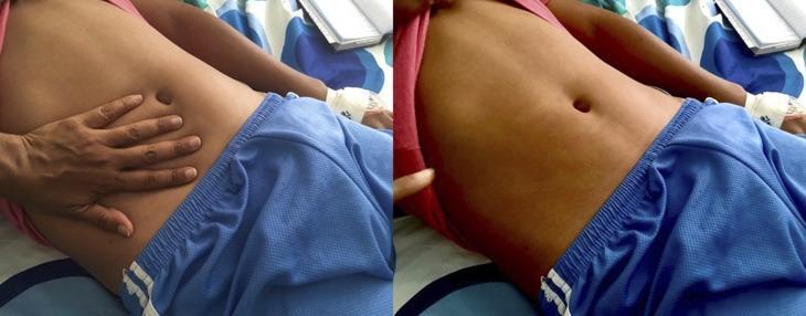
This clinical photograph displays a diffuse, erythematous maculopapular rash on the abdomen of a patient, characteristic of Dengue fever. The image is presented in a two-part sequence to demonstrate clinical blanching. On the left, a clinician's hand is shown applying direct pressure to the right side of the patient's abdomen. On the right, after the hand is removed, a clear, pale handprint-shaped outline remains where the pressure was applied, indicating that the redness fades (blanches) as blood is temporarily displaced from the superficial vessels. This finding confirms the rash is inflammatory rather than hemorrhagic (petechial/purpuric). The rash is widespread across the torso but typically spares the palms and soles in this clinical presentation. This image serves as a teaching tool for medical students and clinicians to distinguish between vascular inflammatory rashes and non-blanching hemorrhagic lesions in the context of viral hemorrhagic fevers and infectious diseases.
This clinical photograph displays a diffuse, erythematous maculopapular rash on the abdomen of a patient, characteristic of Dengue fever. The image is presented in a two-part sequence to demonstrate clinical blanching. On the left, a clinician's hand is shown applying direct pressure to the right side of the patient's abdomen. On the right, after the hand is removed, a clear, pale handprint-shaped outline remains where the pressure was applied, indicating that the redness fades (blanches) as blood is temporarily displaced from the superficial vessels. This finding confirms the rash is inflammatory rather than hemorrhagic (petechial/purpuric). The rash is widespread across the torso but typically spares the palms and soles in this clinical presentation. This image serves as a teaching tool for medical students and clinicians to distinguish between vascular inflammatory rashes and non-blanching hemorrhagic lesions in the context of viral hemorrhagic fevers and infectious diseases.

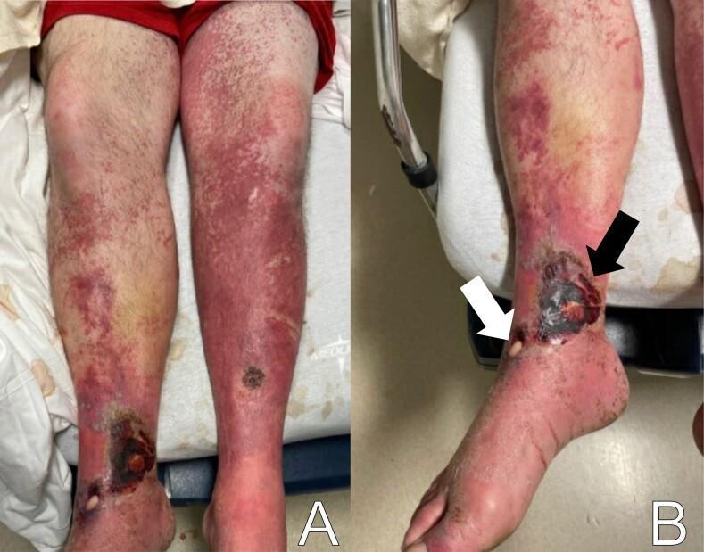
Clinical photograph of the bilateral lower extremities in a 45-year-old male, demonstrating complex dermatological and vascular findings. Panel A shows an extensive, bilateral, erythematous, papular, and non-blanching rash extending from the anterior thighs to the dorsal feet, suggesting a vasculitic or inflammatory etiology. Panel B provides a detailed view of the right ankle, highlighting severe skin lesions. A white arrow indicates a shallow, circular ulceration with a pale, membranous covering. An adjacent larger lesion, indicated by a black arrow, shows dense, dark eschar indicative of tissue necrosis. The surrounding skin exhibits generalized erythema, scaling, and edema consistent with cellulitis or chronic venous insufficiency. These images are educationally significant for distinguishing between different types of lower extremity wounds, including membranous ulcers and necrotic eschar, and identifying the characteristic appearance of a non-blanching purpuric rash.
Clinical photograph of the bilateral lower extremities in a 45-year-old male, demonstrating complex dermatological and vascular findings. Panel A shows an extensive, bilateral, erythematous, papular, and non-blanching rash extending from the anterior thighs to the dorsal feet, suggesting a vasculitic or inflammatory etiology. Panel B provides a detailed view of the right ankle, highlighting severe skin lesions. A white arrow indicates a shallow, circular ulceration with a pale, membranous covering. An adjacent larger lesion, indicated by a black arrow, shows dense, dark eschar indicative of tissue necrosis. The surrounding skin exhibits generalized erythema, scaling, and edema consistent with cellulitis or chronic venous insufficiency. These images are educationally significant for distinguishing between different types of lower extremity wounds, including membranous ulcers and necrotic eschar, and identifying the characteristic appearance of a non-blanching purpuric rash.

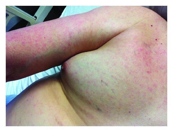
This clinical photograph displays a diffuse, lightly erythematous macular rash on the chest, shoulder, and upper extremity of a patient. The lesions consist of numerous small, blanching or non-blanching red macules that vary in density, appearing most concentrated on the arm and becoming more sparsely distributed across the lateral chest and breast area. The rash is characteristically morbilliform in appearance, suggesting a systemic viral exanthem or drug eruption. Scattered among the macules are a few isolated, more intensely pigmented punctate spots and subtle linear markings on the shoulder region. The overall skin texture remains largely smooth, with no evidence of vesicles, bullae, or desquamation in the visible field. This image is an educational example of common dermatological manifestations of systemic illness, useful for teaching clinical examination and differential diagnosis of acute febrile rashes in dermatology and internal medicine.
This clinical photograph displays a diffuse, lightly erythematous macular rash on the chest, shoulder, and upper extremity of a patient. The lesions consist of numerous small, blanching or non-blanching red macules that vary in density, appearing most concentrated on the arm and becoming more sparsely distributed across the lateral chest and breast area. The rash is characteristically morbilliform in appearance, suggesting a systemic viral exanthem or drug eruption. Scattered among the macules are a few isolated, more intensely pigmented punctate spots and subtle linear markings on the shoulder region. The overall skin texture remains largely smooth, with no evidence of vesicles, bullae, or desquamation in the visible field. This image is an educational example of common dermatological manifestations of systemic illness, useful for teaching clinical examination and differential diagnosis of acute febrile rashes in dermatology and internal medicine.

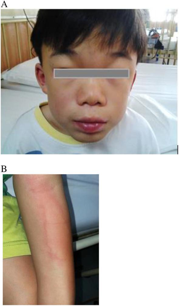
A pair of clinical photographs documenting an infusion-associated reaction in a pediatric patient receiving Enzyme Replacement Therapy (ERT) for Mucopolysaccharidosis (MPS). Panel A is a frontal facial photograph showing marked periorbital edema and swollen lips, accompanied by a diffuse, faint erythematous rash across the cheeks and midface, consistent with angioedema. Panel B focuses on the left forearm, exhibiting a localized urticarial rash characterized by erythematous, blanching wheals with irregular borders. These images illustrate common cutaneous manifestations of a systemic hypersensitivity reaction during intravenous medication administration. The images are highly relevant for medical education concerning the recognition of adverse drug reactions, dermatologic signs of anaphylactoid responses, and the clinical management of metabolic disorders.
A pair of clinical photographs documenting an infusion-associated reaction in a pediatric patient receiving Enzyme Replacement Therapy (ERT) for Mucopolysaccharidosis (MPS). Panel A is a frontal facial photograph showing marked periorbital edema and swollen lips, accompanied by a diffuse, faint erythematous rash across the cheeks and midface, consistent with angioedema. Panel B focuses on the left forearm, exhibiting a localized urticarial rash characterized by erythematous, blanching wheals with irregular borders. These images illustrate common cutaneous manifestations of a systemic hypersensitivity reaction during intravenous medication administration. The images are highly relevant for medical education concerning the recognition of adverse drug reactions, dermatologic signs of anaphylactoid responses, and the clinical management of metabolic disorders.

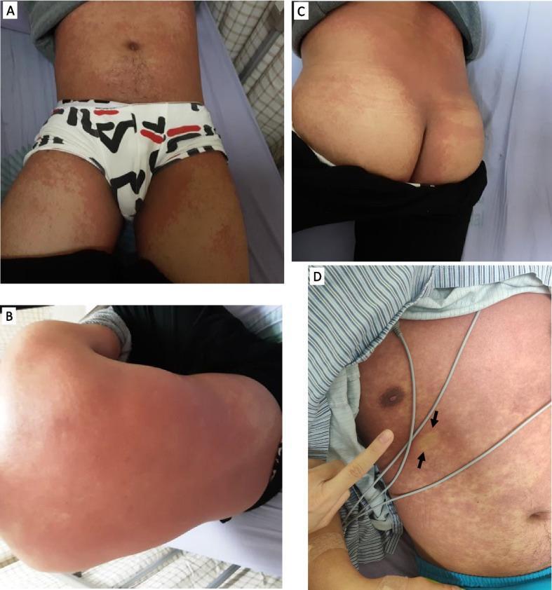
This composite clinical photograph displays a four-panel view (A-D) of an extensive skin rash on a hospitalized patient. Panel A illustrates the anterior trunk and bilateral upper thighs, showing a diffuse, erythematous macular rash with areas of confluence. Panel B shows the posterior trunk, where the rash appears more confluent and intensely erythematous. Panel C depicts the buttocks and lower back, maintaining the same fused macular pattern. Panel D focuses on the anterior chest and abdomen, highlighting the morphology of the rash, which consists of small, reddish, non-pruritic macules. Black arrows in Panel D indicate a specific area of blanching upon digital pressure, a key clinical sign used to differentiate inflammatory erythema from purpura. The distribution is symmetrical and widespread, covering the trunk and limbs while sparing mucous membranes. This visual representation is characteristic of systemic inflammatory responses or drug-related eruptions, such as those associated with atypical infections or hypersensitivity reactions.

This composite clinical photograph displays a four-panel view (A-D) of an extensive skin rash on a hospitalized patient. Panel A illustrates the anterior trunk and bilateral upper thighs, showing a diffuse, erythematous macular rash with areas of confluence. Panel B shows the posterior trunk, where the rash appears more confluent and intensely erythematous. Panel C depicts the buttocks and lower back, maintaining the same fused macular pattern. Panel D focuses on the anterior chest and abdomen, highlighting the morphology of the rash, which consists of small, reddish, non-pruritic macules. Black arrows in Panel D indicate a specific area of blanching upon digital pressure, a key clinical sign used to differentiate inflammatory erythema from purpura. The distribution is symmetrical and widespread, covering the trunk and limbs while sparing mucous membranes. This visual representation is characteristic of systemic inflammatory responses or drug-related eruptions, such as those associated with atypical infections or hypersensitivity reactions.

Searching the Web

blanching rash vs non-blanching rash differential diagnosis

Blanching Rash

A blanching rash is a skin eruption that turns white (fades) briefly when pressure is applied - for example with a finger or a glass tumbler (the "glass test") - and returns to its red/pink color once pressure is released.

Why it blanches

Blanching occurs because the redness is caused by dilated blood vessels (capillaries) in the dermis that are still functionally intact. Pressure temporarily pushes blood out of these vessels, so the skin pales; when released, blood flows back in and the color returns. This tells you the process is vascular/inflammatory (vasodilation), not hemorrhagic - blood has not actually leaked out of the vessels into the tissue.
Blanching test on a maculopapular rash
Clinical photograph demonstrating the blanch test: pressure from a hand leaves a pale handprint that fades back to red, confirming an inflammatory (blanching) rash rather than a hemorrhagic one.

Common causes of blanching rashes

  • Viral exanthems - measles (morbilliform, spreads face to trunk), rubella, roseola, other "childhood" viral rashes
  • Erythematous/maculopapular drug eruptions
  • Urticaria (hives) - blanching wheals, often with angioedema
  • Cellulitis / erysipelas - localized blanching erythema with warmth and tenderness
  • Erythema of infection - e.g., "Horder spots" (faint erythematous blanching maculopapular rash) seen in Q fever, or rose spots of typhoid fever - Goldman-Cecil Medicine
  • Early scarlet fever, sunburn, flushing reactions, some early sepsis-related rashes before petechiae develop

Why the distinction matters clinically

The key reason clinicians check for blanching is to rule out a non-blanching (petechial/purpuric) rash, which does not fade with pressure because blood has actually extravasated into the skin (capillary leak or bleeding). Non-blanching rashes are the red flag finding:
  • Meningococcal septicaemia / meningitis - classically starts as a blanching rash early on, then evolves into a non-blanching petechial or purpuric rash in over 80% of cases. Any fever + non-blanching rash in a child is treated as a medical emergency until proven otherwise (Harrison's Principles of Internal Medicine, p. 1459-1460).
  • Vasculitis (e.g., Henoch-Schonlein purpura)
  • Immune thrombocytopenia (ITP), coagulopathy, or other bleeding/platelet disorders
  • Non-accidental injury in children (petechiae over soft tissue without clear explanation)
Practical rule: if a rash blanches under pressure, it is almost always benign/inflammatory in origin. If it does not blanch (petechiae or purpura), especially with fever, it needs urgent evaluation for sepsis, meningococcal disease, or a bleeding disorder.
Sources: Harrison's Principles of Internal Medicine 22E, p. 1459-1460; Goldman-Cecil Medicine, Cutaneous Manifestations & Physical Examination chapters; Scott-Brown's Otorhinolaryngology Head & Neck Surgery Vol 2.
If you have a specific clinical scenario in mind (a particular patient, rash distribution, or associated symptoms like fever, joint pain, or recent medication), let me know and I can narrow this down further.
This is a shared conversation. Sign in to Orris to start your own chat.