
Development of pharyngeal arches 5m shortnote
Reading File
Searching sources...
Finding Sources
Searching Images
pharyngeal branchial arch development derivatives
Reading File
Reading File
Reading File
Reading File
Loading Image
Reading File
Reading File
I now have all the information needed to write a complete 5-mark short note. Here is the answer:
Development of Pharyngeal Arches
Introduction
The pharyngeal (branchial) arches appear during the 4th and 5th weeks of embryonic development as bilateral, bar-like thickenings on the lateral walls of the foregut. In humans, 6 pairs of arches develop (the 5th is rudimentary and rapidly disappears), separated externally by pharyngeal clefts (grooves) and internally by pharyngeal pouches.
Structure of a Typical Pharyngeal Arch
Each arch has a mesenchymal core covered externally by ectoderm and internally by endoderm. Each arch contains:
- A cartilaginous bar - the skeletal element
- An artery - a pharyngeal arch artery arising from the truncus arteriosus
- A cranial nerve - specific to that arch
- A muscle mass - from paraxial mesoderm
The mesenchyme originates from:
- Neural crest cells (migrate in during week 4 - form skeletal and connective tissue)
- Paraxial mesoderm (forms the muscle primordium)
- Lateral mesoderm / angioblasts (form endothelium of vessels)
Derivatives of Each Arch
First Arch (Mandibular Arch)
| Component | Derivative |
|---|---|
| Cartilage (Meckel's) | Malleus, incus (auditory ossicles); perichondrium → anterior ligament of malleus + sphenomandibular ligament; mandible forms by membranous ossification around it |
| Muscles | Muscles of mastication (temporalis, masseter, pterygoids), mylohyoid, anterior belly of digastric, tensor tympani, tensor veli palatini |
| Nerve | CN V (trigeminal) - mandibular branch |
| Artery | Maxillary artery (terminal part) |
Second Arch (Hyoid Arch)
| Component | Derivative |
|---|---|
| Cartilage (Reichert's) | Stapes, styloid process, stylohyoid ligament, lesser cornu + upper body of hyoid bone |
| Muscles | Muscles of facial expression, stapedius, stylohyoid, posterior belly of digastric, auricular muscles |
| Nerve | CN VII (facial nerve) |
Third Arch
| Component | Derivative |
|---|---|
| Cartilage | Greater cornu + lower body of hyoid bone |
| Muscle | Stylopharyngeus |
| Nerve | CN IX (glossopharyngeal) |
Fourth Arch
| Component | Derivative |
|---|---|
| Cartilage | Superior cornu of thyroid cartilage, thyroid cartilage |
| Muscles | Cricothyroid, levator veli palatini, constrictors of pharynx |
| Nerve | Superior laryngeal branch of CN X (vagus) |
Sixth Arch
| Component | Derivative |
|---|---|
| Cartilage | Cricoid, arytenoid, corniculate, cuneiform cartilages |
| Muscles | Intrinsic muscles of larynx, striated muscles of esophagus |
| Nerve | Recurrent laryngeal branch of CN X (vagus) |
Note: The 5th arch is absent or rudimentary - it leaves no derivatives.
Key Developmental Diagram
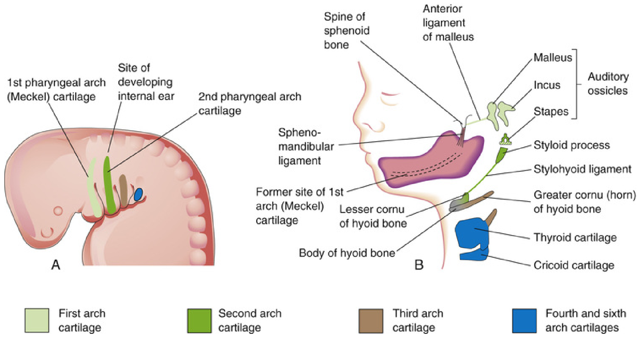
Arch cartilages (A - 4-week embryo) and their definitive derivatives (B - 24-week fetus). First arch (Meckel's) = light green; Second arch (Reichert's) = dark green; Third arch = brown; Fourth and sixth = blue.
Important Clinical Correlates
- First arch syndrome: Abnormal migration of neural crest cells into the first arch leads to mandibulofacial dysostosis (Treacher Collins syndrome) and Pierre Robin sequence - characterized by mandibular hypoplasia, cleft palate, and ear anomalies.
- Nerve rule: Wherever arch-derived muscle migrates, it carries its arch nerve with it. This is why the facial nerve (CN VII) follows muscles of facial expression even as they spread over the head and neck.
- Persistence of Meckel's cartilage: Rare ossification may extend from the styloid process along the stylohyoid ligament, causing pain near the palatine tonsil (Eagle's syndrome).
Sources: - The Developing Human: Clinically Oriented Embryology (Moore & Persaud), p. 463-470 - Langman's Medical Embryology, p. 306-309
This is a shared conversation. Sign in to Orris to start your own chat.