
Blood supply of heart. Coronary Arteries Venous drainage Coronary sinus Explain with diagrams which i have to draw in my mbbs exam. Refer standard anatomy textbook
coronary arteries heart anatomy diagram right left coronary artery branches
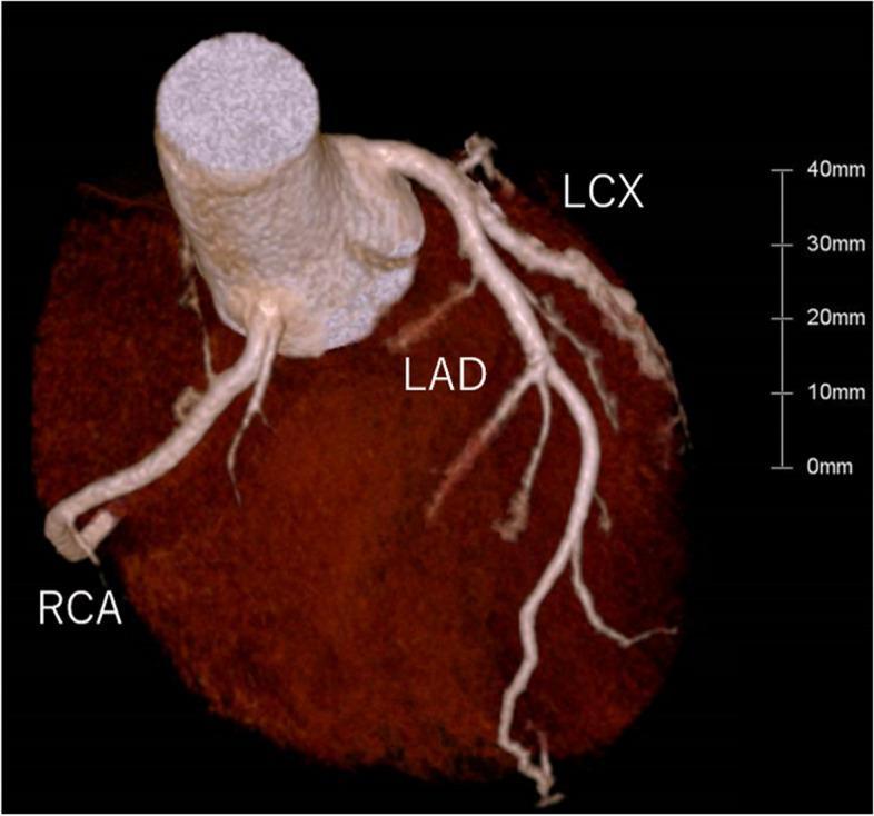
This 3D volume-rendered Computed Tomography Angiography (CTA) image provides a comprehensive visualization of the coronary arterial anatomy. The image displays the major coronary arteries originating from the ascending aorta and coursing over the epicardial surface of the heart. The Left Anterior Descending (LAD) artery is clearly visible descending along the anterior interventricular sulcus, exhibiting multiple diagonal branches. The Left Circumflex (LCX) artery is seen branching from the left main coronary artery and traveling along the left atrioventricular groove. The Right Coronary Artery (RCA) originates from the right coronary sinus and traverses the right atrioventricular groove. All visualized vessels appear patent with smooth luminal contours and no evidence of significant calcified or non-calcified atherosclerotic plaques or stenotic lesions. The myocardium is rendered in a dark, textured brown tone to provide anatomical contrast, and a 40mm scale bar is included for size reference. This diagnostic image is typical for assessing coronary artery disease (CAD) and evaluating coronary anatomy in a clinical setting.
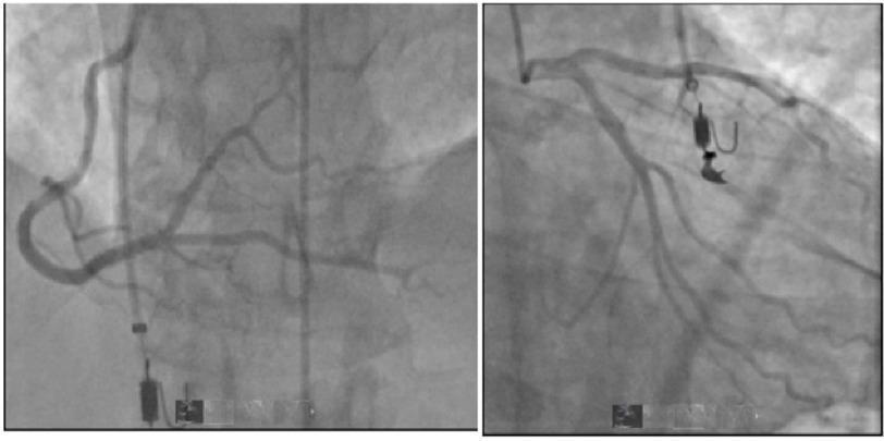
This diagnostic image consists of two grayscale fluoroscopic frames from a coronary angiography procedure, a subset of left heart catheterization. The image on the left shows the right coronary artery (RCA) and its primary branches. The image on the right depicts the left coronary system, including the left main, left anterior descending (LAD), and left circumflex (LCx) arteries. Both panels demonstrate radiopaque catheters selectively engaged in the coronary ostia, with contrast medium filling the lumen to reveal vascular anatomy. The coronary arteries exhibit smooth luminal borders and a patent course without evidence of focal stenosis, significant narrowing, or obstructive coronary artery disease. Small radiopaque surgical or procedural hardware elements are visible in the background of both panels. This material is used to teach cardiovascular assessment, specifically the use of angiography to rule out obstructive atherosclerosis in patients presenting with acute cardiac symptoms like chest pain or ST-segment changes.
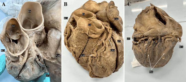
This set of three clinical photographs (A, B, and C) displays a dissected human heart specimen to illustrate coronary artery anatomy and branching patterns. Image A provides an anterosuperior view, highlighting the origins of the Right Coronary Artery (RCA) from the right aortic sinus and the Left Anterior Descending (LAD) and Left Circumflex (LCx) arteries near the Ascending Aorta (AA) and Pulmonary Artery (PA). Image B shows an anterior view of the heart, detailing the LAD descending in the anterior interventricular groove, the LCx wrapping around the left auricle (LAur), and the Left Marginal Artery (LMA) branching from the LCx. Image C presents a posterior view demonstrating a codominant coronary system. In this specimen, the Posterior Descending Artery (PDA) is formed by contributions from both the terminal branches of the RCA and the LCx as they meet at the crux of the heart. These images serve as educational material for cardiovascular anatomy, specifically focusing on variations in coronary dominance and the structural relationship between major epicardial vessels.
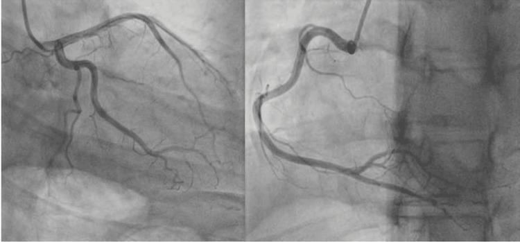
This diagnostic image consists of two side-by-side frames from a coronary angiogram (left heart catheterization). The left panel displays the left coronary artery (LCA) system, including the left main, left anterior descending (LAD), and circumflex arteries with their associated diagonal and marginal branches. The right panel demonstrates the right coronary artery (RCA) in a standard oblique projection. Both images show smooth, regular vessel calibers with well-defined margins and no evidence of significant atherosclerotic stenosis, abrupt narrowing, or filling defects. Contrast distribution is homogenous throughout the arterial tree, indicating normal flow dynamics. This visual serves as a clinical example of normal coronary anatomy in a patient presenting with cardiac symptoms (e.g., chest pain or ST-elevation), used to rule out obstructive coronary artery disease or spontaneous coronary artery dissection (SCAD) in the differential diagnosis of myopericarditis.
coronary sinus venous drainage heart great cardiac vein middle small cardiac vein
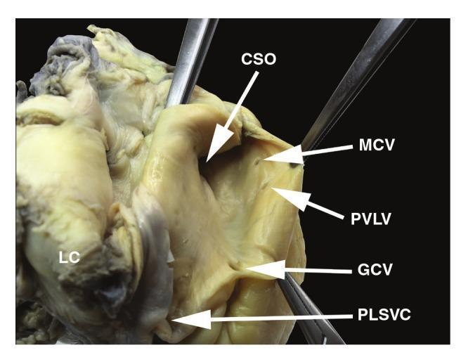
This clinical photograph of a dissected human heart specimen displays the internal anatomy of the coronary sinus and its associated venous tributaries. The primary feature is a significantly enlarged, funnel-shaped coronary sinus ostium (CSO). Positioned inferior to the CSO is the much smaller, rounded ostium of the great cardiac vein (GCV). The middle cardiac vein (MCV) and the posterior vein of the left ventricle (PVLV) are visible as smaller openings superior to the GCV. A critical anatomical variant, a persistent left superior vena cava (PLSVC), is shown draining into the coronary sinus from an inferior position. The specimen highlights the absence of the Thebesian valve at the CSO and the absence of the Vieussens valve at the GCV ostium. This image illustrates complex cardiac venous remodeling often associated with congenital anomalies like PLSVC, which can significantly alter the diameter and structure of the coronary sinus, holding importance for electrophysiological procedures and cardiac surgery.
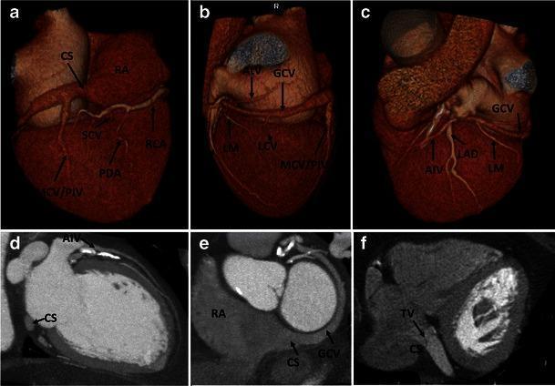
A multi-panel figure illustrating normal human coronary venous anatomy using Cardiac Computed Tomography (CCT). Panels (a-c) present 3D volume-rendered reconstructions of the heart. Panel (a) shows the posterior view, highlighting the coronary sinus (CS) in the atrioventricular groove, draining into the right atrium (RA), alongside the middle cardiac vein (MCV/PIV), small cardiac vein (SCV), and right coronary artery (RCA). Panels (b) and (c) provide anterior and lateral views, demonstrating the great cardiac vein (GCV) transitioning from the anterior interventricular vein (AIV), running parallel to the left anterior descending artery (LAD). Panels (d) and (e) are multiplanar reformat (MPR) images showing the cross-sectional relationship between the CS, the RA, and the ventricular chambers. Panel (f) is an axial CCT slice identifying the valve of Thebesius (TV) at the ostium of the coronary sinus. The collection demonstrates the spatial orientation of the cardiac venous system relative to major coronary arteries and cardiac chambers for educational use in radiology and cardiology.
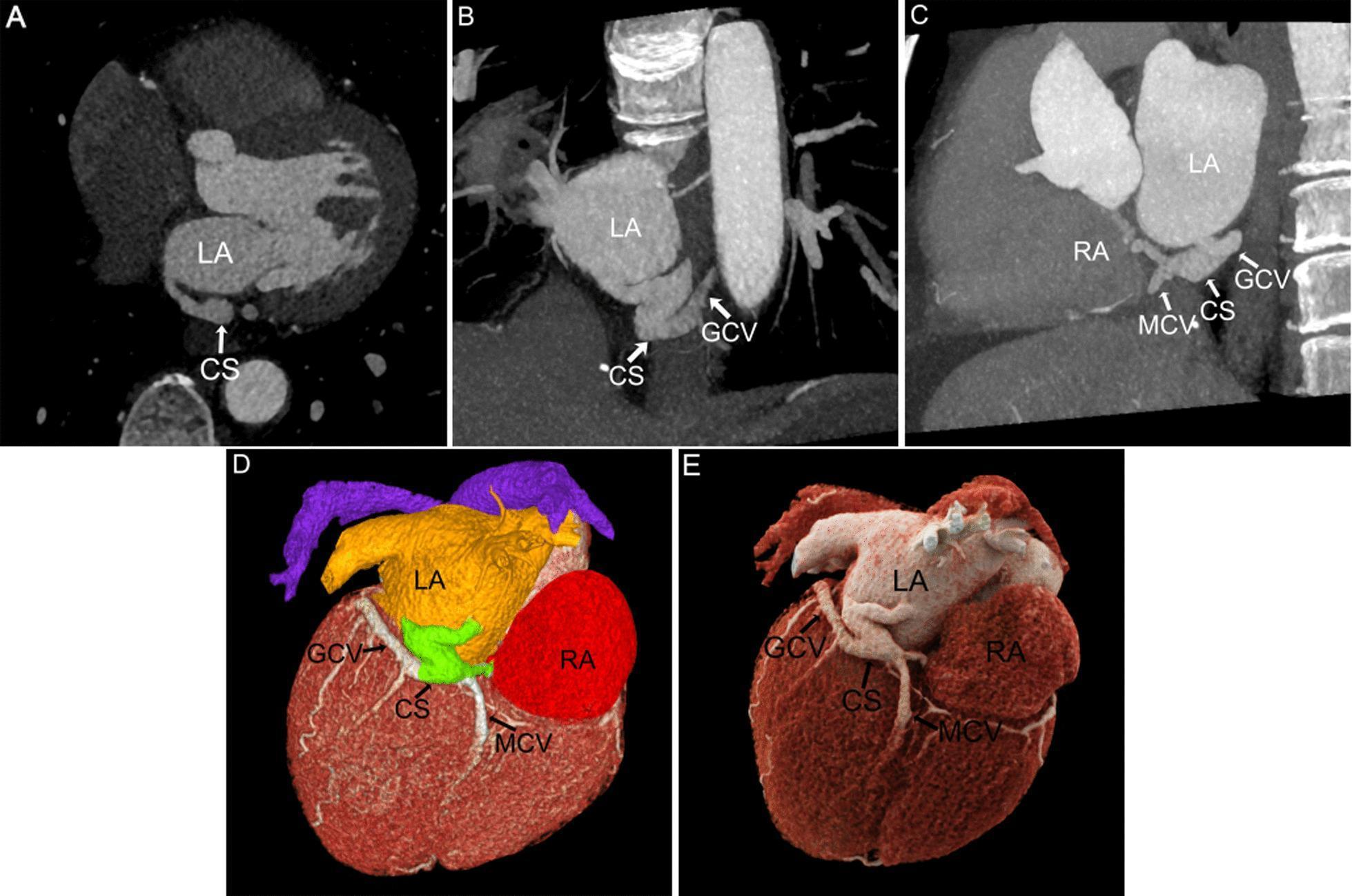
This composite diagnostic image demonstrates Cardiac Computed Tomography (CCT) reconstructions of a Type V Unroofed Coronary Sinus (UCSS). The figure displays five distinct post-processing modalities: (A) Multiplanar Reconstruction (MPR) axial view showing the coronary sinus (CS) inferior to the left atrium (LA); (B, C) Maximum Intensity Projection (MIP) views illustrating a 'Z'-shaped vascular communication between the LA and CS, and the convergence of the CS into the right atrium (RA). These images also detail the positioning of the great cardiac vein (GCV) and middle cardiac vein (MCV). (D, E) Volume Rendering (VR) and Cinematic Rendering (CR) provide 3D spatial context. Panel D utilizes color-coding (Yellow: LA, Red: RA, Green: CS) to differentiate cardiac chambers and the venous system, while Panel E offers high-fidelity realistic rendering of the heart's posterior surface. This image serves to educate on the utility of CCT post-processing for diagnosing complex congenital venous anomalies and understanding the three-dimensional relationship between the coronary sinus and the atrial chambers.
Blood Supply of the Heart
Overview
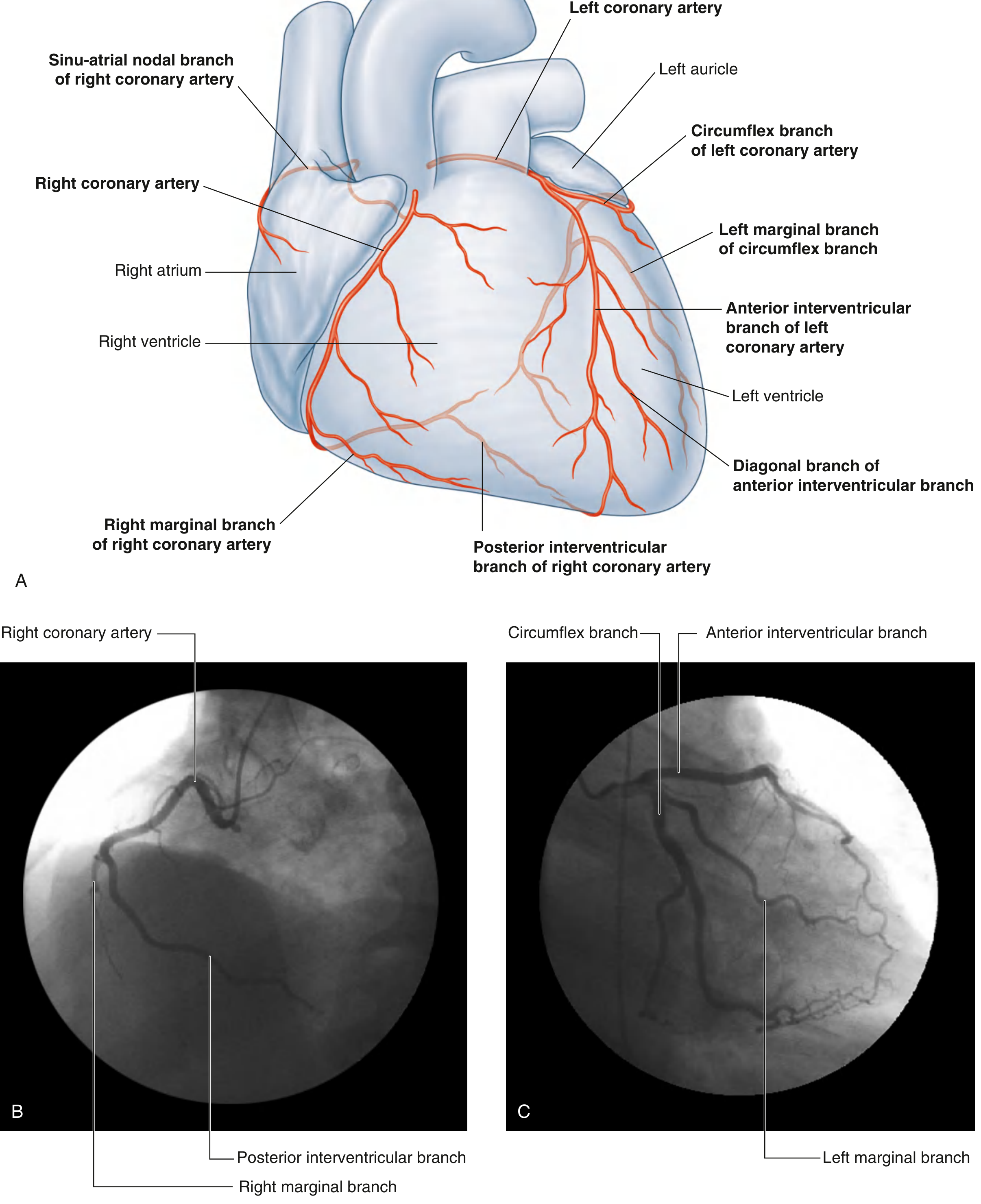
I. CORONARY ARTERIES
Right Coronary Artery (RCA)
| Branch | Course & Supply |
|---|---|
| Sinu-atrial (SA) nodal branch | Early atrial branch; passes around SVC to supply SA node |
| Right marginal branch | Along inferior (acute) margin toward apex |
| AV nodal branch | Small branch before final division |
| Posterior interventricular branch (PDA) | Runs in posterior interventricular sulcus |
- Right atrium and right ventricle
- SA and AV nodes
- Interatrial septum
- Portion of left atrium
- Posteroinferior 1/3 of interventricular septum
- Posterior portion of left ventricle
Left Coronary Artery (LCA)
- Descends in the anterior interventricular sulcus toward the apex
- May give off diagonal branches across the anterior surface of the left ventricle
- Courses left in the coronary sulcus, onto the base/diaphragmatic surface
- Gives off the left marginal artery (obtuse marginal branch) along the rounded left margin
- Most of left atrium and left ventricle
- Anterior 2/3 of interventricular septum (including AV bundle and its branches)
- Anterior portion of right ventricle
Dominance Pattern
| Type | Frequency | Key Feature |
|---|---|---|
| Right dominant (most common) | ~85% | Posterior interventricular branch arises from RCA |
| Left dominant | ~8% | Posterior interventricular branch arises from circumflex |
| Co-dominant (balanced) | ~7% | Both contribute to posterior supply |
Diagram to Draw in Exam - Coronary Arteries (Anterior View)
              ASCENDING AORTA
              /              \
    LEFT CORONARY        RIGHT CORONARY
    ARTERY (LCA)         ARTERY (RCA)
         |                     |
     (Short stem)          SA nodal branch
      /      \                 |
  LAD        Circumflex    Right marginal
  (AIV)      branch        branch
    |           |               |
Diagonal    Left            Posterior
branches   marginal        interventricular
           artery          branch (PDA)
                                |
                            AV nodal branch
II. VENOUS DRAINAGE OF THE HEART
Coronary Sinus
- A large venous channel (~3 cm long)
- Located in the coronary sulcus on the posterior surface of the heart between left atrium and left ventricle
- Opens into the right atrium between the opening of the inferior vena cava and the right AV orifice
- Protected by the valve of Thebesius (Thebesian valve) at its opening
Tributaries of the Coronary Sinus
| Vein | Course | Drains |
|---|---|---|
| Great cardiac vein | Begins in anterior interventricular sulcus (as anterior interventricular vein), runs with LAD, turns into left coronary sulcus | Left ventricle, left atrium, interventricular septum |
| Middle cardiac vein | Posterior interventricular sulcus, runs with PDA | Both ventricles posteriorly |
| Small cardiac vein | Right side, runs in coronary sulcus | Right atrium and right ventricle |
| Posterior vein of left ventricle | Posterior surface of LV | Left ventricle |
| Oblique vein of Marshall (oblique vein of left atrium) | Runs on posterior surface of left atrium | Left atrium |
Veins that Drain DIRECTLY into the Right Atrium (bypass coronary sinus)
- Anterior cardiac veins (anterior veins of right ventricle) - small veins from anterior right ventricular surface that cross the coronary sulcus and open directly into the right atrium
- Venae cordis minimae (Veins of Thebesius / smallest cardiac veins) - drain directly into all four cardiac chambers; most numerous in the right atrium and right ventricle
Diagram to Draw in Exam - Venous Drainage (Posterior View)
         LEFT ATRIUM        RIGHT ATRIUM
              |                   |
        Oblique vein         <-- Coronary Sinus opens here
        of left atrium           (between IVC & right AV orifice)
              |                         ^
              +-----> CORONARY SINUS <--+
              (posterior coronary sulcus)
                           ^
            _______________^_______________
           |               |               |
    GREAT          MIDDLE CARDIAC    SMALL CARDIAC
    CARDIAC VEIN   VEIN (with PDA)   VEIN (right side)
    (with LAD                               |
    & circumflex)              Posterior vein of LV
III. CORONARY SINUS - Details
| Feature | Detail |
|---|---|
| Location | Posterior coronary sulcus, between LA and LV |
| Length | ~3 cm |
| Drainage | Into right atrium |
| Position of opening | Between IVC orifice and right AV orifice |
| Valve at opening | Valve of Thebesius (Thebesian valve) |
| Main tributaries | Great, middle, small cardiac veins + posterior LV vein + oblique vein of Marshall |
IV. Clinical Correlations (for viva/MCQs)
| Point | Fact |
|---|---|
| Coronary arteries are functional end arteries | Anastomoses exist but are insufficient for collateral circulation if occluded acutely |
| LAD occlusion | Anterior wall MI - most common and dangerous |
| RCA occlusion | Inferior wall MI + SA/AV node ischemia (bradycardia) |
| Circumflex occlusion | Lateral wall MI |
| Clinical name: LCA short stem | "Left main stem" |
| Clinical name: AIV | "LAD (Left anterior descending)" |
| Clinical name: Posterior interventricular artery | "PDA (posterior descending artery)" |
| SA node supply | RCA in ~60%, LCA (circumflex) in ~40% |
| AV node supply | RCA in ~80-90% |
Summary Diagram for Exam (Overall Blood Supply)
         AORTA
        /      \
      LCA       RCA
      |           |
  LAD + Cx      RA branch
      |         RMB
  LV (ant),    SA node
  IVS (ant),   AV node
  LV (lat)     RV, RA
               IVS (post)
               LV (post inf)
               [in right dominant]

VENOUS DRAINAGE:
Ant. interventricular v. → Great cardiac v. → Coronary sinus
Post. interventricular v. = Middle cardiac v. → Coronary sinus
Small cardiac v. → Coronary sinus
Anterior cardiac veins → directly to RA
Venae cordis minimae → directly to chambers
Coronary sinus → RIGHT ATRIUM
- Gray's Anatomy for Students (pp. 238-243) - primary reference
- Color Atlas of Human Anatomy Vol. 2: Internal Organs (pp. 58-59)