
Classification of chronic lympgoprolifertive disorder with special emphasis of leukemic manifestations and diagnostic approach through immunophenotyping and molecular studies 30 marks question from tejinder only and who latest classification
WHO 2022 classification chronic lymphoproliferative disorders CLL leukemic manifestations
https://www.pathologyoutlines.com/topic/lymphomaWHOHAEM5ICCB…
chronic lymphocytic leukemia CLL peripheral blood smear smudge cells
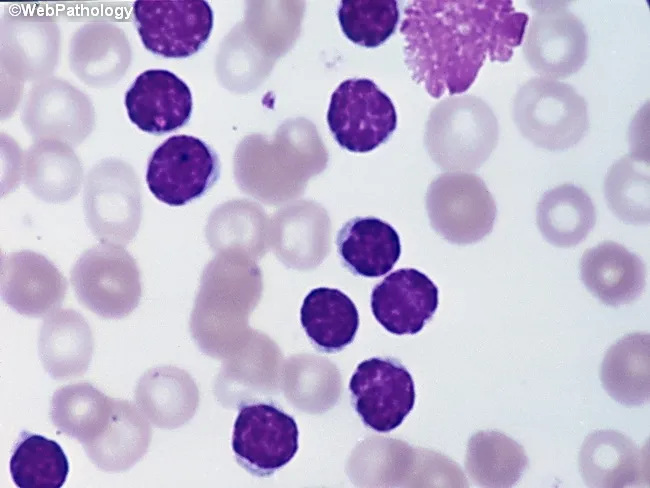
Peripheral blood smear from a patient with suspected chronic lymphocytic leukemia (CLL) shows a predominance of small, mature-appearing lymphocytes with scant cytoplasm and clumped nuclear chromatin, accompanied by fragile cells that readily disrupt during smear preparation, producing numerous smudge or basket cells visible on the slide. The image depicts a single smudge cell in the upper right, with numerous small darkly staining lymphocytes occupying the field. The smear was prepared with Wright-Giemsa stain and examined under light microscopy at high magnification (approximately 1000x total magnification with oil immersion). Clinically, CLL is defined by absolute mature lymphocytosis of ≥5.0 × 10^9/L sustained for ≥3 months, often asymptomatic and detected incidentally on complete blood count. Morphology alone is insufficient for diagnosis; immunophenotyping showing co-expression of CD5, CD19, CD23 with weak surface immunoglobulin and light chain restriction supports CLL diagnosis. Smudge cells result from fragile leukemic cells and reflect underlying cytoskeletal fragility, though they are not specific. The image illustrates typical features: monomorphic lymphocytes, scant cytoplasm, condensed chromatin, and absent prominent nucleoli. This finding has diagnostic significance, guiding further workup, including flow cytometry, cytogenetics, and molecular studies, to distinguish CLL from other chronic lymphoproliferative disorders such as small lymphocytic lymphoma (SLL), mantle cell lymphoma, prolymphocytic leukemia, and reactive lymphocytosis.
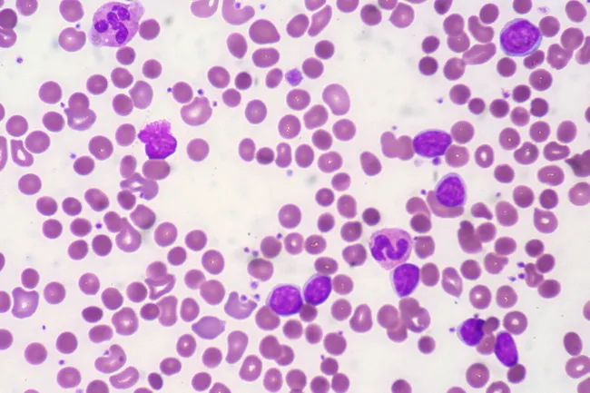
Peripheral blood smear from a patient with known chronic lymphocytic leukemia (CLL) showing autoimmune hemolytic anemia (AIHA). Brightfield microscopy of Wright-Giemsa stained smear at 1000x (oil immersion) reveals two dominant cell populations: numerous small mature lymphocytes typical of CLL and red blood cells showing spherocytic deformation. A smudge cell is evident just left of center, a classical clue for CLL. Red cells display reduced central pallor consistent with spherocytes, indicating intravascular or extravascular hemolysis driven by autoantibodies. The background shows mild polychromasia and rare nucleated RBCs, consistent with a robust reticulocyte response; the corrected reticulocyte count in this case is markedly elevated (>7%). Platelets are not significantly abnormal. These features collectively support autoimmune destruction of erythrocytes in the setting of CLL. Diagnostic significance lies in recognizing AIHA as a common autoimmune complication in CLL, with a clinical picture of sudden Hb decline and compensatory erythropoiesis. Differential considerations include drug-induced hemolysis, other causes of hemolytic anemia, or marrow infiltration by CLL. Clinical correlation includes therapy implications (steroids, rituximab) and close monitoring of hemolysis markers. This image is a valuable teaching resource for hematology morphology, AIHA in CLL, and differential diagnosis in anemia.
hairy cell leukemia peripheral blood TRAP stain

This AI-generated infographic functions as a complex visual mnemonic for Hairy Cell Leukemia (HCL). The image depicts a morbidly obese male figure with light-purple skin and long, flowing dark hair and a beard, shown suspended over a mechanical trap in a barren desert landscape. Each element of the illustration symbolizes a key diagnostic or clinical feature of the disease. The figure's significant 'fat' build represents massive splenomegaly, a hallmark clinical finding. The 'purple' skin tone corresponds to the characteristic appearance of malignant lymphocytes on hematoxylin and eosin (H&E) staining. The 'long hair' serves as a mnemonic for the filamentous, hair-like cytoplasmic projections visible on peripheral blood smears or bone marrow biopsies. The 'trap' represents the Tartrate-Resistant Acid Phosphatase (TRAP) stain used for definitive diagnosis. Finally, the 'dry desert' environment symbolizes the 'dry tap' (unsuccessful bone marrow aspiration) frequently encountered due to extensive bone marrow fibrosis associated with HCL. This visual aid is designed for medical students to enhance long-term recall of hematologic pathology through associative learning.
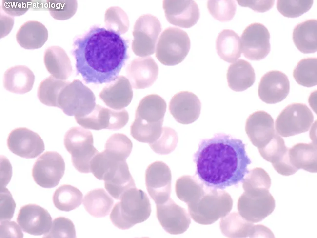
This is a high-magnification brightfield photomicrograph of a peripheral blood smear stained with Wright-Giemsa. The image highlights circulating hairy cells, which are larger than normal lymphocytes (approximately 1.5-2x). Hairy cells exhibit oval to bean-shaped nuclei with finely dispersed chromatin and inconspicuous nucleoli, and a moderate amount of pale, flocculent cytoplasm bearing fine cytoplasmic projections (hairy processes). The cytoplasm appears lightly basophilic and extends as delicate, hairlike extensions into the surrounding specimen. The background contains numerous mature erythrocytes and occasional platelets; there is no prominent nuclear atypia in these cells. Notably, the hairlike cytoplasmic projections are more readily appreciated in well-prepared, thin areas of the smear and are less evident in aspirate smears. This morphology is classically associated with hairy cell leukemia (HCL) and serves as a visual cue prompting confirmatory testing by flow cytometry for CD11c, CD25, CD103, and annexin A1, as well as tartrate-resistant acid phosphatase (TRAP) staining. Clinically, recognizing this pattern supports diagnosis in the appropriate hematologic context and guides subsequent therapeutic decisions. This image is valuable for education in hematopathology, cytology, and differential diagnosis of atypical lymphoid cells. Equivalent images may illustrate variably circulating hairy cells and aid differential diagnosis in practice.
Classification of Chronic Lymphoproliferative Disorders (CLPD) - 30 Marks
INTRODUCTION
- Low proliferation rate
- Prolonged cell survival (often due to BCL-2 anti-apoptotic activity)
- Indolent clinical course (with some exceptions)
- Leukemic manifestation (blood/bone marrow involvement) common in many subtypes
CLASSIFICATION
A. WHO HAEM5 (2022) and ICC (2022) - Current Classification
I. B-CELL CHRONIC LYMPHOPROLIFERATIVE DISORDERS
1. Chronic Lymphocytic Leukemia / Small Lymphocytic Lymphoma (CLL/SLL)
- Peripheral blood monoclonal B-lymphocytes ≥ 5,000/µL sustained for at least 3 months
- CLL immunophenotype (see below)
- If < 5,000/µL B-lymphocytes without lymphadenopathy/organomegaly = Monoclonal B-cell lymphocytosis (MBL)
- Presentation with lymphadenopathy rather than blood involvement = SLL (same disease biologically)
- Small lymphocytes with condensed "soccer ball" chromatin, scant cytoplasm, inconspicuous nucleoli
- Smudge/basket cells - pathognomonic artifact on smear due to cytoskeletal fragility
- Occasional prolymphocytes (up to 10-54% = atypical CLL; >55% = transformation to B-PLL)
- Bone marrow: Nodular, interstitial, mixed, or diffuse infiltration (diffuse = worse prognosis)
- Lymph node: Diffuse effacement with proliferation centers (pseudo-follicles) - pathognomonic
| Marker | CLL |
|---|---|
| CD5 | + (key) |
| CD19 | + |
| CD20 | + (dim) |
| CD23 | + (key) |
| Surface Ig | + (dim) |
| CD10 | - |
| Cyclin D1 | - |
| FMC7 | - |
| CD38 | +/- (adverse) |
| ZAP-70 | +/- (adverse) |
| CD49d | +/- (adverse) |
| CD200 | + (helps distinguish from MCL) |
- CD5+ (1 point)
- CD23+ (1 point)
- FMC7- (1 point)
- Dim sIg (1 point)
- CD22/CD79b dim or absent (1 point)
| Abnormality | Frequency | Significance |
|---|---|---|
| del(13q14) | ~55% | Favorable; MiR-15a/16-1 deletion |
| del(11q22-23) | ~18% | Adverse; ATM gene |
| Trisomy 12 | ~16% | Intermediate |
| del(17p13.1) | ~7% | Very poor prognosis; TP53 loss |
| t(14;18) | Rare |
- Mutated IGHV (≥2% deviation from germline): Favorable, reflects post-germinal center origin
- Unmutated IGHV (<2% deviation): Adverse, resembles naive B-cells, associated with ZAP-70+, CD38+
- Low-count MBL: <500/µL, not detectable on CBC, different BCR gene usage
- High-count MBL: 500-5000/µL, detectable on CBC; 1-2% per year risk of progression to CLL
| Stage | Features | Risk |
|---|---|---|
| 0 | Lymphocytosis only | Low |
| I | + Lymphadenopathy | Intermediate |
| II | + Spleen/liver enlargement | Intermediate |
| III | + Anemia (Hb <11 g/dL) | High |
| IV | + Thrombocytopenia (<100,000/µL) | High |
2. B-Cell Prolymphocytic Leukemia (B-PLL)
- Morphology: Medium-sized cells (~2x normal lymphocyte), round nuclei, prominent central nucleolus, pale blue cytoplasm; classic "fried egg" appearance
- >55% prolymphocytes in peripheral blood required for diagnosis
- Splenomegaly prominent; minimal lymphadenopathy
- Age ~70 years; male predominance (M:F 1.6:1)
- CD19+, CD20+ (bright), CD22+, surface IgM+ (bright)
- CD23 absent (distinguishes from CLL)
- CD5+ in ~1/3 cases
- FMC7+
- ~50% have TP53 mutation/deletion
- ~20% have t(11;14) → confusion with mantle cell lymphoma
- 11q23 and 13q14 deletions
3. Hairy Cell Leukemia (HCL)
- Middle-aged to elderly; massive splenomegaly
- Pancytopenia + monocytopenia (characteristic triad)
- "Dry tap" on bone marrow aspiration (~10% of dry taps)
- Small lymphocytes with oval/"kidney-shaped" nuclei, abundant pale cytoplasm
- Hairy (villous) cytoplasmic projections - best seen on phase contrast or SEM
- BM: "Fried egg"/"honeycomb" pattern on trephine biopsy
- CD103+ (most specific)
- CD11c+ (bright)
- CD25+
- Annexin A1+ (most specific for HCL; absent in HCL-v and SMZL)
- CD19+, CD20+ (bright), CD22+
- CD5-, CD10-, CD23-
- TRAP (Tartrate-resistant acid phosphatase) - positive; isoenzyme 5b
- BRAF V600E mutation in >95% of classic HCL - pathognomonic
- MAP2K1 mutation in HCL-variant (BRAF-negative)
- No IGHV somatic hypermutation pattern typical of CLL
4. Splenic Marginal Zone Lymphoma (SMZL)
- Villous lymphocytes in peripheral blood (short polar villi, unlike hairy cells)
- Splenomegaly + BM + PB involvement
- Median age: mid-50s; M=F
- CD19+, CD20+ (bright), surface Ig+
- CD5-, CD10-, CD103-, CD43-
- CD23-/+; CD11c-/+
- Del 7q21 (~40%) - FLNC gene
- NOTCH2 mutations (~25%)
- 8p deletions; MYD88 mutations in some
5. Lymphoplasmacytic Lymphoma (LPL) / Waldenström Macroglobulinemia (WM)
- Clonal B-cells showing plasmacytoid/plasma cell differentiation
- IgM paraprotein in serum (>30 g/L for WM diagnosis)
- BM, spleen, liver involvement
- Hyperviscosity syndrome characteristic
- CD19+, CD20+, CD22+, surface Ig+ (often IgM)
- CD5-, CD10-, CD23-
- Cytoplasmic IgM+ (plasma cell component)
- MYD88 L265P mutation: ~90% of LPL/WM cases; diagnostic hallmark
- CXCR4 mutations (25-40%): Predict ibrutinib resistance
6. Mantle Cell Lymphoma (MCL) - Leukemic Phase
- CD5+, CD19+, CD20+ (bright), surface Ig+ (bright)
- CD23- (differentiates from CLL)
- Cyclin D1+ (nuclear) - hallmark
- FMC7+, CD200-
- t(11;14)(q13;q32) - IGH-CCND1 rearrangement in virtually all cases
- Secondary mutations: TP53, CDKN2A, SOX11 (aggressive form)
7. Follicular Lymphoma (FL) with Leukemic Phase
- CD10+ (germinal center marker) - key positive
- CD19+, CD20+, surface Ig+
- CD5-, CD23+/-, Cyclin D1-
- BCL2+, BCL6+
- t(14;18)(q32;q21) - IGH-BCL2 rearrangement (~85%)
II. T-CELL / NK-CELL CHRONIC LYMPHOPROLIFERATIVE DISORDERS
1. T-Cell Large Granular Lymphocytic (T-LGL) Leukemia
- Persistent (>6 months) clonal expansion of large granular lymphocytes (LGLs) in blood
- Associated with rheumatoid arthritis, autoimmune cytopenias
- Neutropenia (most prominent), often with normal lymphocyte count
- Indolent course
- CD3+, CD8+ (most common), CD57+, CD16+
- CD4- (usually); CD56-
- TCR αβ+ (most)
- Loss of CD5, CD7 common
- TCR gene clonal rearrangement (Southern blot / PCR) - diagnostic
- STAT3 mutations (~40%); STAT5b mutations (rarer, more aggressive)
2. Chronic NK-Cell Lymphoproliferative Disorder (Chronic CLPD-NK)
- Persistent NK cell lymphocytosis (>2x10⁹/L for >6 months)
- Must exclude reactive causes
- Indolent; usually incidental finding
- CD3-, CD56+, CD16+, CD57+
- CD2+, CD7+/-
- Cytotoxic molecules (TIA-1, granzyme B) positive
- No TCR gene rearrangement (clonality assessed by NK-specific methods)
3. T-Cell Prolymphocytic Leukemia (T-PLL)
- Previously called "T-CLL" - this term is now obsolete
- Aggressive disease; short survival without therapy
- CD3+, CD2+, CD7+, CD5+
- CD4+ (most); CD8+ (25%); CD4+CD8+ (rare)
- CD52+ (important - alemtuzumab therapeutic target)
- TCL1A overexpressed
- inv(14)(q11;q32) or t(14;14)(q11;q32) = TCL1A rearrangement (~80%)
- t(X;14)(q28;q11) = MTCP1 rearrangement
- ATM mutations/deletions (11q22-23)
4. Sézary Syndrome (Leukemic CTCL)
- Triad: Erythroderma + lymphadenopathy + circulating Sézary cells
- Sézary cell: cerebriform nuclei (hyperconvoluted)
- Defined by Sézary count ≥1000/µL in blood
- CD3+, CD4+, CD26- (key), CD7-
- Loss of CD26 and CD7 is characteristic
- CD45RO+, CLA+
- TCR clonal rearrangement
- Complex karyotype
- T helper cell gene expression
5. Adult T-Cell Leukemia/Lymphoma (ATLL)
- Caused by HTLV-1 retrovirus
- Geographic clustering: Japan, Caribbean, Africa
- CD3+, CD4+, CD25+ (IL-2R; constitutive)
- CD7-, CD8-
- FoxP3+ (Treg phenotype)
DIAGNOSTIC APPROACH - IMMUNOPHENOTYPING
Flow Cytometry Panel for Leukemic CLPDs
- CD45, CD19, CD5, CD23, CD20, κ/λ light chains, CD3, CD4, CD8
- CD10, CD103, CD11c, CD25, CD38, CD200, FMC7, CD79b, CD22, Cyclin D1, Annexin A1
- CD56, CD16, CD57, CD26, CD7, TCRαβ/γδ
Key Immunophenotypic Patterns (Consolidated Table)
| Entity | CD5 | CD10 | CD23 | CD103 | Cyclin D1 | sIg | MYD88 | Key molecular |
|---|---|---|---|---|---|---|---|---|
| CLL | + | - | + | - | - | Dim | - | IGHV mut status; del13q |
| B-PLL | +/- | - | - | - | - | Bright | - | TP53, t(11;14) |
| MCL | + | - | - | - | + | Bright | - | t(11;14) |
| FL | - | + | +/- | - | - | + | - | t(14;18) |
| HCL | - | - | - | + | - | + | - | BRAF V600E |
| SMZL | - | - | -/+ | - | - | Bright | - | del7q, NOTCH2 |
| LPL/WM | - | - | - | - | - | IgM | + (L265P) | MYD88 L265P |
| T-LGL | CD3+CD8+ | - | - | - | - | - | - | STAT3 mut |
| T-PLL | CD3+CD4+ | - | - | - | - | - | - | inv(14)/TCL1A |
| Sézary | CD3+CD4+CD26- | - | - | - | - | - | - | TCR clonal |
| ATLL | CD3+CD4+CD25+ | - | - | - | - | - | - | HTLV-1 |
MOLECULAR STUDIES
1. FISH (Fluorescence In Situ Hybridization)
2. PCR-Based Clonality
- IGH VDJ rearrangement → confirms B-cell clonality
- TCR gene rearrangement (β or γ chains) → confirms T-cell clonality
- BIOMED-2 protocol - standardized multiplex PCR for clonality testing
3. IGHV Somatic Hypermutation Analysis
- Sequencing of VH region
- Mutated (≥2% divergence from germline): Post-GC origin; better prognosis in CLL
- Unmutated: Pre-GC; aggressive CLL
4. Specific Mutations (NGS / Sanger)
| Gene | Disease | Significance |
|---|---|---|
| BRAF V600E | HCL | Pathognomonic; therapeutic (vemurafenib) |
| MYD88 L265P | LPL/WM | ~90% specific; therapeutic (ibrutinib) |
| TP53 | CLL, B-PLL | Chemo resistance; BTK inhibitor preferred |
| IGHV | CLL | Key prognostic; determines therapy response |
| STAT3/STAT5b | T-LGL | Confirms clonality; STAT3 correlates with neutropenia |
| NOTCH2 | SMZL | Clonal marker |
| ATM | T-PLL, CLL | DNA repair deficiency |
5. Cytogenetics (Karyotype)
- Stimulated karyotype preferred in CLL (unmutated or complex karyotype = worse prognosis)
- Complex karyotype (≥3 abnormalities): High-risk CLL
6. HTLV-1 Serology
- Mandatory in ATLL workup
- PCR for HTLV-1 proviral DNA in blood
DIFFERENTIAL DIAGNOSIS ALGORITHM
Persistent lymphocytosis on CBC
         ↓
Flow cytometry (B vs T/NK)
         ↓
B-cell:                          T/NK cell:
↓                                ↓
CD5+ → CD23+ → CLL               CD3+/CD8+ → T-LGL
      → CD23- → MCL (Cyclin D1+) CD3+/CD4+ → T-PLL, Sézary, ATLL
CD5- → CD10+ → FL                CD3-/CD56+ → NK-CLPD, Aggressive NK
      → CD10- → CD103+ → HCL
               → CD103- → MYD88 → LPL/WM
                         → NOTCH2 → SMZL
SPECIAL POINTS FOR EXAM (30-MARK ANSWER)
-
Richter Transformation in CLL: 5-10% of CLL transforms to DLBCL (Richter syndrome) or Hodgkin lymphoma; sudden clinical deterioration, elevated LDH, constitutional symptoms
-
CLL vs SLL: Identical immunophenotype; distinction by >5000 circulating B-cells (CLL) vs predominantly nodal disease with <5000/µL (SLL); clinically managed identically
-
B-PLL vs CLL: B-PLL has NO proliferation centers on LN biopsy, absent CD23, bright CD20/sIg, and >55% prolymphocytes
-
Annexin A1 is a definitive marker differentiating HCL from HCL-variant and SMZL
-
CD200: Positive in CLL and HCL, negative in MCL; helps in difficult CD5+ cases
-
WHO HAEM5 2022 key changes:
- B-PLL eliminated as a distinct entity (heterogeneous group)
- "Hairy cell leukemia variant" renamed → "Splenic B-cell lymphoma/leukemia with prominent nucleoli"
- Cold agglutinin disease added as new entity
- MGRS defined as new entity
- EBV+ T/NK lymphoproliferative disorders better characterized
-
ICC 2022 vs WHO 2022 key differences for CLPDs:
- ICC retains B-PLL; WHO HAEM5 dissolves it
- ICC has expanded MBL subcategorization (low count vs clonal B-cell expansion)
- Both agree on CLL/SLL criteria (no changes from 2016)
SOURCES
- Goldman-Cecil Medicine, 27th Ed (2022) - Table 169: Typical Immunophenotype of CLL vs other CLPDs
- Harrison's Principles of Internal Medicine, 22nd Ed (2025) - Table 115-2: Immunophenotype of Small Lymphocyte Tumors
- Henry's Clinical Diagnosis and Management by Laboratory Methods - Chapter 35: Flow Cytometry in Lymphoma
- WHO HAEM5 vs ICC 2022 B-cell classification comparison
- CAP 2022 Leukemic Lymphoproliferative Neoplasms - Foucar