
Crhon disease with infalllmatiom
"Crohn disease"[MeSH Terms] AND "inflammation"[MeSH Terms]
Crohn disease inflammation pathology histology
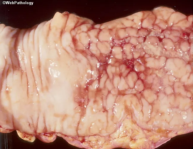
Macroscopic gross image of a resected bowel segment displaying marked mucosal disease. The right-hand mucosa shows a cobblestone pattern with discrete raised islands interspersed by shallow to deep ulcerations; a sharp demarcation separates involved from uninvolved mucosa on the left. These features arise from focal inflammatory changes producing mucosal edema, friability, and fissuring, yielding the characteristic cobblestone appearance. The mucosa appears erythematous with vascular congestion but without visible serosal perforation in this view. The pattern is suggestive of inflammatory bowel disease, most consistent with Crohn disease presenting as patchy skip-lesion involvement of the colon. The observed gross morphology supports clinical correlation and histopathologic confirmation to distinguish Crohn colitis from other causes of segmental ulceration, such as intestinal tuberculosis or ischemic colitis. Depth of ulceration cannot be determined from this surface image alone. This specimen provides essential gross-pathology context for surgical pathology, gastroenterology, and education, and would be indexed under inflammatory bowel disease, cobblestone mucosa, Crohn disease, skip lesions, mucosal ulceration, and colonic involvement. Keywords for search include colon, colonoscopy correlation, macroscopic bowel pathology, Crohn disease pattern, transmural inflammation suspicion, and gross mucosal pattern. Additional histology will optimize differential diagnosis and guide therapy in clinical practice.
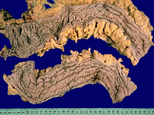
Comprehensive Description: Gross pathology photograph of a resected colon segment demonstrating Crohn disease (Crohn colitis). Modality: Gross external view of a macroscopic specimen; not microscopic imaging. Anatomic location: colon with terminal ileum involvement described; in this specimen, segments show transmural inflammation with deep longitudinal ulcers oriented horizontally; serpentine ulcers connected by short transverse ulcers; cobblestone mucosa due to pseudopolyps; mucosal edema and possible hemorrhage. Pathologic features: segmental, discontinuous involvement (skip areas) with sharp demarcation between diseased and normal mucosa; strictures from muscularis propria hypertrophy and fibrosis; long-standing disease produces luminal narrowing; fissures between mucosal folds; transmural inflammation may lead to perforation or fistula tracts; creeping fat may envelop the serosa in advanced disease. Digital imaging of external surface highlights creeping fat and mesenteric fat wrapping. Clinical significance: pattern helps differentiate Crohn disease from ulcerative colitis; transmural involvement, fissuring ulcers, and creeping fat correlate with fistulization and stricturing risk; surgical planning and prognosis are informed by gross features. Differential considerations include ulcerative colitis (continuous colitis) and intestinal tuberculosis; clinical correlation with radiologic and endoscopic findings is essential for accurate diagnosis and management. This image is valuable for surgical education, pathology training, and correlating gross features with imaging and histology in practice.
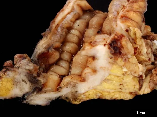
Gross pathology photograph of a sigmoid colectomy specimen from a young male with Crohn disease presenting with a colovesical fistula. The specimen is opened longitudinally to expose the colonic mucosa. The left lower quadrant bowel reveals a conspicuous fistulous tract extending medially toward the urinary bladder. The mucosal surface shows a cobblestone pattern with deep, irregular ulcers and fissuring along the length of the sigmoid colon, consistent with transmural Crohn disease. The serosa is thickened with creeping fat and mesenteric inflammatory change; there is surrounding inflammatory exudate. The fistulous tract is conjoined with granulation tissue and inflammatory debris; there may be partial luminal narrowing from stricture formation. The adjacent bladder wall may show involvement where the fistula communicates, though histology is needed for confirmation. Scale bar indicates 1 cm. Overall the specimen demonstrates Crohn disease features: chronic transmural inflammation, fissuring ulcers, cobblestone mucosa, fistulization, and fistulous connection to the urinary tract. Clinically this configuration causes symptoms such as pneumaturia and fecaluria; surgical resection is indicated. This finding highlights increased colorectal cancer risk in Crohn disease versus the general population and underscores the need for ongoing surveillance in young patients. Correlation with histologic sampling would confirm granulomatous enterocolitis and exclude alternative etiologies.
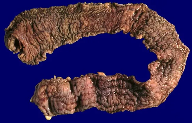
Gross photograph of a total colectomy specimen from a young male with ulcerative colitis showing pancolitis. The colon is laid flat, mucosal surface exposed, with diffuse hyperemia and irregular mucosal pattern. The mucosa displays multiple shallow and deeper ulcers with overlying pseudopolyps; the luminal contour remains relatively preserved except for mucosal edema. There is no appreciable wall thickening or stenosis; serosal surface appears smooth. The tissue demonstrates continuous inflammatory involvement along the entire colon, from cecum to rectum, without skip lesions, consistent with ulcerative colitis rather than Crohn disease. There are no discrete masses or signs of transmural inflammation; no perforation noted. The presence of pseudopolyps indicates chronic disease with mucosal regeneration. The external view shows a swollen, corrugated mucosal surface with corrugated transverse folds. The image is a macroscopic documentation suitable for gastroenterology and surgical pathology, useful for teaching, histologic correlation, and differential diagnosis between ulcerative colitis pancolitis and Crohn colitis, ischemic or infectious colitis. Clinically, this specimen supports a prognosis requiring colectomy due to refractory disease or dysplasia. The photograph provides a reference for pathology atlases and educational resources. Correlate with histology is essential for confirming ulcerative colitis versus Crohn disease and guiding patient management.
Crohn's Disease: Inflammation - Comprehensive Overview
What Is Crohn's Disease?
- Schwartz's Principles of Surgery, 11th ed. - "Crohn's disease is a transmural inflammatory process that can affect any part of the gastrointestinal tract."
Pathology and Inflammatory Features
Gross Pathology (What You See Macroscopically)
| Feature | Description |
|---|---|
| Skip lesions | Discontinuous patches of inflammation separated by normal-appearing bowel - a hallmark of CD |
| Cobblestone mucosa | Deep longitudinal fissures + transverse ulcers between inflamed mucosal islands |
| Fat wrapping / creeping fat | Mesenteric fat encroaches onto the serosal surface; virtually pathognomonic of CD |
| Strictures | Luminal narrowing from fibrosis after chronic transmural inflammation |
| Fistulas | Transmural inflammation erodes through bowel wall into adjacent structures |
| Aphthous ulcers | Earliest endoscopic lesion - small, shallow erosions overlying lymphoid follicles |
Microscopic / Histological Features
- Transmural inflammation - all layers (mucosa → serosa) involved
- Non-caseating granulomas - present in 50-60% of cases; pathognomonic when found
- Crypt abscesses and distortion
- Pyloric gland metaplasia (indicates repeated bouts of inflammation and repair)
- Submucosal fibrosis
Spread of Inflammation
- Superficial disease - mucosal/submucosal inflammation
- Advanced disease - full-thickness (transmural) involvement → adhesions, abscesses, fistulas, and rarely free perforation
Sites of Involvement
| Location | Frequency |
|---|---|
| Terminal ileum + cecum (ileocecal) | Most common (~60%) |
| Small bowel alone | 80% have small bowel involvement |
| Colon alone | ~20% |
| Perianal/anorectal | 5-10% |
| Stomach/duodenum | Uncommon; usually with intestinal disease |
| Esophagus | Rare |
- Schwartz's Principles of Surgery: "The disease affects the small bowel in 80% of cases and colon alone in 20%."
Clinical Presentation by Inflammation Type
- Aggressive inflammatory disease - active mucosal and transmural inflammation
- Fibrostenotic disease - chronic inflammation leads to fibrosis and strictures
- Fistulizing disease - transmural inflammation erodes through to adjacent structures
Symptoms Due to Active Inflammation
- Abdominal pain (crampy, usually right lower quadrant in ileocecal disease)
- Diarrhea (>10 bowel movements/day during a flare)
- Fever
- Weight loss
- Nausea, vomiting
- Bloating
- Perianal pain, swelling, drainage (fistulas/abscesses)
"Symptoms of Crohn's disease depend on the severity of inflammation and/or fibrosis and the location of inflammation in the gastrointestinal tract. Acute inflammation may produce diarrhea, crampy abdominal pain, and fever." - Schwartz's Principles of Surgery
Measuring Disease Activity (Inflammation Severity)
| Tool | Notes |
|---|---|
| CDAI (Crohn's Disease Activity Index) | Score <150 = remission; drop ≥100 = treatment response |
| CRP (C-reactive protein) | Elevated in active disease; tracks inflammatory activity |
| Fecal calprotectin | Sensitive marker of intestinal inflammation; correlates with relapse and biologic response |
| Fecal lactoferrin | Another neutrophil-derived marker of mucosal inflammation |
| Endoscopy | Gold standard for mucosal healing; "symptoms alone should not guide therapeutic decisions" |
| MRI/CT enterography | Shows mural thickening, hyperenhancement, "comb sign" (engorged peri-enteric vessels) |
Extraintestinal Manifestations of Inflammation
| System | Manifestation |
|---|---|
| Joints | Arthritis (20x more common than general population), sacroiliitis, ankylosing spondylitis |
| Skin | Erythema nodosum (5-15%), pyoderma gangrenosum |
| Eyes | Uveitis, episcleritis |
| Liver/Biliary | Fatty liver (40-50%), primary sclerosing cholangitis, pericholangitis |
| Growth | Growth retardation in children |
CD vs Ulcerative Colitis: Key Inflammatory Differences
| Feature | Crohn's Disease | Ulcerative Colitis |
|---|---|---|
| Depth | Transmural (all layers) | Mucosa + submucosa only |
| Distribution | Discontinuous, skip lesions | Continuous from rectum |
| Rectum | Often spared | Almost always involved |
| Granulomas | Yes (non-caseating) | No |
| Fistulas/abscesses | Common | Rare |
| Any GI segment | Yes (mouth to anus) | Colon only |
Management of Inflammation
Medical Therapy (Step-up Approach)
| Severity | Options |
|---|---|
| Mild | Mesalamine (limited efficacy), budesonide (for ileocecal disease) |
| Moderate | Corticosteroids (short-term), azathioprine/6-MP (maintenance), methotrexate |
| Severe/Refractory | Anti-TNF-α biologics: infliximab, adalimumab; Anti-integrin: vedolizumab; Anti-IL-12/23: ustekinumab |
- PPIs are first-line for gastric/duodenal Crohn's
- Anti-TNF therapy (infliximab) is effective for fistulizing disease and refractory luminal inflammation
- Strictures with an inflammatory component may improve with optimal medical therapy (anti-fibrotic strictures require endoscopic dilation or surgery)
Surgical Indications
- Bowel obstruction not responsive to medical management
- Intra-abdominal abscesses
- Fistulas refractory to medical therapy
- Free perforation
- Dysplasia/malignancy
- ~75% of CD patients will require at least one abdominal operation in their lifetime
Complications of Chronic Inflammation
- Strictures - from fibrosis after transmural inflammation; cause small bowel obstruction (SBO)
- Fistulas - enteroenteric, enterovesical (colovesical fistula → pneumaturia), enterocutaneous, rectovaginal
- Abscesses - intra-abdominal or perianal
- Malabsorption - protein-losing enteropathy, vitamin B12 deficiency (terminal ileum), bile acid malabsorption
- Short bowel syndrome - after repeated surgical resections
- Colorectal cancer - elevated risk with long-standing colonic CD
- Sleisenger and Fordtran's Gastrointestinal and Liver Disease (Crohn Disease chapters)
- Schwartz's Principles of Surgery, 11th ed. (Chapter 28)
- Maingot's Abdominal Operations (Chapters on perianal Crohn's and IBD)
Diet
"Crohn disease"[MeSH Terms] AND "diet therapy"[MeSH Terms]
PMID: 39107691
PMID: 37513532
Diet in Crohn's Disease
Why Nutrition Matters in Crohn's Disease
- Reduced oral intake (pain, nausea, fear of eating)
- Malabsorption (especially with ileal disease - vitamin B12, fat-soluble vitamins, bile acids)
- Protein-losing enteropathy
- Increased metabolic demands during inflammation
- Surgical bowel resections leading to short bowel
"Nutritional support in all patients with Crohn disease is paramount. Ideally, preservation of oral feeding in any form is crucial for maintenance of the absorptive and protective mechanisms provided by the GI mucosal lumen." - Mulholland and Greenfield's Surgery, 7th ed.
Dietary Therapeutic Strategies
1. Exclusive Enteral Nutrition (EEN) - First-Line in Pediatrics
-
In pediatric CD: EEN has similar efficacy to oral corticosteroids for inducing clinical remission, but is more effective at achieving mucosal healing - making it the preferred first-line induction therapy in children (endorsed by NASPGHAN).
-
It avoids the growth suppression caused by steroids in children.
-
In adults: EEN has not shown superior benefit over corticosteroids for remission induction.
-
Drawback: poor palatability, restrictive, and may reduce gut bacterial diversity (decreased Bifidobacterium, Ruminococcus, Faecalibacterium).
-
Yamada's Textbook of Gastroenterology, 7th ed.; Mulholland and Greenfield's Surgery, 7th ed.
2. Crohn's Disease Exclusion Diet (CDED) + Partial Enteral Nutrition (PEN)
- Gluten
- Dairy products
- Red/processed meats
- Refined sugars and processed foods
- Emulsifiers and additives
- Sleisenger and Fordtran's GI and Liver Disease
3. CD-TREAT Diet (Crohn's Disease Treatment-with-Eating)
- Gluten
- Dairy
- Alcohol
- Refined sugars
- Yamada's Textbook of Gastroenterology, 7th ed.
4. Mediterranean Diet (MD)
- Rich in fruits, vegetables, whole grains, legumes, olive oil, fish; low in red/processed meat.
- A 2021 RCT (referenced in Harrison's Principles of Internal Medicine, 22E) compared the Specific Carbohydrate Diet (SCD) to the Mediterranean Diet in adult CD - both achieved comparable clinical remission rates.
- The Mediterranean diet is more sustainable and palatable long-term.
5. Specific Carbohydrate Diet (SCD)
- Eliminates all grains, lactose, sucrose, and processed foods; allows meats, fish, eggs, certain fruits/vegetables, and specific cheeses.
- Promising results in pediatric CD, but evidence quality is moderate.
- Referenced in Harrison's 22E and Sleisenger's GI Disease.
6. Low-FODMAP Diet
-
Reduces symptoms (bloating, gas, diarrhea) in patients with quiescent or mildly active CD.
-
A RCT showed 52% symptom relief vs 16% on control diet (p=0.007).
-
Important caveat: Low-FODMAP reduces symptoms but its effect on actual mucosal inflammation in CD is unknown. It may also reduce Bifidobacteria and Faecalibacterium prausnitzii - beneficial gut bacteria.
-
Best used for symptom management, not as primary anti-inflammatory therapy.
-
Yamada's Textbook of Gastroenterology
7. High-Fiber Diet
- During acute flares with strictures or obstruction, fiber should be restricted to avoid blockage - low-residue diet is preferred during flares.
Nutritional Support: When Oral Diet Is Not Enough
| Support Type | When Used | Notes |
|---|---|---|
| Partial enteral nutrition (PEN) | Supplement to solid food diets; maintenance | Used with CDED; improves outcomes |
| Exclusive enteral nutrition (EEN) | Active CD induction (especially pediatric) | 6-8 weeks; mucosal healing |
| Total parenteral nutrition (TPN) | When enteral route is not possible (post-op, severe obstruction, fistulas) | Complication rates higher than enteral; use short-term |
"Total parenteral nutrition (TPN) was useful in patients with active Crohn disease; however, complication rates exceed those for enteral nutrition." - Sabiston Textbook of Surgery
Key Nutritional Deficiencies to Monitor and Supplement
| Nutrient | Why Deficient | Supplement |
|---|---|---|
| Vitamin B12 | Terminal ileum disease/resection | IM injection or high-dose oral |
| Iron | Chronic blood loss, inflammation | Oral or IV iron |
| Vitamin D | Malabsorption, reduced sun exposure | Oral supplementation |
| Folic acid | Reduced intake, methotrexate use | Oral |
| Zinc | High stool losses during diarrhea | Oral zinc |
| Calcium | Steroid use, reduced dairy intake | Oral + vitamin D |
| Magnesium | Chronic diarrhea, malabsorption | Oral or IV |
| Fat-soluble vitamins (A, D, E, K) | Bile acid malabsorption (ileal disease) | Water-soluble formulations |
Practical Dietary Guidance by Disease State
During Active Flare
- Low-residue / low-fiber diet - reduces stool frequency and risk of obstruction with strictures
- Avoid raw vegetables, seeds, nuts, corn, popcorn
- Small, frequent meals
- Adequate hydration
- Enteral nutrition support if oral intake is severely compromised
During Remission (Maintenance)
- Mediterranean-style diet - anti-inflammatory, sustainable, diverse
- High-fiber foods (if tolerated and no strictures) - support microbiome
- Adequate protein intake (1.2-1.5 g/kg/day) for tissue repair
- Avoid ultra-processed foods, food additives, emulsifiers, excess red meat
- Limit alcohol (mucosal irritant)
- Avoid smoking - smoking significantly worsens CD course, increases relapse rates, and increases need for surgery
Foods to Generally Avoid or Limit
- Fried/greasy foods
- Carbonated drinks
- Spicy foods (can aggravate symptoms)
- High-lactose dairy (if lactose intolerant)
- Alcohol
- Excess caffeine
Recent Evidence Summary
| Study | Finding |
|---|---|
| Systematic Review 2024, PMID 39107691 | Mediterranean diet and CDED show promising results for remission induction in mild-moderate CD; low-FODMAP improves symptoms but evidence is mixed |
| Meta-Analysis 2023, PMID 37513532 | High-fiber diet improved remission rates in CD patients (11 studies, 2,389 patients) |
| Network Meta-Analysis 2024, PMID 39686992 | Dietary therapies effective in active pediatric CD |
Bottom Line
| Goal | Best Dietary Approach |
|---|---|
| Induce remission (pediatric) | EEN (first-line) or CD-TREAT |
| Induce remission (adult) | CDED + PEN, Mediterranean diet |
| Maintain remission | Mediterranean diet, high-fiber diet, CDED |
| Symptom relief only | Low-FODMAP diet |
| Nutritional support | Supplement B12, D, iron, zinc, calcium |
| Active flare | Low-residue diet + enteral support if needed |