
Answer the following questions as per bds 3rd year oriented for university exam pattern of general medicine: as a long note Describe Myocardial Infarction: etiology, pathogenesis, clinical features, investigations, management. * Write in detail about Infective Endocarditis + antibiotic prophylaxis. * Rheumatic fever & Rheumatic heart disease: pathogenesis and features. * Congestive Cardiac Failure: causes, clinical features, management.
myocardial infarction ECG changes ST elevation
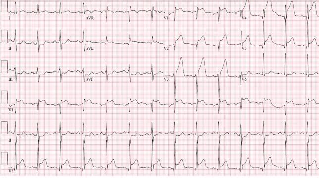
A 12-lead electrocardiogram (ECG) demonstrating an acute anterolateral ST-elevation myocardial infarction (STEMI). The tracing shows significant, convex-upward ST-segment elevation in the precordial leads (V1–V6), consistent with an anterior wall injury. Leads I and aVL also display ST-elevation, indicating lateral involvement. These changes are accompanied by hyperacute, peaked T-waves most prominent in V2 through V4. There is evidence of reciprocal ST-segment depression in the inferior leads (II, III, and aVF). The QRS complexes appear widened in the leads with maximal ST elevation. The heart rhythm is sinus. This ECG pattern is clinically significant for proximal left anterior descending (LAD) artery occlusion, necessitating urgent reperfusion therapy. The visual presentation focuses on teachable hallmarks of cardiac ischemia, including the relationship between anatomical lead distribution and corresponding vascular territories, as well as the identification of reciprocal changes in opposing leads.
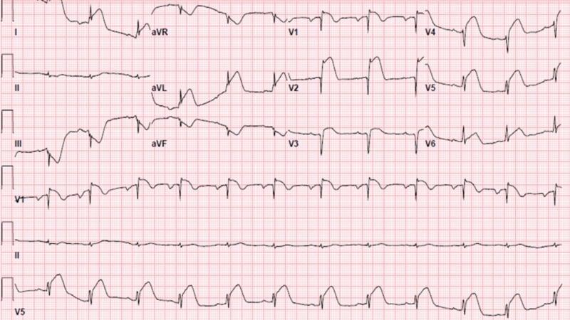
This diagnostic image is a 12-lead electrocardiogram (ECG) demonstrating signs of an acute ST-segment elevation myocardial infarction (STEMI). The primary finding is significant ST-segment elevation in the anterior precordial leads (V1, V2, and V3), with the highest amplitude visible in lead V2. The ST segments in these leads exhibit a convex or 'tombstoning' morphology, transitioning directly into prominent T waves. Complementary reciprocal changes are observed in the inferior leads (II, III, and aVF), characterized by ST-segment depression and T-wave inversion, particularly notable in lead III. Lead aVL also shows some ST-segment elevation. The rhythm is sinus, and these findings are classically associated with an acute proximal left anterior descending (LAD) artery occlusion. This ECG serves as a critical clinical example for medical students and clinicians to identify acute anterior wall myocardial injury and understand the concept of reciprocal ST-segment changes in inferior leads during an anterior infarction.
infective endocarditis Osler nodes Janeway lesions clinical features
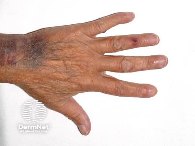
Clinical photography of a human hand highlighting digital Osler nodes associated with infective endocarditis. Modality: clinical photograph; perspective: dorsal hand view with fingers extended to expose distal finger pads. The ring finger bears a localized tender subcutaneous nodule at the fingertip/pulp, with additional subtler nodules on the adjacent middle and little fingers. The nodules are erythematous to violaceous, slightly raised, and may be palpably firm; surrounding skin appears otherwise unremarkable apart from subtle venous markings and age-related signaling. This image documents cutaneous manifestations that support a diagnosis of infective endocarditis in the appropriate clinical context. Osler nodes (Osler’s nodules) are immune-complex–mediated microvasculitis presenting as painful nodules on the digits; they contrast with Janeway lesions, which are usually painless. Clinically relevant features include tenderness, localized swelling, and distal finger involvement. The photograph is useful for education about dermatologic findings in infectious diseases and for medical-legal documentation of exam signs. In a patient with fever, new heart murmur, or positive blood cultures, these lesions increase pretest probability of infective endocarditis and guide downstream evaluation, including echocardiography and serology. Differential and educational use cases include distinguishing infectious endocarditis cutaneous signs from rheumatoid nodules, gouty tophi, or from digital trauma. Educational reference for clinicians.
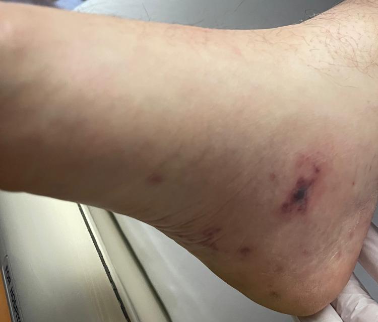
Clinical photograph of the medial aspect of the right foot and ankle demonstrating immunologic and vascular cutaneous manifestations of infective endocarditis. The primary finding is a well-circumscribed, tender, erythematous to violaceous (red-purple) nodular lesion located inferior to the medial malleolus, consistent with an Osler node. The lesion exhibits a central dark purpuric area surrounded by a diffuse inflammatory halo. Additionally, several smaller, discrete, non-blanching erythematous macules are visible in the surrounding area, which may represent Janeway lesions. The image serves as an educational reference for identifying peripheral stigmata of bacteremia, specifically related to Staphylococcus aureus endocarditis. Key concepts illustrated include the distinction between painful, immune-mediated Osler nodes and painless, embolic Janeway lesions in the context of Duke criteria for diagnosing valvular vegetations.
rheumatic heart disease mitral valve pathology
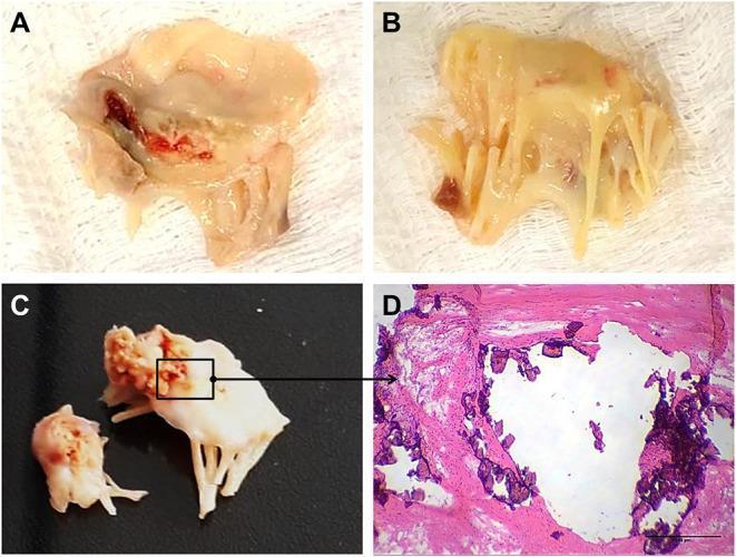
A multi-panel figure illustrating the gross and microscopic pathology of rheumatic heart valve disease (RHVD) in human mitral valves. Panels A and B present clinical photographs of excised mitral valve tissue from a 49-year-old female, showing marked leaflet thickening, retraction, and an opaque, yellowish appearance consistent with chronic fibrosis. Panel A specifically shows focal areas of reddish-dark discoloration, suggesting hemorrhage or tissue damage. Panel C displays gross pathology from a 61-year-old male, highlighting advanced calcification with rigid, cauliflower-like deposits on the valve structure. Panel D is a high-magnification light microscopy image (Hematoxylin and Eosin stain) of the anterior mitral valve leaflet. It demonstrates nodular calcification, appearing as irregular, dark purple hematoxylin-positive deposits embedded within the eosinophilic (pink) fibrotic collagenous matrix. A scale bar of 500 μm is provided. This figure serves as an educational resource for identifying the progression from chronic inflammation and fibrosis to end-stage valvular calcification in rheumatic heart disease.
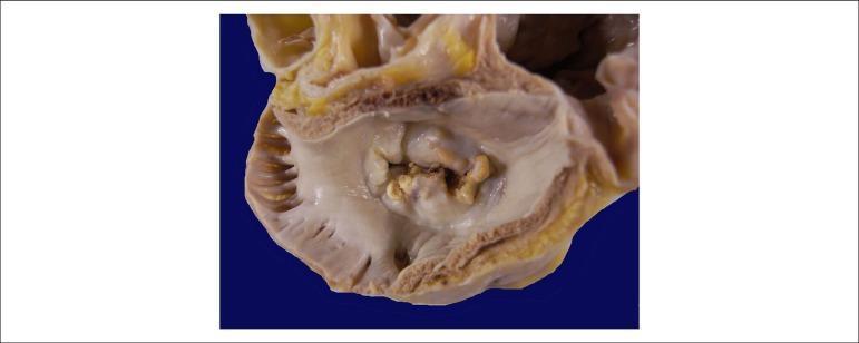
This clinical photograph shows a gross pathological specimen of a human heart, specifically an atrial view of the mitral valve. The image demonstrates hallmark signs of chronic rheumatic heart disease. Key features include significant thickening and fibrosis of the valve leaflets, resulting in a 'fish-mouth' or 'buttonhole' appearance of the valvular orifice. There is evidence of extensive commissural fusion and multiple focal areas of yellowish-white calcification along the leaflet margins and within the valve body. These structural changes characterize severe mitral stenosis, where the restricted opening of the valve impairs blood flow from the left atrium to the left ventricle. The surrounding atrial endocardium appears thickened, consistent with chronic pressure overload. This visual is highly representative for educational purposes in cardiology and pathology to illustrate the long-term sequelae of rheumatic fever on cardiac valves.
General Medicine Long Notes - BDS 3rd Year (University Exam Pattern)
QUESTION 1: MYOCARDIAL INFARCTION
Describe Myocardial Infarction - Etiology, Pathogenesis, Clinical Features, Investigations, Management
DEFINITION
ETIOLOGY
- Age (males >45 years, females >55 years)
- Male sex
- Positive family history (first-degree relative with MI before age 55 in males / 65 in females)
- Genetic predisposition
- Hypertension (HTN)
- Hyperlipidemia (elevated LDL, low HDL)
- Diabetes mellitus
- Cigarette smoking
- Obesity and physical inactivity
- Metabolic syndrome
- Coronary artery spasm (Prinzmetal's angina)
- Coronary arteritis (e.g., Kawasaki disease, SLE)
- Cocaine use (causes vasospasm + thrombosis)
- Embolism to coronary artery (from prosthetic valves, IE)
- Severe anemia with reduced oxygen delivery
- Congenital coronary artery anomalies
PATHOGENESIS
- Platelet adhesion to exposed subendothelial collagen via von Willebrand factor and GP Ib receptors
- Platelet activation and release of ADP, thromboxane A2, serotonin (amplification)
- Platelet aggregation via GP IIb/IIIa receptors + fibrinogen
- Activation of coagulation cascade forming fibrin clot
- Result: Occlusive thrombus in the coronary lumen
- Complete occlusion stops blood flow distal to the obstruction
- Small collateral flow is insufficient to prevent necrosis
- Cardiac muscle requires ~1.3 mL O2/100g/min just to remain alive
- In the central core of infarction (zero collateral flow): cells die within 20-40 minutes
- Subendocardial muscle is most susceptible because it has higher O2 consumption and vessels are compressed during systole
- ATP depletion, anaerobic glycolysis, lactic acid accumulation
- Intracellular Na+ and Ca2+ overload
- Cell swelling, membrane disruption
- Irreversible cell death (coagulative necrosis) - starts at 20-40 min, complete by 4-6 hours
- Release of cardiac enzymes (Troponin, CK-MB) into bloodstream
- Zone of necrosis (core): dead myocytes
- Zone of injury (periphery): stunned, potentially salvageable
- Zone of ischemia (outer): ischemic but viable
- STEMI (ST Elevation MI): Full-thickness (transmural) infarct - complete occlusion
- NSTEMI (Non-ST Elevation MI): Partial thickness (subendocardial) - incomplete occlusion
| Time | Changes |
|---|---|
| 0-6 hours | Wavy fiber change, coagulative necrosis begins |
| 1-3 days | Neutrophil infiltration, early coagulative necrosis |
| 4-7 days | Macrophage phagocytosis, granulation tissue |
| 1-3 weeks | Fibroblast proliferation, collagen deposition |
| >6 weeks | Dense fibrous scar |
CLINICAL FEATURES
-
Chest pain - The cardinal symptom
- Severe, crushing, constricting, or squeezing
- Retrosternal in location
- Radiates to left arm (most classic), jaw, neck, epigastrium, or right arm
- Duration: >30 minutes (distinguishes from angina which is <20 min)
- NOT relieved by nitrates (unlike angina)
-
Sweating (diaphoresis) - profuse, cold, clammy
-
Nausea and vomiting - due to vagal stimulation
-
Dyspnea - due to LV failure/pulmonary edema
-
Sense of impending doom (angor animi)
-
Palpitations - due to arrhythmias
| System | Finding |
|---|---|
| General | Anxious, pale, sweating, cold extremities |
| Pulse | Tachycardia (often), may be bradycardic (inferior MI) |
| BP | Initially elevated (pain), may fall (cardiogenic shock) |
| JVP | Elevated in RV failure or inferior MI |
| Heart sounds | S3 gallop (LV failure), S4 (pre-existing), pericardial friction rub |
| Lungs | Fine basal crepitations (pulmonary edema) |
- Arrhythmias (most common): VF (leading cause of early death), VT, AF, heart block (especially in inferior MI - RCA occlusion)
- Cardiogenic shock: BP <90 mmHg, cold clammy skin, urine output <20 mL/hr
- Left ventricular failure / pulmonary edema
- Mechanical complications: Papillary muscle rupture (acute MR), VSP (VSD), free wall rupture, cardiac tamponade
- Pericarditis: Day 1-3 (early fibrinous); Dressler syndrome: 2-10 weeks post-MI (immune-mediated)
- Ventricular aneurysm: Persistent ST elevation, thrombus, heart failure
- Re-infarction
INVESTIGATIONS
- Hyperacute T waves: First change (minutes to hours) - tall peaked T waves
- ST segment elevation (>1mm in limb leads, >2mm in chest leads): Indicates acute transmural injury
- Pathological Q waves: Width >0.04 sec, depth >25% of R wave - indicates transmural necrosis (develops over hours to days)
- T wave inversion: Develops as injury evolves
| Leads with changes | Artery occluded | Area of MI |
|---|---|---|
| V1-V4 | LAD | Anterior |
| V5-V6, I, aVL | LCx | Lateral |
| II, III, aVF | RCA | Inferior |
| V1-V2 (tall R, ST depression) | RCA/LCx | Posterior |
| Marker | Rises | Peaks | Returns to normal | Notes |
|---|---|---|---|---|
| Troponin I/T | 3-4 hrs | 24-48 hrs | 7-14 days | Most sensitive & specific - "gold standard" |
| CK-MB | 4-6 hrs | 24 hrs | 48-72 hrs | Good for re-infarction |
| Myoglobin | 1-2 hrs | 4-6 hrs | 24 hrs | Earliest but non-specific |
| LDH | 24 hrs | 3-6 days | 8-14 days | Useful late presentation |
- Identifies wall motion abnormalities (hypokinesis, akinesis, dyskinesis)
- Assesses EF, pericardial effusion, mechanical complications
- Bedside echo is invaluable in emergency setting
- CBC: Leukocytosis (neutrophilia) within 24-48 hours
- ESR, CRP: Elevated (inflammatory response)
- Blood glucose (hyperglycemia common)
- Lipid profile (taken at admission - levels fall after 24 hrs)
- Renal function (before thrombolysis/contrast)
- Coagulation profile (PT, aPTT)
- Cardiomegaly (if pre-existing LV dysfunction)
- Pulmonary venous congestion, Kerley B lines, pulmonary edema
- May be normal early on
- Definitive investigation to identify culprit vessel
- Performed urgently for STEMI (door-to-balloon time <90 min)
- PCI performed at same sitting
MANAGEMENT
A. IMMEDIATE / EMERGENCY MANAGEMENT ("MONA" + "REACT")
- Aspirin 300 mg stat (chewed) - antiplatelet
- Clopidogrel/Ticagrelor - P2Y12 inhibitor (dual antiplatelet therapy)
- Morphine 2-4 mg IV - pain relief, reduces preload, allays anxiety
- Oxygen - only if SpO2 <94% (avoid hyperoxia in non-hypoxic patients)
- Nitrates (GTN sublingual) - for pain relief if SBP >90 mmHg; avoid in inferior MI with RV involvement
- 12-lead ECG and cardiac monitoring
- IV access, blood samples
B. REPERFUSION THERAPY (for STEMI - most critical intervention)
- Treatment of choice if available within 90 min of first medical contact
- Balloon angioplasty + stenting of culprit artery
- Superior to thrombolysis
- Door-to-balloon time should be <90 min
- Streptokinase 1.5 million units IV over 60 min
- tPA (Alteplase), Tenecteplase - more fibrin-specific
- Contraindications: recent surgery, stroke, active bleeding, uncontrolled HTN
C. ANTICOAGULATION
- Heparin (LMWH - enoxaparin, or UFH) - prevents re-thrombosis
- Fondaparinux - in NSTEMI/UA
D. ADDITIONAL MEDICAL THERAPY
| Drug | Rationale |
|---|---|
| Beta-blockers (metoprolol, carvedilol) | Reduce HR, BP, myocardial O2 demand; prevent arrhythmias; reduce mortality |
| ACE inhibitors (ramipril, lisinopril) | Prevent LV remodeling; reduce mortality post-MI especially with reduced EF |
| Statins (atorvastatin 40-80 mg) | Plaque stabilization, lipid lowering; start within 24 hrs |
| Aldosterone antagonist (eplerenone) | Post-MI HF with EF <40% |
E. LONG-TERM MANAGEMENT (Secondary Prevention)
- Dual antiplatelet therapy (aspirin + P2Y12 inhibitor) for 12 months
- Statin indefinitely
- ACE inhibitor / ARB
- Beta-blocker (especially if EF reduced)
- Lifestyle modification: smoking cessation, diet, exercise, weight loss, BP and DM control
- Cardiac rehabilitation
QUESTION 2: INFECTIVE ENDOCARDITIS + ANTIBIOTIC PROPHYLAXIS
Write in Detail about Infective Endocarditis + Antibiotic Prophylaxis
DEFINITION
CLASSIFICATION
- Acute IE: Aggressive, rapid onset (days) - often due to S. aureus; destroys normal valves
- Subacute IE (SBE): Indolent course over weeks to months - often viridans streptococci; occurs on abnormal valves
- Native Valve IE (NVE): On normal or diseased native valves
- Prosthetic Valve IE (PVE): On artificial valves
- Early PVE: <60 days after surgery
- Late PVE: >60 days
- IVDU-associated IE: Right-sided (tricuspid valve most common) - S. aureus
EPIDEMIOLOGY AND PREDISPOSING CONDITIONS
- Rheumatic heart disease (developing countries - most common predisposing condition)
- Congenital heart disease (VSD, PDA, bicuspid aortic valve)
- Mitral valve prolapse with regurgitation
- Degenerative valvular disease (elderly)
- Prosthetic cardiac valves (highest risk)
- Previous IE
- Hypertrophic cardiomyopathy
- IV drug use (IDU)
- Indwelling central venous catheters / hemodialysis access
- Poor dental hygiene
- Immunosuppression (HIV, diabetes, malignancy)
- Recent dental/surgical/urological procedures
MICROBIOLOGY
| Organism | Type of IE | Notes |
|---|---|---|
| Viridans streptococci (S. sanguis, S. mutans, S. mitis) | Subacute NVE | Oral flora; dental procedures trigger; most common in developing countries |
| Staphylococcus aureus | Acute NVE, PVE, IVDU-IE | Most common overall in developed countries; aggressive; MRSA increasingly common |
| Staphylococcus epidermidis | PVE (especially early) | Coagulase-negative; nosocomial |
| Enterococcus faecalis | NVE (elderly, GI/GU procedures) | |
| HACEK organisms (Haemophilus, Aggregatibacter, Cardiobacterium, Eikenella, Kingella) | Subacute, culture-negative | |
| Streptococcus bovis (gallolyticus) | NVE | Associated with colon cancer |
| Fungi (Candida, Aspergillus) | Prosthetic valves, IDU, immunosuppressed | High mortality |
| Culture-negative IE | ~5-10% | Coxiella burnetii, Bartonella, Brucella, Tropheryma whipplei |
PATHOGENESIS
- Endothelial damage: Turbulent blood flow (from valvular disease, congenital defects) damages the endothelium
- Non-bacterial thrombotic endocarditis (NBTE): Sterile platelet-fibrin thrombus deposits at the site of injury
- Bacteremia: Organisms enter the bloodstream during dental procedures, catheterization, IV drug use, or even routine activities (chewing, brushing teeth)
- Bacterial adherence: Organisms adhere to the NBTE via surface adhesins (fibronectin-binding proteins in S. aureus, dextran in viridans streptococci)
- Vegetation formation: Bacteria proliferate within the fibrin-platelet matrix, protected from host defenses and antibiotics; further fibrin deposition enlarges the vegetation
- Consequences: Vegetation fragments embolize; local valve destruction; immune complex deposition causing remote organ damage
CLINICAL FEATURES
- Fever (most common symptom - present in >90%)
- Chills, rigors, night sweats
- Fatigue, malaise, anorexia, weight loss
- Dyspnea (due to valvular regurgitation / heart failure)
- Arthralgia, myalgia
| Sign | Description | Mechanism |
|---|---|---|
| Osler's nodes | Painful, tender, red/purple nodules on finger and toe pads | Immune complex deposition (microemboli) |
| Janeway lesions | Non-tender, flat, irregular erythematous macules on palms and soles | Septic emboli (vasculitis) |
| Splinter hemorrhages | Longitudinal dark red lines under nails | Microemboli to nail bed vessels |
| Roth spots | Oval retinal hemorrhages with pale centers | Immune complex vasculitis |
| Petechiae | On skin, conjunctivae, oral mucosa | Microemboli / vasculitis |
| Clubbing | Digital clubbing | Chronic hypoxemia / long-standing IE |
- New or changing heart murmur (most important sign - present in ~85%)
- Aortic regurgitation (soft decrescendo diastolic murmur)
- Mitral regurgitation (pansystolic murmur at apex)
- Signs of heart failure (elevated JVP, crepitations, S3 gallop)
- Pericardial rub (pericarditis)
- Stroke / TIA (left-sided IE - brain embolism)
- Splenic infarct (pain in left hypochondrium)
- Renal infarct (hematuria, loin pain)
- Pulmonary embolism (right-sided IE - S. aureus, IVDU)
- Mycotic aneurysm (weakening of arterial wall by septic emboli)
- Immune complex glomerulonephritis (hematuria, proteinuria)
- Focal embolic glomerulonephritis
DIAGNOSIS: DUKE CRITERIA
- Positive blood cultures: Typical organisms (viridans streptococci, S. bovis, HACEK, S. aureus, enterococci) from two separate cultures; OR persistently positive (>12 hours apart)
- Evidence of endocardial involvement on echocardiography:
- Oscillating vegetation on valve or supporting structures
- Abscess
- New partial dehiscence of prosthetic valve
- New valvular regurgitation
- Predisposing heart condition or IV drug use
- Fever >38°C
- Vascular phenomena: arterial emboli, Janeway lesions, septic pulmonary infarcts, mycotic aneurysm
- Immunological phenomena: glomerulonephritis, Osler's nodes, Roth spots, positive RF
- Positive blood culture not meeting major criteria
- Positive echocardiogram not meeting major criteria
- Definitive IE: 2 major, OR 1 major + 3 minor, OR 5 minor criteria
- Possible IE: 1 major + 1 minor, OR 3 minor criteria
INVESTIGATIONS
-
Blood cultures (most important):
- 3 sets from 3 different venepuncture sites, before antibiotics
- Aerobic and anaerobic bottles
- 45-minute intervals between each
- Positive in ~90% of cases
-
Echocardiography:
- TTE (transthoracic echo): First line; sensitive for large vegetations
- TEE (transoesophageal echo): More sensitive (>90%); for prosthetic valves, TTE-negative cases, intracardiac abscess
-
CBC: Normocytic normochromic anemia, leukocytosis, elevated ESR, CRP
-
Urinalysis: Hematuria, proteinuria, red cell casts (glomerulonephritis)
-
Renal function: Creatinine may be elevated
-
Rheumatoid factor: May be positive in subacute IE
-
Serology: For culture-negative organisms (Coxiella, Bartonella)
-
Chest X-ray: Cardiomegaly, pulmonary edema, or septic emboli
-
ECG: New heart block (aortic root abscess spreading to AV node)
MANAGEMENT
- Must be bactericidal, prolonged (4-6 weeks IV)
- Blood cultures must be obtained before starting
- Empirical therapy adjusted according to culture results
- Native valve: Amoxicillin + Flucloxacillin + Gentamicin IV
- If MRSA suspected/nosocomial: Vancomycin + Gentamicin
- Prosthetic valve: Vancomycin + Gentamicin + Rifampicin
| Organism | Treatment |
|---|---|
| Viridans streptococci | Penicillin G or Amoxicillin IV x 4 weeks (± Gentamicin 2 weeks) |
| MSSA | Flucloxacillin (Nafcillin) IV x 4-6 weeks |
| MRSA | Vancomycin IV x 4-6 weeks (Daptomycin alternative) |
| Enterococcus | Ampicillin + Gentamicin x 4-6 weeks |
| PVE | As above + Rifampicin (for staphylococci) |
- Heart failure due to severe valvular regurgitation (most common indication)
- Persistent bacteremia/fever despite 5-7 days of appropriate antibiotics
- Perivalvular abscess (aortic root abscess)
- Prosthetic valve endocarditis with dehiscence
- Large mobile vegetations (>10mm) with high embolic risk
- Fungal endocarditis
- Recurrent emboli despite treatment
- Daily clinical assessment, repeat blood cultures
- Weekly CRP, CBC, renal function
- Repeat echo if clinical deterioration
ANTIBIOTIC PROPHYLAXIS FOR IE
- Prosthetic cardiac valves (mechanical or bioprosthetic)
- Transcatheter prosthetic valves
- Cardiac valve repair with devices (annuloplasty rings, clips)
- Previous IE
- Congenital heart disease:
- Unrepaired cyanotic CHD (including palliative shunts)
- Completely repaired CHD with prosthetic material/device in first 6 months
- Repaired CHD with residual defects at the prosthetic patch site
- Cardiac transplant recipients who develop cardiac valvulopathy
- Left ventricular assist devices (LVADs)
- Uncomplicated ASD, VSD, PDA (after complete repair)
- Isolated MVP without regurgitation
- Coronary artery disease
- Implanted pacemakers/defibrillators
- Previous CABG
- Dental procedures involving gingival tissue manipulation
- Manipulation of periapical region of teeth
- Perforation of oral mucosa (extractions, scaling)
- Respiratory tract procedures with incision/biopsy of respiratory mucosa
- Procedures on infected skin/musculoskeletal tissue
| Situation | Drug | Adult Dose | Timing |
|---|---|---|---|
| Standard (oral) | Amoxicillin | 2 g PO | 30-60 min before procedure |
| Unable to take oral | Amoxicillin or Ampicillin | 2 g IV/IM | |
| Penicillin-allergic | Azithromycin or Clarithromycin | 500 mg PO | |
| Penicillin-allergic + unable oral | Cefazolin or Ceftriaxone | 1 g IM/IV | |
| Clindamycin - NO longer recommended (C. diff risk) |
QUESTION 3: RHEUMATIC FEVER AND RHEUMATIC HEART DISEASE
Pathogenesis and Features
DEFINITION
EPIDEMIOLOGY
- Most common in children aged 5-15 years; no sex predilection
- Prevalent in developing countries (~100 cases/100,000 children/year)
- Associated with overcrowding, poverty, poor sanitation
- Occurs primarily in cooler months (mirroring streptococcal pharyngitis epidemiology)
- 1/3 of patients have mild or asymptomatic preceding streptococcal infection
- Recurrence is common without prophylaxis; risk decreases with time
PATHOGENESIS
- S. pyogenes (Group A beta-hemolytic streptococcus) infects the pharynx
- Specific "rheumatogenic" M protein types (types 1, 3, 5, 6, 18) are responsible
- Important: Only pharyngeal infection (NOT skin infection) triggers RF
- 2-4 weeks after pharyngitis, the immune response goes awry
- Streptococcal M protein antigens share structural similarity with human heart tissue proteins:
- Cardiac myosin
- Sarcolemmal membrane proteins
- Laminin (heart valve endothelium)
- Antibodies produced against streptococcal antigens (anti-streptolysin O, anti-M protein) cross-react with cardiac proteins
- CD4+ T cells also cross-react with cardiac antigens
- This autoimmune inflammation causes damage to the heart
- Fibrinous pericarditis
- Pericardial friction rub
- "Bread and butter" pericarditis appearance
- Aschoff bodies = pathognomonic lesion of RF
- Focal areas of fibrinoid necrosis surrounded by
- Large macrophages with owl-eye nuclei ("Anitschkow cells" / caterpillar cells)
- Lymphocytes and plasma cells
- Located in perivascular regions of myocardium
- Myocardial dysfunction, heart block
- Inflammation along the valve leaflets
- Small (1-2 mm), flat, firm, wartlike vegetations along the lines of valve closure
- Mitral valve most commonly affected (75%), then aortic (25%)
- Order of valve involvement: Mitral > Aortic > Tricuspid > Pulmonary ("MATP")
- Repeated episodes of valve inflammation cause:
- Leaflet thickening and fibrosis
- Commissural fusion
- Chordae tendineae shortening, thickening, and fusion
- Calcification (in advanced disease)
- Result: Predominantly Mitral Stenosis (MS) - "fish-mouth" valve appearance
- Also: Mitral regurgitation, aortic stenosis, aortic regurgitation
CLINICAL FEATURES
Diagnostic Criteria: REVISED JONES CRITERIA (2015 AHA)
- Pancarditis affecting all 3 layers
- Most serious manifestation (only one that causes permanent damage)
- Murmurs: New murmur - mitral regurgitation (pansystolic at apex, most common), Carey-Coombs murmur (mid-diastolic - due to mitral valvulitis)
- Signs of heart failure (tachycardia, elevated JVP, hepatomegaly, pulmonary congestion)
- Pericardial friction rub
- Tachycardia out of proportion to fever
- Conduction abnormalities (prolonged PR interval)
- Migratory, fleeting arthritis
- Large joints mainly: knees, ankles, wrists, elbows
- Hot, swollen, tender joints
- Shifts from joint to joint (migratory)
- Very painful (exquisitely tender)
- Resolves completely without deformity
- Responds dramatically to aspirin (this response is so characteristic it is "diagnostic")
- Also called St. Vitus' dance
- Involuntary, purposeless, non-repetitive movements of the face, limbs
- Emotional lability
- Muscular weakness
- Late manifestation (2-6 months after streptococcal infection)
- Pure chorea alone is sufficient for diagnosis of RF (single major criterion)
- Self-limiting, resolves in weeks to months
- Pink/red macular rash with serpiginous (wavy) borders
- Clear center (rings) - margin extends outward
- Seen on trunk and proximal limbs (NOT the face)
- Non-pruritic, evanescent (comes and goes)
- Specific for RF (though uncommon)
- Hard, painless, freely movable nodules
- Located over bony prominences and tendons: elbows, knees, wrists, spine, scalp
- 3mm to 2cm in size
- Associated with severe carditis
- Similar histologically to Aschoff bodies
- Fever (>38.5°C)
- Elevated ESR (>60 mm/hr), CRP (>3 mg/dL)
- Prolonged PR interval on ECG
- Arthralgia (only if arthritis is not used as major criterion)
- Elevated or rising ASO (antistreptolysin O) titer - most commonly used
- Elevated anti-DNase B, anti-hyaluronidase
- Positive throat culture for GAS
- Positive rapid antigen test
INVESTIGATIONS
- Throat culture for Group A Streptococcus
- ASO titer (Anti-Streptolysin O) - elevated in ~80%; useful retrospectively
- Anti-DNase B - more sensitive for skin strains
- CBC - leukocytosis, normocytic anemia
- ESR, CRP - markedly elevated
- ECG - prolonged PR interval (1st degree AV block), ST changes
- Chest X-ray - cardiomegaly, pulmonary congestion
- Echocardiography - subclinical carditis, valvular lesions; most sensitive for RHD
MANAGEMENT
-
Antibiotic therapy (eradicate GAS):
- Benzathine Penicillin G 1.2 million units IM single dose (gold standard)
- OR Penicillin V oral 10 days
- Amoxicillin 250 mg TID x 10 days (preferred in children)
- If penicillin-allergic: Erythromycin 250 mg QID x 10 days
-
Anti-inflammatory therapy:
- Arthritis without carditis: Aspirin 80-100 mg/kg/day in 4-5 doses (dramatic response expected)
- Carditis (moderate-severe): Corticosteroids (Prednisolone 2 mg/kg/day, max 80 mg) for 2-4 weeks, then taper
-
Heart failure management: Digoxin, diuretics, ACE inhibitors
-
Sydenham's chorea: Haloperidol or valproate; penicillin prophylaxis
-
Bed rest: During acute phase
| Risk Category | Duration of Prophylaxis |
|---|---|
| RF without carditis | 5 years or until age 21 (whichever is longer) |
| RF with mild carditis (no residual valve disease) | 10 years or until age 21 |
| RF with persistent valvular disease | Until age 40, or lifelong in high-risk |
- Benzathine Penicillin G 1.2 million units IM every 3-4 weeks - most effective (drug of choice)
- Penicillin V 250 mg orally BD
- Sulfadiazine 1g daily (adults)
- If penicillin-allergic: Erythromycin 250 mg BD
QUESTION 4: CONGESTIVE CARDIAC FAILURE (CCF)
Causes, Clinical Features, Management
DEFINITION
CLASSIFICATION
- Left heart failure: Reduced forward output + pulmonary congestion
- Right heart failure: Systemic venous congestion
- Biventricular (congestive) failure: Both sides involved
- HFrEF (Heart Failure with reduced EF, EF <40%): Systolic dysfunction - ventricle cannot contract adequately
- HFpEF (Heart Failure with preserved EF, EF >50%): Diastolic dysfunction - ventricle cannot relax/fill adequately
| Class | Symptoms |
|---|---|
| I | No symptoms with ordinary activity |
| II | Mild symptoms with moderate exertion |
| III | Symptoms with minimal exertion, comfortable only at rest |
| IV | Symptoms at rest |
CAUSES
-
Ischemic Heart Disease (most common cause in Western countries):
- Myocardial infarction (acute or chronic)
- Coronary artery disease with chronic ischemia
-
Hypertension (most common cause globally):
- Pressure overload leads to LV hypertrophy, then failure
- Diastolic dysfunction initially
-
Valvular Heart Disease:
- Aortic stenosis/regurgitation (pressure/volume overload)
- Mitral regurgitation (volume overload)
- Mitral stenosis (impaired LV filling)
-
Cardiomyopathies:
- Dilated cardiomyopathy (systolic dysfunction)
- Hypertrophic cardiomyopathy (diastolic dysfunction)
- Restrictive cardiomyopathy (diastolic dysfunction)
-
Myocarditis (viral, autoimmune)
-
Arrhythmias: Chronic AF, VT
- Left heart failure (most common cause of RHF) - backed-up pressure from LHF
- Pulmonary hypertension (primary or secondary)
- Cor Pulmonale: RHF due to lung disease (COPD, pulmonary fibrosis)
- Pulmonary stenosis / regurgitation
- Tricuspid valve disease
- RV infarction (inferior MI)
- ASD, VSD, large PDA (congenital left-to-right shunts)
- Severe anemia
- Thyrotoxicosis
- Arteriovenous fistula
- Paget's disease of bone
- Beriberi (thiamine deficiency)
- Pregnancy
- Infection (especially pneumonia)
- Myocardial infarction
- Arrhythmias (especially fast AF)
- Non-compliance with medications or diet (excessive salt/fluid intake)
- Pulmonary embolism
- Hypertensive crisis
- Anemia
- Thyrotoxicosis
- Pregnancy
- NSAIDs (cause fluid retention)
PATHOPHYSIOLOGY
-
Frank-Starling mechanism: Increased preload (ventricular filling) → increased stroke volume; but eventually the overstretched ventricle fails
-
Neurohormonal activation:
- Sympathetic Nervous System (SNS): Increased catecholamines → tachycardia, increased contractility, vasoconstriction; short-term beneficial but long-term toxic to myocardium, causes arrhythmias
- RAAS (Renin-Angiotensin-Aldosterone System): Reduced renal perfusion → renin → angiotensin II → vasoconstriction, aldosterone release → Na+/H2O retention → increased preload; long-term causes cardiac fibrosis and hypertrophy
- ADH (vasopressin): Water retention → hyponatremia (dilutional), edema
-
Ventricular Remodeling:
- LV dilation and eccentric hypertrophy (volume overload)
- Concentric hypertrophy (pressure overload)
- Progressive loss of myocytes, fibrosis, collagen deposition
- Reduced EF, further cardiac dysfunction - a vicious cycle
CLINICAL FEATURES
LEFT HEART FAILURE
- Dyspnea on exertion (DOE): Earliest and most common symptom; initially on exertion, later at rest
- Orthopnea: Dyspnea on lying flat; relieved by sitting up; caused by redistribution of blood from legs to central circulation; graded in terms of pillows
- Paroxysmal Nocturnal Dyspnea (PND): Patient wakes from sleep after 1-3 hours with severe breathlessness; sits up, opens window; due to re-absorption of peripheral edema + reduction in adrenergic drive during sleep
- Cardiac Asthma: Bronchospasm due to pulmonary congestion
- Acute Pulmonary Edema: Severe form - frothy, pink-tinged sputum; extreme distress
- Fatigue and weakness: Reduced cardiac output
- Nocturia: Nocturnal fluid redistribution from periphery; renal perfusion improves at night
- Tachycardia (compensatory)
- Displaced apex beat (cardiomegaly, LV dilation) - displaced laterally and inferiorly
- S3 gallop (ventricular filling in dilated LV) - "Ken-tuc-ky" rhythm; indicates systolic failure
- S4 gallop (atrial kick against stiff LV) - diastolic dysfunction
- Mitral regurgitation murmur (functional - due to LV dilation)
- Pulsus alternans (alternating strong and weak beats - severe LV failure)
- Fine basal crepitations (bilateral, due to pulmonary edema; do NOT clear with coughing)
- Pleural effusion (typically bilateral, larger on right)
- Cheyne-Stokes respiration (in severe LV failure - alternating hyperpnea/apnea)
RIGHT HEART FAILURE
- Ankle/leg swelling (pitting edema)
- Abdominal swelling, fullness (ascites, hepatomegaly)
- Anorexia, nausea (hepatic and gut congestion)
- Fatigue
- JVP elevated (most important sign) - raised >3 cm above sternal angle
- Hepatojugular reflux (HJR) positive - pressing on liver causes JVP to rise
- Pitting edema: Ankle/pretibial (ambulant patients); sacral (bedridden patients); anasarca (generalized edema)
- Tender hepatomegaly: Smooth, pulsatile (if TR present)
- Ascites: Transudate (protein <25g/L)
- Pleural effusion (usually bilateral)
- Raised S2 (P2 component): If pulmonary hypertension present
- Right ventricular heave (parasternal left sternal edge)
- Tricuspid regurgitation murmur (functional)
- Splenomegaly (chronic passive congestion)
- Icterus (jaundice): Cardiac cirrhosis in long-standing RHF
INVESTIGATIONS
-
ECG:
- LVH (tall R in V5/V6, deep S in V1, strain pattern)
- AF (common precipitant and consequence)
- Old MI, LBBB, RVH
- Prolonged QRS (LBBB - indicates dyssynchrony, candidate for CRT)
-
Chest X-ray (CXR) - "ABCDE" of CCF:
- Alveolar edema (bat-wing/butterfly shadows)
- B-lines/Kerley B lines (horizontal lines at lung bases - interstitial edema)
- Cardiomegaly (CTR >0.5)
- Diversion (upper lobe blood diversion/venous prominence in upper zones)
- Effusions (pleural effusions - blunting of costophrenic angles)
-
Echocardiography (most important investigation):
- Assesses EF (systolic vs diastolic failure)
- Identifies cause (valvular, cardiomyopathy, regional wall motion abnormality)
- LV size, wall thickness
- Diastolic function (tissue Doppler)
- Pulmonary artery pressure
-
BNP / NT-proBNP:
- Most useful biomarker for diagnosis and monitoring of HF
- BNP >100 pg/mL or NT-proBNP >300 pg/mL = HF likely
- Elevated due to ventricular wall stress
- Used to guide therapy and predict prognosis
-
Blood tests:
- CBC (anemia as cause/aggravant)
- Renal function (cardiorenal syndrome, pre-treatment)
- Electrolytes (hyponatremia = poor prognosis; K+ important with diuretics)
- LFTs (hepatic congestion)
- TFTs (thyrotoxicosis as cause)
- Fasting glucose, lipids
- Troponin (if acute MI suspected)
-
Urinalysis: Proteinuria, microalbuminuria
MANAGEMENT
A. GENERAL / NON-PHARMACOLOGICAL
- Fluid restriction: <1.5-2 L/day
- Salt restriction: <2g sodium/day
- Daily weight monitoring: Alert if >2 kg gain in 2 days (fluid retention)
- Rest (acute decompensation); graded exercise program (stable CHF - proven to improve outcomes)
- Smoking cessation, alcohol restriction (alcoholic cardiomyopathy)
- Treat underlying cause (revascularization, valve surgery, arrhythmia control)
- DVT prophylaxis (hospitalized patients)
B. PHARMACOLOGICAL MANAGEMENT
- Loop diuretics (first choice): Furosemide 20-80 mg/day oral or IV
- Acts on Na+/K+/2Cl- cotransporter in thick ascending loop of Henle
- Rapid symptom relief; reduces congestion
- SE: Hypokalemia, dehydration, ototoxicity
- Thiazide diuretics (metolazone): Added to loop diuretics for diuretic resistance
- Aldosterone antagonists (spironolactone/eplerenone): Added for potassium-sparing and mortality benefit (see below)
- ACE inhibitors: Lisinopril, ramipril, enalapril
- Block RAAS → reduce angiotensin II → reduce afterload + preload
- Reduce LV remodeling
- Reduce mortality by ~25% in HFrEF
- Start low, titrate up
- SE: Dry cough (switch to ARB), hyperkalemia, renal impairment, angioedema
- ARBs: Candesartan, losartan, valsartan - if ACE inhibitor intolerant
- ARNI (Sacubitril/Valsartan): Superior to ACE inhibitor in HFrEF; increases natriuretic peptides while blocking RAAS
- Carvedilol, bisoprolol, metoprolol succinate
- Block SNS overactivation → reduce HR, prevent arrhythmias, allow LV remodeling reversal
- Improve EF and reduce mortality by ~35%
- Start at very low dose, titrate slowly
- DO NOT start in acutely decompensated HF
- SE: Bradycardia, hypotension, bronchospasm (avoid in asthma)
- Spironolactone / Eplerenone
- Block aldosterone → reduce fibrosis and adverse remodeling; also K+-sparing
- Add in persistent HFrEF (EF <35%) despite ACE-I + beta-blocker
- Reduce mortality ~30%
- SE: Hyperkalemia, gynecomastia (spironolactone)
- Dapagliflozin, empagliflozin
- Reduce HF hospitalizations and CV mortality in both HFrEF and HFpEF
- Osmotic diuresis; reduce myocardial fibrosis and inflammation
- Now considered "4th pillar" of HFrEF therapy
- Positive inotrope (inhibits Na+/K+ ATPase) + rate control (AV node effect)
- Use in: Symptomatic HFrEF despite optimal therapy; HF with AF for rate control
- Narrow therapeutic window; monitor serum levels
- SE: Arrhythmias, nausea, visual disturbances (yellow vision), bradycardia
- Toxicity worsened by hypokalemia (common with loop diuretics)
- Alternative to RAAS blockade if intolerant (e.g., renal failure, hyperkalemia)
- Particularly beneficial in African-American patients
| Drug class | Reduces mortality | Primary benefit |
|---|---|---|
| ACE-I / ARB / ARNI | Yes | Reduces remodeling, afterload |
| Beta-blocker | Yes | Reduces arrhythmias, remodeling reversal |
| Aldosterone antagonist | Yes | Reduces fibrosis, K+ retention |
| SGLT2 inhibitor | Yes | Reduces hospitalizations, metabolic |
C. DEVICE THERAPY
- ICD (Implantable Cardioverter-Defibrillator): EF ≤35% despite optimal therapy + expected survival >1 year - for sudden cardiac death prevention
- CRT (Cardiac Resynchronization Therapy): LBBB with QRS >150ms + EF ≤35%; improves LV synchrony, EF, and quality of life
- CRT-D: Combines both
D. MANAGEMENT OF ACUTE DECOMPENSATED HF (Acute Pulmonary Edema)
- Lay patient upright (sitting position - reduces preload)
- Morphine 2-4 mg IV (reduces anxiety, dilates veins)
- Nitrates (IV GTN - powerful vasodilator, reduces preload and afterload)
- Oxygen (high-flow; CPAP/BiPAP in respiratory failure)
- Pump (IV Furosemide 40-80 mg - diuresis)
- Treat precipitating cause (AF → rate control; HTN → IV antihypertensives)
- If cardiogenic shock: Inotropes (dobutamine, dopamine); IABP; urgent revascularization
E. ADVANCED / END-STAGE HF
- Heart transplantation (EF <25%, refractory symptoms, no response to medical therapy)
- Left Ventricular Assist Devices (LVAD): Bridge to transplant or destination therapy
- Palliative care (end-stage)
SUMMARY TABLE - KEY HIGH-Yield Points for Exam
| Topic | Key Point |
|---|---|
| MI most common cause | Atherosclerotic coronary artery disease |
| Earliest biomarker in MI | Myoglobin (rises 1-2 hrs) |
| Most specific biomarker | Troponin I/T |
| STEMI reperfusion: time target | Primary PCI within 90 min (door-to-balloon) |
| MI prophylaxis drugs | Aspirin + Statin + Beta-blocker + ACE inhibitor |
| IE most common organism (developing countries) | Viridans streptococci |
| IE most common overall (developed countries) | S. aureus |
| IE pathognomonic sign | Roth spots (retinal) + Osler's nodes + Janeway lesions |
| Duke criteria for definitive IE | 2 major OR 1 major + 3 minor OR 5 minor |
| IE prophylaxis: drug of choice | Amoxicillin 2g PO 30-60 min before procedure |
| RF: most common major criterion | Migratory polyarthritis |
| RF: most diagnostic histological lesion | Aschoff body |
| RF: most serious manifestation | Carditis |
| RF: most common valvular sequela | Mitral stenosis |
| RF secondary prophylaxis: drug of choice | Benzathine Penicillin G 1.2 million units IM every 3-4 weeks |
| CCF most common cause | Ischemic heart disease (globally); Hypertension |
| CCF hallmark biomarker | BNP / NT-proBNP |
| CCF 4 pillars of HFrEF treatment | ACE-I, Beta-blocker, Aldosterone antagonist, SGLT2 inhibitor |
| Acute pulmonary edema mnemonic | LMNOP (Lay up, Morphine, Nitrates, O2, Pump/Furosemide) |
Now give short notes for this as per university exam pattern for 5 markers: * Angina pectoris * Hypertensive crisis * Infective endocarditis prophylaxis * Cardiac arrest management
SHORT NOTES - General Medicine (BDS 3rd Year)
5-Mark University Exam Pattern
SHORT NOTE 1: ANGINA PECTORIS
Definition
Types
- Most common form
- Predictable episodic chest discomfort brought on by exertion, cold, emotion, heavy meals
- Relieved by rest within 1-5 minutes or by sublingual nitroglycerin
- Due to fixed atherosclerotic coronary stenosis
- Threshold may vary by time of day (worse in morning)
- Increasingly frequent pain with progressively less exertion, or occurring at rest
- Part of the Acute Coronary Syndrome spectrum
- Due to plaque rupture/erosion with superimposed thrombus
- Most cases show evidence of myocyte injury
- Medical emergency - requires hospitalization and aggressive management
- Occurs at rest, not on exertion
- Due to coronary artery vasospasm (can involve normal vessels)
- ST elevation during episode (unlike stable angina)
- Responds promptly to vasodilators (nitrates, calcium channel blockers)
Clinical Features
- Central, substernal crushing/squeezing/heaviness (rarely described as sharp)
- Radiation to left arm (ulnar aspect), jaw, neck, back, or epigastrium
- Levine's sign: Patient places clenched fist on sternum to describe pain
- Duration: 2-5 minutes (stable); >20 min (unstable)
- Associated: dyspnea, diaphoresis, nausea
| Grade | Description |
|---|---|
| I | Angina only with strenuous exertion |
| II | Slight limitation - angina walking fast, climbing stairs quickly |
| III | Marked limitation - angina walking 1-2 blocks on level ground |
| IV | Inability to do any activity without angina; may occur at rest |
Investigations
- ECG (resting): May be normal between episodes; ST depression or T-wave flattening during pain (Prinzmetal: ST elevation)
- Exercise stress test (TMT): >1mm ST depression = positive; most common investigation
- Stress echocardiography: Wall motion abnormalities during ischemia
- Nuclear scan (Thallium/MIBI): Perfusion defects in ischemic zones
- Coronary angiography: Gold standard - defines anatomy; performed when revascularization is planned
- Cardiac biomarkers: Normal in stable angina (elevated in unstable angina/NSTEMI)
- Lipid profile, blood glucose, CBC
Management
- Risk factor modification (smoking cessation, BP control, DM control, weight reduction, diet)
- Graded exercise program
- Avoid precipitating factors
| Drug | Role |
|---|---|
| Sublingual GTN (Nitroglycerin) | Acute relief - venodilator, reduces preload |
| Beta-blockers (metoprolol, atenolol) | First-line for stable angina - reduce HR, O2 demand |
| Calcium channel blockers (amlodipine) | Vasodilation; choice for Prinzmetal angina |
| Long-acting nitrates (ISDN, ISMN) | Prophylaxis; tolerance develops - nitrate-free interval needed |
| Aspirin 75 mg | Antiplatelet - all IHD patients |
| Statins (atorvastatin) | Plaque stabilization, reduce events |
| ACE inhibitors | If concurrent HF, DM, or hypertension |
| Ranolazine | Late Na+ channel blocker - refractory angina |
- PCI (Percutaneous Coronary Intervention): For single/two-vessel disease; better symptom relief than medical therapy
- CABG (Coronary Artery Bypass Grafting): For left main, three-vessel, or diabetic patients
SHORT NOTE 2: HYPERTENSIVE CRISIS
Definition
Classification
| Feature | Hypertensive Emergency | Hypertensive Urgency |
|---|---|---|
| BP | ≥180/120 mmHg | ≥180/120 mmHg |
| Target organ damage | Present (active, ongoing) | Absent |
| Management | IV drugs, hospital admission | Oral drugs, outpatient |
| Rate of BP reduction | Reduce by 25% in 1st hour; then to 160/100 in next 2-6 hrs | Gradual over 24-48 hours |
Hypertensive Emergency - Target Organ Damage (CHELPS)
- Cardiac: Acute LV failure / pulmonary edema, acute MI, unstable angina
- Hypertensive encephalopathy: Headache, confusion, seizures, coma
- Eclampsia / severe preeclampsia
- Left ventricular failure
- Pheochromocytoma crisis
- Stroke: Hemorrhagic or ischemic
- Acute aortic dissection
- Acute kidney injury (oliguria, hematuria)
- Hypertensive retinopathy (grade 3-4): Flame hemorrhages, papilledema
Clinical Features
- Severe headache (occipital)
- Visual disturbances (blurring, diplopia, loss of vision)
- Nausea and vomiting
- Altered consciousness, confusion
- Chest pain or dyspnea (if cardiac involvement)
- Focal neurological deficits (if stroke)
- Oliguria (if renal involvement)
- Severe anxiety/diaphoresis (pheochromocytoma)
- Extremely high BP (may need repeat measurements, both arms)
- Papilledema on fundoscopy (confirms hypertensive emergency)
- Neurological signs (if encephalopathy or stroke)
- S3 gallop, pulmonary crepitations (if LV failure)
- Arteriovenous nicking, flame hemorrhages, exudates on fundoscopy
Investigations
- Urinalysis (hematuria, proteinuria, casts)
- Renal function (creatinine, urea)
- ECG (LVH, acute ischemia, arrhythmia)
- Chest X-ray (pulmonary edema, aortic widening in dissection)
- CBC (microangiopathic hemolytic anemia - MAHA)
- Fundoscopy (grade of hypertensive retinopathy)
- CT brain (if encephalopathy/stroke suspected)
- Echocardiography (LV function)
- Thyroid function, urinary catecholamines (to find secondary cause)
Management
Hypertensive Urgency:
- No IV drugs required
- Oral agents: Amlodipine, labetalol, captopril, clonidine
- Reduce BP gradually over 24-48 hours
- Close outpatient follow-up
Hypertensive Emergency - IV Drug Options:
| Drug | Mechanism | Onset | Special Indication |
|---|---|---|---|
| Nicardipine IV | CCB (dihydropyridine) | 5-15 min | Most emergencies - first choice |
| Labetalol IV | Alpha + beta blocker | 5-10 min | Aortic dissection, perioperative; avoid in asthma/COPD |
| Sodium Nitroprusside IV | cGMP ↑ → vasodilation | Immediate | Hypertensive encephalopathy; caution - cyanide toxicity, monitor closely |
| IV Nitroglycerin | Nitrate - venodilatation | 2-5 min | Cardiac ischemia, pulmonary edema |
| Enalaprilat IV | ACE inhibitor | 15-30 min | LV failure |
| Hydralazine IV/IM | Direct vasodilator | 10-20 min | Eclampsia |
| Phentolamine IV | Alpha blocker | Immediate | Pheochromocytoma crisis |
| MgSO4 IV | NMDA antagonist | Minutes | Eclampsia/preeclampsia |
- General: Reduce MAP by no more than 25% in first hour, then to 160/100-110 in 2-6 hrs; then normalize over 24-48 hrs
- Exception - Aortic dissection: Target SBP <120 mmHg within minutes (labetalol + nitroprusside)
- Ischemic stroke (no thrombolysis): Lower only if BP >220/120 mmHg
SHORT NOTE 3: INFECTIVE ENDOCARDITIS PROPHYLAXIS
Definition
Rationale
- Invasive dental procedures (scaling, extraction, periodontal surgery) cause transient bacteremia with oral viridans group streptococci (S. sanguis, S. mutans)
- These bacteria can adhere to damaged/abnormal valves, initiating IE
- No randomized controlled trials prove efficacy, but observational data support a temporal association between invasive dental procedures and IE, and show reduced incidence with prophylaxis - Goldman-Cecil Medicine
- Important for dental students: You will be responsible for identifying high-risk patients and administering prophylaxis before invasive dental work
Which Patients Need Prophylaxis? (AHA 2021 / ESC Guidelines)
- Prosthetic cardiac valves (mechanical or bioprosthetic)
- Transcatheter implanted prosthetic valves
- Cardiac valve repair with devices (annuloplasty rings, clips)
- Previous infective endocarditis (highest risk of recurrence)
- Congenital Heart Disease (CHD):
- Unrepaired cyanotic CHD (including palliative shunts and conduits)
- Completely repaired CHD with prosthetic material/device - within first 6 months after procedure
- Repaired CHD with residual defects adjacent to prosthetic patch/device
- Cardiac transplant recipients who develop cardiac valvulopathy
- Left ventricular assist devices (LVADs)
- Isolated secundum ASD, VSD, PDA after complete repair (>6 months)
- Mitral valve prolapse without regurgitation
- Coronary artery disease / previous CABG
- Bicuspid aortic valve without surgery
- Implanted pacemakers or defibrillators (ICD)
- Hypertrophic cardiomyopathy (without valve abnormality)
Which Procedures Require Prophylaxis?
- Dental extractions
- Subgingival scaling / root planing / probing
- Periodontal surgery
- Procedures involving manipulation of gingival tissue
- Procedures involving the periapical region of teeth
- Any procedure that perforates the oral mucosa
- Tonsillectomy and adenoidectomy
- Surgery on infected skin, skin structures, or musculoskeletal tissue
- Prophylaxis NOT routinely recommended solely to prevent IE
Antibiotic Regimens (AHA 2021 Updated Guidelines)
| Situation | Antibiotic | Dose | Route | Timing |
|---|---|---|---|---|
| Standard (can take oral) | Amoxicillin | 2 g | PO | 30-60 min before |
| Unable to take oral | Ampicillin or Cefazolin/Ceftriaxone | 2 g / 1 g | IM or IV | |
| Penicillin-allergic (oral) | Azithromycin or Clarithromycin | 500 mg | PO | |
| Penicillin-allergic (unable oral) | Cefazolin or Ceftriaxone | 1 g | IM/IV | |
| Penicillin-allergic + beta-lactam allergic | Doxycycline | 100 mg | PO |
- Clindamycin is NO longer recommended as an alternative (risk of Clostridioides difficile infection)
- Doxycycline added as a new alternative for penicillin-allergic patients
- 90% of patients labeled "penicillin-allergic" test negative on skin testing - amoxicillin can be used after allergy evaluation
- "Shared decision making" with patient education is emphasized
Role of Oral Hygiene
SHORT NOTE 4: CARDIAC ARREST MANAGEMENT
Definition
Causes (4Hs and 4Ts - Reversible Causes)
| 4 Hs | 4 Ts |
|---|---|
| Hypoxia | Tension pneumothorax |
| Hypovolemia | Tamponade (cardiac) |
| Hypo/Hyperkalemia (metabolic) | Thrombosis (pulmonary embolism) |
| Hypothermia | Thrombosis (coronary - MI) |
Cardiac Arrest Rhythms
- Ventricular Fibrillation (VF): Most common initial rhythm; chaotic electrical activity - NO organized contraction
- Pulseless Ventricular Tachycardia (pVT): Rapid but organized VT with no palpable pulse
Sequence of Management: AHA 2020 Guidelines (CAB - C First)
STEP 1: RECOGNITION & ACTIVATION
- Check safety of scene
- Check for response (tap shoulders, shout)
- Check for pulse (<10 seconds) + check breathing simultaneously
- If no pulse/no normal breathing: Call for help / activate emergency response system (call 112/108 or have someone call)
- Get AED/defibrillator
STEP 2: HIGH-QUALITY CPR (Cardiopulmonary Resuscitation)
- Rate: 100-120 compressions/min
- Depth: At least 5 cm (2 inches) in adults; 1/3 chest depth in children
- Recoil: Allow full chest recoil between compressions (do not lean on chest)
- Interruptions: Minimize pauses; keep interruptions <10 seconds
- Position: Heel of hand on lower half of sternum; arms straight; firm surface
- 30:2 ratio (30 compressions : 2 breaths) for single or two rescuers without advanced airway
- Once advanced airway placed (ETT / supraglottic): continuous compressions + 1 breath every 6 seconds (10 breaths/min) - asynchronous
- Head-tilt chin-lift to open airway; jaw thrust if cervical injury suspected
- Each breath over 1 second; visible chest rise
STEP 3: DEFIBRILLATION (for VF/pVT)
- Attach AED/monitor as soon as available
- If VF/pVT: Deliver shock immediately
- Biphasic: 120-200 J (manufacturer recommendation)
- Monophasic: 360 J
- After shock: Resume CPR immediately for 2 minutes before re-checking rhythm
- Do not delay CPR to set up defibrillator
STEP 4: ADVANCED CARDIAC LIFE SUPPORT (ACLS)
- Bag-valve-mask ventilation with 100% O2
- Advanced airway: Endotracheal intubation (gold standard) or supraglottic airway (LMA, i-gel)
- Confirm placement: Clinical assessment + waveform capnography (PETCO2)
- PETCO2 >10 mmHg indicates adequate CPR quality; <10 mmHg after 20 min = poor prognosis for ROSC
- Establish IV access (antecubital vein first)
- If IV not obtained quickly: Intraosseous (IO) access (tibia)
- All drugs can be given IO
| Drug | Indication | Dose |
|---|---|---|
| Epinephrine (Adrenaline) | All rhythms (VF/pVT after 3rd shock; PEA/asystole immediately) | 1 mg IV/IO every 3-5 min |
| Amiodarone | Refractory VF/pVT (shock-resistant) | 300 mg IV bolus; then 150 mg if needed |
| Lidocaine | Refractory VF/pVT (if amiodarone unavailable) | 1-1.5 mg/kg IV |
| Sodium Bicarbonate | Hyperkalemia, TCA overdose, severe pre-existing acidosis | 1 mEq/kg IV |
| Calcium gluconate | Hyperkalemia, hypocalcemia, CCB overdose | 10 mL of 10% IV |
| Magnesium sulfate | Torsades de pointes, hypomagnesemia | 2g IV push |
| Vasopressin | Was used, but removed from 2020 guidelines; no advantage over epinephrine | |
| Atropine | NOT recommended for PEA/asystole (per current guidelines) | - |
Algorithm Summary:
Cardiac Arrest Confirmed
        ↓
Start CPR (30:2) + Attach Monitor/Defibrillator
        ↓
    Check Rhythm
   ↙            ↘
Shockable        Non-Shockable
(VF/pVT)        (PEA/Asystole)
   ↓                    ↓
Shock (1x)         CPR 2 min
   ↓               Epinephrine ASAP
CPR 2 min          + Find/treat reversible causes
Epinephrine after    (4Hs & 4Ts)
  3rd shock
   ↓
Amiodarone if
 persistent VF
STEP 5: POST-RESUSCITATION CARE (After ROSC)
- ROSC: Return of Spontaneous Circulation = palpable pulse, rising PETCO2
- Targeted Temperature Management (TTM): Cool to 32-36°C for 24 hours in comatose survivors (prevents neurological injury from reperfusion)
- Urgent coronary angiography if STEMI or suspected coronary cause
- Hemodynamic stabilization (vasopressors if hypotensive)
- Treat precipitating cause (thrombolysis for PE, PCI for MI)
- Neurological assessment - prognosis at 72 hours minimum
| Topic | High-yield Fact |
|---|---|
| Angina duration | 2-5 min (stable); relieved by rest/GTN |
| Prinzmetal angina | Caused by vasospasm; occurs at rest; ST elevation during episode |
| Hypertensive emergency | BP ≥180/120 + target organ damage |
| Hypertensive urgency | BP ≥180/120 but NO organ damage |
| Drug of choice - hypertensive emergency | IV Nicardipine (or labetalol) |
| Aortic dissection | IV Labetalol; target SBP <120 rapidly |
| Eclampsia | IV MgSO4 + IV Hydralazine/Labetalol |
| IE prophylaxis - drug of choice | Amoxicillin 2g PO 30-60 min before |
| IE prophylaxis - NOT recommended | Clindamycin (2021 update - C. diff risk) |
| CPR ratio (no advanced airway) | 30 compressions : 2 breaths |
| CPR rate | 100-120 compressions/min |
| CPR depth (adult) | ≥5 cm |
| Epinephrine dose in cardiac arrest | 1 mg IV every 3-5 min |
| Shockable rhythms | VF and pulseless VT |
| Drug for refractory VF | Amiodarone 300 mg IV bolus |
| PETCO2 in CPR | >10 mmHg = adequate CPR; <10 mmHg after 20 min = poor prognosis |
Further questions are from liver unit so answer them according for bds 3rd year general medicine student for university exam pattern based in india : * Define viral hepatitis. Classify and describe clinical features, diagnosis & management. * Hepatitis B: mode of transmission, clinical features, complications & prevention. * Compare Hepatitis A vs Hepatitis B. * Describe chronic hepatitis and its complications.
hepatitis B serological markers HBsAg HBeAg timeline acute chronic
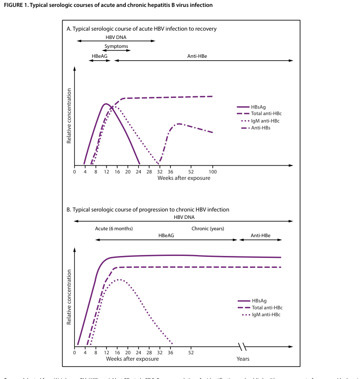
Summary : This figure presents two line plots showing the typical serologic courses of acute and chronic hepatitis B virus (HBV) infection, comparing the progression and recovery of serologic markers over time. line plot: # Panel A: Typical serologic course of acute HBV infection to recovery ## Title & Axes : • Title: "A. Typical serologic course of acute HBV infection to recovery" • X-axis: "Weeks after exposure" (tick labels: 0, 4, 8, 12, 16, 20, 24, 28, 32, 36, 52, 100) • Y-axis: "Relative concentration" (no explicit tick labels) • Additional timeline annotations: "HBV DNA", "Symptoms", "HBeAG", "Anti-HBe" (horizontal arrows above plot) ## Data Points : • HBsAg (solid line): Rises sharply after exposure, peaks around 8–12 weeks, then declines and disappears by ~24–28 weeks. • Total anti-HBc (dashed line): Rises after exposure, peaks around 16–20 weeks, remains elevated beyond 100 weeks. • IgM anti-HBc (dotted line): Rises after exposure, peaks around 12–16 weeks, declines and disappears by ~28–32 weeks. • Anti-HBs (long dashed line): Appears after ~24–28 weeks, rises and remains elevated beyond 100 weeks. ## Design Encodings : • HBsAg: solid purple line • Total anti-HBc: dashed purple line • IgM anti-HBc: dotted purple line • Anti-HBs: long dashed purple line • All lines plotted against the same axes; no log scale or error bars. ## Distribution & Trends : • HBsAg and IgM anti-HBc both show transient peaks and then decline. • Total anti-HBc and anti-HBs persist long-term after recovery. # Panel B: Typical serologic course of progression to chronic HBV infection ## Title & Axes : • Title: "B. Typical serologic course of progression to chronic HBV infection" • X-axis: "Weeks after exposure" (tick labels: 0, 4, 8, 12, 16, 20, 24, 28, 32, 36, 52), then "Years" (break in axis) • Y-axis: "Relative concentration" (no explicit tick labels) • Additional timeline annotations: "HBV DNA", "Acute (6 months)", "Chronic (years)", "HBeAG", "Anti-HBe" (horizontal arrows above plot) ## Data Points : • HBsAg (solid line): Rises after exposure, remains persistently elevated for years. • Total anti-HBc (dashed line): Rises after exposure, remains persistently elevated for years. • IgM anti-HBc (dotted line): Rises after exposure, peaks around 12–16 weeks, declines and disappears by ~28–32 weeks. ## Design Encodings : • HBsAg: solid purple line • Total anti-HBc: dashed purple line • IgM anti-HBc: dotted purple line • All lines plotted against the same axes; no log scale or error bars. ## Distribution & Trends : • HBsAg and total anti-HBc remain persistently elevated in chronic infection. • IgM anti-HBc is transient and disappears after the acute phase. # Analysis : • In acute HBV infection (Panel A), HBsAg and IgM anti-HBc are transient, while total anti-HBc and anti-HBs persist after recovery, indicating resolution and immunity. • In chronic HBV infection (Panel B), HBsAg and total anti-HBc remain persistently elevated for years, while IgM anti-HBc is only present during the acute phase, indicating ongoing infection without resolution. • The presence and duration of serologic markers distinguish acute recovery from progression to chronic infection.
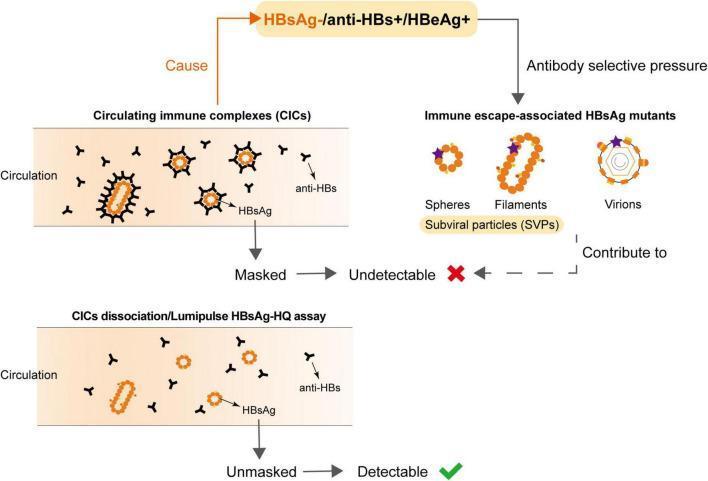
This pathophysiology diagram illustrates the mechanisms underlying undetectable Hepatitis B surface antigen (HBsAg) in patients who are HBeAg-positive and anti-HBs-positive. The diagram is divided into two primary pathways: the formation of Circulating Immune Complexes (CICs) and the emergence of HBsAg mutants. In the CIC pathway, HBsAg particles are shown in circulation bound by Y-shaped anti-HBs antibodies; this 'masking' of epitopes renders the antigen undetectable by standard assays. A lower panel demonstrates that chemical dissociation or specialized assays (e.g., Lumipulse HBsAg-HQ) can unmask these antigens, making them detectable. The second pathway shows 'Immune escape-associated HBsAg mutants' arising from antibody selective pressure. These are depicted as spheres, filaments (together termed subviral particles or SVPs), and complete virions with altered surface epitopes (marked by purple stars). Both CIC formation and mutant emergence contribute to the diagnostic challenge of occult Hepatitis B infection (OBI). This diagram is intended for intermediate to advanced medical education regarding HBV serology, diagnostic pitfalls, and immune escape mechanisms.
LIVER UNIT - General Medicine Long Notes
BDS 3rd Year | University Exam Pattern (India)
QUESTION 1: VIRAL HEPATITIS
Define Viral Hepatitis. Classify and Describe Clinical Features, Diagnosis & Management.
DEFINITION
CLASSIFICATION
| Feature | Hep A (HAV) | Hep B (HBV) | Hep C (HCV) | Hep D (HDV) | Hep E (HEV) |
|---|---|---|---|---|---|
| Virus type | Picornavirus (RNA) | Hepadnavirus (DNA) | Flavivirus (RNA) | Viroid (RNA) | Calicivirus (RNA) |
| Transmission | Fecal-oral | Parenteral / sexual | Parenteral | Parenteral (co/superinfection with HBV) | Fecal-oral |
| Incubation | 15-50 days (avg 28 days) | 45-180 days (avg 60-90 days) | 15-150 days | Similar to HBV | 15-60 days |
| Chronicity | Never | ~5-10% adults; 90% neonates | 70-85% | Yes | No (except transplant recipients) |
| Fulminant failure | <1% | 0.1-0.5% | Rare | High (co-infection) | ~1-3% (20-25% in pregnancy) |
| Vaccine available | Yes | Yes | No | Prevented by HBV vaccine | No (except in China/India) |
| Carrier state | No | Yes | Yes | Yes | No |
PHASES OF VIRAL HEPATITIS
- No symptoms; virus replicating in liver
- Specific to each virus (see table above)
- Flu-like symptoms: malaise, fatigue, myalgia, fever (low grade)
- Anorexia, nausea, vomiting - most prominent
- Aversion to food, smoking, alcohol
- Right upper quadrant discomfort / mild hepatic tenderness
- Urticaria and arthralgia (especially HBV - due to immune complexes)
- Dark urine (bilirubinuria) appears just before jaundice
- Appearance of jaundice (scleral icterus first, then skin)
- Systemic symptoms often improve paradoxically with onset of jaundice
- Pale/clay-colored stools (acholic stools) - bile not entering intestine
- Dark tea-colored urine (conjugated bilirubin)
- Pruritus (bile salt deposition)
- Tender hepatomegaly (~70%)
- Splenomegaly (~20%)
- Jaundice resolves; appetite returns
- Fatigue may persist weeks to months
- Liver function tests normalize
CLINICAL FEATURES
- Fatigue and malaise (most constant symptom)
- Anorexia and nausea/vomiting
- Fever (more prominent in HAV)
- Right upper quadrant pain
- Jaundice, dark urine, pale stools
- Pruritus
- Arthralgias (HBV - serum sickness-like)
- Jaundice (scleral icterus, skin yellowing)
- Tender hepatomegaly (soft liver with rounded edges)
- Splenomegaly
- Lymphadenopathy (mild)
- In fulminant hepatitis: Confusion, asterixis (liver flap), bleeding tendency, extreme jaundice
- Skin: Urticaria, purpura, vasculitis
- Joints: Arthritis, arthralgias
- Kidneys: Membranous GN (HBV), membranoproliferative GN (HCV)
- Hematology: Cryoglobulinemia (HCV), aplastic anemia (HAV)
- Neurological: Peripheral neuropathy, Guillain-Barré (rare)
DIAGNOSIS
- Serum Transaminases (ALT, AST): Markedly elevated (often 500-2000 IU/mL); ALT typically > AST in viral hepatitis (opposite in alcoholic hepatitis)
- Serum Bilirubin: Elevated (conjugated + unconjugated); >34 µmol/L causes clinical jaundice
- Alkaline Phosphatase (ALP): Mildly elevated (markedly elevated suggests cholestasis/obstruction)
- Serum Albumin: Low in severe/chronic disease
- Prothrombin Time (PT/INR): Prolonged in severe disease; important prognostic marker
| Test | Meaning |
|---|---|
| Anti-HAV IgM | Acute HAV infection (diagnostic) |
| Anti-HAV IgG | Past infection / immunity to HAV |
| HBsAg (Hepatitis B surface antigen) | Active HBV infection (appears first); persists >6 months = chronic |
| Anti-HBs | Immunity (post-infection recovery or vaccination) |
| IgM Anti-HBc | Acute HBV infection (most reliable marker for acute HBV) |
| IgG Anti-HBc | Past HBV exposure (lifelong marker) |
| HBeAg | Active viral replication; high infectivity |
| Anti-HBe | Low infectivity; seroconversion = good prognosis |
| HBV DNA (PCR) | Viral load; guides antiviral therapy |
| Anti-HCV | Exposure to HCV (appears 2-8 weeks post infection; not protective) |
| HCV RNA (PCR) | Active HCV replication; confirms infection |
| Anti-HDV | Hepatitis D infection (only in HBsAg-positive patients) |
- Bilirubin in urine (conjugated hyperbilirubinemia) - a very early sign
- Urobilinogen elevated
- Mild leukopenia with relative lymphocytosis (atypical lymphocytes)
- Thrombocytopenia in severe disease
- Ultrasound abdomen: Hepatomegaly, echogenicity; excludes biliary obstruction; splenomegaly; ascites
- Not required for routine viral hepatitis but helps exclude obstructive jaundice
- For chronic hepatitis - to assess grade of necroinflammation and stage of fibrosis
- Not required in acute viral hepatitis
MANAGEMENT
A. GENERAL PRINCIPLES (All forms of acute viral hepatitis)
- Rest: Bed rest during acute phase; activity as tolerated (no evidence for strict bed rest)
- Diet: High-calorie, high-carbohydrate diet; small frequent meals (nausea is limiting factor)
- Avoid: Alcohol (hepatotoxic), hepatotoxic drugs (paracetamol high dose, NSAIDs, tetracyclines), fatty foods
- Hydration: Oral rehydration; IV fluids if vomiting is severe
- Isolation: Enteric precautions for HAV/HEV; universal precautions for HBV/HCV
- Antipruritics: Cholestyramine for pruritus
- Antiemetics (metoclopramide, ondansetron) for nausea
- Antipyretics (paracetamol at standard doses - safe for liver if used appropriately)
- Prolonged PT/INR >1.5x normal
- Bilirubin >10 mg/dL with encephalopathy
- Unable to maintain oral hydration
- Signs of fulminant hepatic failure
B. SPECIFIC ANTIVIRAL THERAPY
- Tenofovir (TDF or TAF) - first-line; potent, high barrier to resistance
- Entecavir - first-line; potent, high barrier to resistance
- Pegylated Interferon alpha-2a (48 weeks) - for selected patients (finite duration, immune modulation)
- Older agents (lamivudine, adefovir, telbivudine) - not preferred due to resistance
- Goal: Suppress HBV DNA to undetectable; HBeAg seroconversion; prevent cirrhosis/HCC
- Pan-genotypic regimens: Sofosbuvir/Velpatasvir (12 weeks), Glecaprevir/Pibrentasvir (8 weeks)
- Older: Simeprevir, ombitasvir, elbasvir
- Achieve Sustained Virologic Response (SVR) in >95% of patients
- SVR at 12 weeks (SVR12) = "functional cure"
- Older regimens: Pegylated IFN + Ribavirin (24-48 weeks; SVR ~50%)
- Pegylated Interferon for 48 weeks (only approved treatment; limited efficacy)
- Bulevirtide (newer entry inhibitor) - approved in some countries
C. FULMINANT HEPATIC FAILURE MANAGEMENT
- ICU admission
- Lactulose for hepatic encephalopathy
- Vitamin K, FFP for coagulopathy
- IV glucose (hypoglycemia prevention)
- Avoid sedatives; monitor ICP
- Liver transplantation is definitive treatment
D. PREVENTION
- HAV and HBV: Active vaccination (see Hepatitis B section)
- HBV: Passive immunization with HBIG post-exposure
- HCV: No vaccine; harm reduction (safe injection practices, blood screening)
- General: Hand hygiene, safe water, food safety, blood/body fluid precautions
QUESTION 2: HEPATITIS B
Mode of Transmission, Clinical Features, Complications & Prevention
INTRODUCTION
MODE OF TRANSMISSION
- Intravenous drug use (sharing contaminated needles/syringes) - major route in developed countries
- Blood transfusions (now rare in countries with blood screening)
- Needle-stick injuries in healthcare workers (risk ~30% per needle-stick from HBsAg+ source)
- Unsafe injections (reuse of syringes)
- Tattooing, body piercing, acupuncture with unsterile equipment
- Most common route in developed countries (India: also significant)
- Heterosexual and homosexual contact
- HBV concentration in semen and vaginal secretions is high
- During childbirth (perinatal): Exposure to infected maternal blood during delivery
- In utero (transplacental): Less common
- Postnatal: Via breast milk (uncommon)
- In developing countries (Southeast Asia, Sub-Saharan Africa, India), this is the predominant route
- Risk: ~90% if mother is HBeAg positive (highly infective); ~10-40% if HBeAg negative
- Close household contact (sharing razors, toothbrushes)
- Child-to-child transmission in endemic areas
- Dental procedures - exposure to infected blood/saliva (occupational risk for dental professionals)
- Casual contact (hugging, shaking hands)
- Fecal-oral route (unlike HAV)
- Aerosol/respiratory droplets
- Contaminated food or water
Relevance to Dentistry: HBV is an occupational hazard for dental professionals due to needle-stick injury and contact with blood/saliva. All dental healthcare workers must be vaccinated. Standard precautions (gloves, masks, glasses) are mandatory.
CLINICAL FEATURES
ACUTE HEPATITIS B
- Malaise, fatigue, anorexia, nausea, vomiting
- Low-grade fever
- Arthralgia and urticaria (serum sickness-like; due to immune complex deposition - more common in HBV than HAV)
- Right hypochondrial discomfort
- Dark urine
- Jaundice (scleral icterus → skin)
- Hepatomegaly (tender)
- Splenomegaly (~20%)
- Lymphadenopathy
- Pale stools, dark urine
CHRONIC HEPATITIS B (Infection lasting >6 months)
- Immune tolerant phase: High viral replication (HBeAg+, high HBV DNA), normal ALT, minimal liver damage (especially in perinatally infected)
- Immune active phase (immune clearance): Active inflammation, elevated ALT, liver damage occurring - can progress to cirrhosis
- Inactive carrier phase: HBeAg seroconversion to anti-HBe; low HBV DNA; normal ALT; minimal liver injury; low risk of progression
- Reactivation phase: HBeAg-negative hepatitis; ongoing replication via HBV mutants
- Often asymptomatic for years/decades
- Fatigue, right upper quadrant discomfort
- Jaundice (if flaring or decompensated)
- Features of cirrhosis if advanced
COMPLICATIONS
- 5-10% of acutely infected adults develop chronic infection
- ~20-30% of patients with chronic HBV develop cirrhosis over 10-30 years
- Higher risk: HBeAg positive, high HBV DNA, co-infection with HCV/HDV/HIV, alcohol use
- Clinical features: Jaundice, splenomegaly, ascites, esophageal varices, hepatic encephalopathy
- Child-Pugh score and MELD score assess severity
- HBV is the leading cause of HCC worldwide (80% of HCC globally)
- Risk increases 100-fold in chronic HBV carriers
- HBV can cause HCC even without cirrhosis (direct oncogenic mechanism: viral DNA integrates into host genome)
- Surveillance: 6-monthly ultrasound + AFP in chronic HBV patients
- Presents as: Weight loss, RUQ pain, progressive hepatomegaly, worsening jaundice
- 0.1-0.5% of acute HBV; higher with co-infection (HDV)
- Features: Hepatic encephalopathy, profound coagulopathy (INR >1.5), hypoglycemia, renal failure
- Mortality >80% without liver transplantation
- Polyarteritis nodosa (PAN): Systemic vasculitis
- Membranous glomerulonephritis: Proteinuria, nephrotic syndrome
- Essential mixed cryoglobulinemia
- Serum sickness: Arthralgias, urticaria, fever (prodromal phase)
- Aplastic anemia (rare)
- Hepatitis D can only infect persons who are HBsAg positive
- Co-infection (simultaneous HBV+HDV): Severe acute hepatitis, usually self-limited
- Superinfection (HDV in chronic HBV carrier): High risk of fulminant hepatitis and rapid cirrhosis
- Ascites
- Esophageal varices (variceal bleeding - 10-15% mortality per episode)
- Spontaneous Bacterial Peritonitis (SBP)
- Hepatorenal syndrome
- Hepatic encephalopathy
PREVENTION
A. ACTIVE IMMUNIZATION (Vaccination - most effective prevention):
- Contains HBsAg produced by recombinant yeast (S. cerevisiae)
- Nearly 100% efficacy in immunocompetent individuals
- Provides long-lasting immunity (>20 years); may be lifelong
- Induces Anti-HBs antibody (protective level: ≥10 mIU/mL)
- Standard (adults and children): 3 doses - 0, 1, 6 months (intramuscular deltoid)
- Newborns: First dose at birth (within 24 hours); then 1 and 6 months
- Accelerated schedule: 0, 1, 2 months (for rapid protection in high-risk individuals)
- Combined vaccine (DPT-HBV-Hib): Given at 6, 10, 14 weeks in India's national immunization programme (NIP)
- Healthcare workers (including dental professionals) - mandatory
- Newborns of HBsAg-positive mothers
- Sexual partners of HBV-infected persons
- IV drug users
- Dialysis patients
- Travelers to endemic areas
- Unvaccinated adults
B. PASSIVE IMMUNIZATION (Post-exposure prophylaxis):
- Contains high-titer anti-HBs antibodies
- Used for immediate (passive) protection after exposure
- Efficacy: ~90% in prevention of neonatal infections; ~75% in occupational exposure
- Newborn of HBsAg-positive mother: HBIG 0.5 mL IM + HBV vaccine - both within 12 hours of birth
- Needle-stick injury in unvaccinated healthcare worker: HBIG + HBV vaccine within 24 hours
- Sexual exposure to HBsAg-positive person (within 14 days)
C. OTHER PREVENTIVE MEASURES:
- Screening of blood donors for HBsAg (all donated blood in India screened)
- Universal precautions in healthcare settings
- Sterile injection practices; avoid reuse of syringes
- Safe sex / barrier contraception
- Not sharing razors, toothbrushes, nail cutters
- Proper sterilization in dental practice
- Screening pregnant women for HBsAg (universal in India's RMNCH+A programme)
- Antiviral therapy (tenofovir) in highly viremic pregnant women (HBV DNA >200,000 IU/mL) to prevent vertical transmission
QUESTION 3: COMPARE HEPATITIS A vs HEPATITIS B
Detailed Comparison Table
| Feature | HEPATITIS A (HAV) | HEPATITIS B (HBV) |
|---|---|---|
| Virus | Picornavirus (ssRNA) | Hepadnavirus (partially dsDNA) |
| Genome | Single-stranded RNA | Partially double-stranded DNA |
| Transmission | Fecal-oral route | Parenteral, sexual, vertical (mother-to-child) |
| Common sources | Contaminated food/water, poor sanitation | Blood, semen, vaginal secretions, saliva, breast milk |
| Risk groups | Children in endemic areas; travelers; poor sanitation | IV drug users, HCWs, neonates, sexual partners of carriers |
| Endemic regions | Developing countries (India, Africa, SE Asia) | Worldwide; especially SE Asia, sub-Saharan Africa |
| Incubation period | 15-50 days (average 28 days) | 45-180 days (average 60-90 days) |
| Onset | Acute, abrupt | Insidious |
| Prodrome (pre-icteric) | Fever, nausea, malaise (prominent) | Malaise, arthralgia, urticaria (serum sickness-like) |
| Icteric illness | Common (70% in adults) | Only 30% develop jaundice |
| Severity | Generally mild, self-limiting | Variable; can be severe |
| Fulminant hepatitis | <1% (mostly in underlying liver disease) | 0.1-0.5% |
| Chronicity | NEVER - no chronic carrier state | Yes - 5-10% of adults; 90% of neonates |
| Carrier state | No | Yes (HBsAg persistently positive >6 months) |
| Cirrhosis | Does NOT cause cirrhosis | Major cause of cirrhosis (20-30% of chronic HBV) |
| Hepatocellular carcinoma | No association | Major cause - #1 cause of HCC worldwide |
| DIAGNOSTIC MARKERS | ||
| Acute infection | Anti-HAV IgM (diagnostic) | IgM Anti-HBc, HBsAg (earliest) |
| Past infection/immunity | Anti-HAV IgG | Anti-HBs + IgG Anti-HBc |
| Active infection | - | HBsAg (>6 months = chronic) |
| Viral replication | - | HBeAg, HBV DNA |
| Recovery marker | - | Anti-HBs, Anti-HBe |
| Vaccination immunity | Anti-HAV IgG | Anti-HBs only (no Anti-HBc) |
| Liver enzymes | ALT/AST markedly elevated | ALT/AST elevated; higher in acute phase |
| Treatment - Acute | Supportive only | Supportive (antivirals NOT needed in acute) |
| Treatment - Chronic | Not applicable | Tenofovir / Entecavir; Pegylated IFN |
| Vaccine | Inactivated HAV vaccine (2 doses) | Recombinant HBsAg vaccine (3 doses) |
| Passive immunization | Normal human IG (within 2 weeks of exposure) | HBIG (within 12-24 hrs of exposure) |
| Vaccine schedule (India) | 2 doses: 0 and 6-12 months | 3 doses: 0, 1, 6 months (or birth, 6, 10, 14 wks via NIP) |
| Occupational risk (dentistry) | Low (fecal-oral) | HIGH - needle-stick, blood/saliva exposure |
| Prognosis | Excellent - complete recovery in >99% | Good if acute; chronic disease risk of cirrhosis/HCC |
| Prevention emphasis | Hand hygiene, safe water, food safety | Vaccination (mandatory for HCWs), safe practices |
Key Similarities:
- Both cause acute hepatitis with similar clinical presentation (jaundice, elevated transaminases)
- Both are reportable diseases
- Both have effective vaccines
- Both predominantly affect developing countries including India
- Both cause tender hepatomegaly, anorexia, nausea, dark urine, pale stools
Key Differences (High-Yield for Exam):
- HAV: self-limiting, no chronic state, no cancer
- HBV: can become chronic, causes cirrhosis and HCC
- HAV: fecal-oral; HBV: blood/sexual/vertical
- HAV: common in children; HBV: neonatal infection in endemic areas
- HAV prodrome: fever prominent; HBV prodrome: arthralgia + urticaria (immune complex)
QUESTION 4: CHRONIC HEPATITIS AND ITS COMPLICATIONS
Describe Chronic Hepatitis and Its Complications
DEFINITION
ETIOLOGY / CAUSES OF CHRONIC HEPATITIS
| Cause | Features |
|---|---|
| Chronic Hepatitis B | Most common cause globally; 5-10% of acute HBV progress to chronic |
| Chronic Hepatitis C | 70-85% of HCV infections become chronic; major cause in developed countries |
| Chronic Hepatitis D | Only in HBV-infected individuals; accelerates progression |
| Autoimmune Hepatitis (AIH) | Young women; anti-smooth muscle antibody (ASMA), ANA positive; hypergammaglobulinemia |
| Non-Alcoholic Steatohepatitis (NASH) | Associated with obesity, DM type 2, metabolic syndrome; increasingly common |
| Alcoholic hepatitis | Chronic heavy alcohol intake; AST:ALT >2:1 |
| Drug-induced chronic hepatitis | Isoniazid, methotrexate, methyldopa, nitrofurantoin |
| Wilson's disease | Copper metabolism disorder; young patients |
| Alpha-1 antitrypsin deficiency | Genetic; liver + lung disease |
| Primary Biliary Cholangitis (PBC) | Middle-aged women; anti-mitochondrial antibody (AMA) |
HISTOLOGICAL GRADING & STAGING
- Grading (severity of necroinflammation): Grade 1-4 (Knodell HAI or Metavir score)
- Staging (degree of fibrosis): Stage F0-F4 (Metavir)
- F0 = No fibrosis
- F1 = Portal fibrosis without septa
- F2 = Portal fibrosis with few septa
- F3 = Numerous septa without cirrhosis
- F4 = Cirrhosis
CLINICAL FEATURES OF CHRONIC HEPATITIS
- Fatigue and weakness (most common)
- Right upper quadrant discomfort / ache
- Anorexia, nausea
- Low-grade fever (in active phases)
- Jaundice (mild or intermittent; becomes persistent in advanced disease)
- Weight loss
- Arthralgia (especially HBV - immune complexes)
- Hepatomegaly (tender or non-tender, firm)
- Mild splenomegaly
- Jaundice (mild)
- Palmar erythema, spider angiomata (early signs of portal hypertension)
- Jaundice (persistent, deepening)
- Spider naevi (>5 = significant; upper body, face, arms)
- Palmar erythema (thenar and hypothenar)
- Leukonychia (white nails)
- Clubbing
- Dupuytren's contracture (especially alcoholic)
- Parotid enlargement (alcoholic)
- Gynaecomastia + testicular atrophy (hyperestrogenism from impaired estrogen metabolism)
- Caput medusae (dilated periumbilical veins - portal hypertension)
- Splenomegaly
- Ascites (shifting dullness, fluid thrill)
- Peripheral edema (hypoalbuminemia)
- Flapping tremor (Asterixis) - hepatic encephalopathy
INVESTIGATIONS
- ALT, AST: Elevated (may fluctuate; may normalize in burnt-out cirrhosis)
- Bilirubin: Elevated in advanced disease
- Albumin: Low (marker of synthetic function)
- PT/INR: Prolonged (poor prognostic marker)
- Gamma-globulins: Elevated (especially autoimmune hepatitis)
- CBC: Pancytopenia (hypersplenism), anemia
- HBsAg, HBV DNA, HBeAg, Anti-HCV, HCV RNA (as detailed in Q1)
- ANA, ASMA (anti-smooth muscle antibody) → Autoimmune hepatitis
- AMA (anti-mitochondrial antibody) → PBC
- Anti-LKM1 → AIH type 2
- Serum IgG levels
- Serum copper, ceruloplasmin (Wilson's disease)
- Alpha-1 antitrypsin level and phenotype
- Ultrasound abdomen: Liver echogenicity, size, portal vein diameter, splenomegaly, ascites
- CT/MRI: Better detail; for HCC screening
- FibroScan (Transient Elastography): Non-invasive fibrosis assessment
- Gold standard for diagnosis, grading, and staging
- Guides treatment decisions
- Tumor marker for HCC surveillance
- Every 6 months in cirrhotic patients + abdominal ultrasound
COMPLICATIONS OF CHRONIC HEPATITIS / CIRRHOSIS
1. PORTAL HYPERTENSION
- Dilated portosystemic collateral veins at gastroesophageal junction
- Variceal hemorrhage: Life-threatening emergency (10-15% mortality per episode)
- Presents as: Sudden massive hematemesis (bright red blood), melena, hypovolemic shock
- Management: Emergency endoscopy (band ligation), IV terlipressin, IV octreotide, Sengstaken-Blakemore tube (balloon tamponade), TIPS, prophylactic beta-blockers (propranolol/carvedilol)
- Accumulation of fluid in peritoneal cavity
- Mechanism: Portal hypertension + hypoalbuminemia + hyperaldosteronism (RAAS activation)
- Presents as: Increasing abdominal girth, ankle edema, shifting dullness, fluid thrill
- SAAG (serum-ascites albumin gradient) >1.1 g/dL = portal hypertension-related ascites
- Management: Salt restriction (2g/day), diuretics (spironolactone first, then furosemide), large-volume paracentesis with albumin infusion, TIPS, liver transplantation
- Portal congestion → spleen enlarges
- Hypersplenism: Pancytopenia (anemia, leucopenia, thrombocytopenia)
- Low platelet count - risk for bleeding
- Shunting of portal blood (containing gut-derived toxins especially NH3 - ammonia) directly into systemic circulation bypassing liver
- Triggers: GI bleeding, infection, high protein intake, constipation, electrolyte imbalance, sedatives
- Features: Asterixis (hepatic flap), confusion, altered sleep-wake cycle, stupor, coma
- Grade I-IV (West Haven criteria)
- Investigations: Serum ammonia elevated; EEG shows triphasic waves
- Management: Identify + treat precipitant; Lactulose (reduces ammonia by acidification of gut), Rifaximin (non-absorbable antibiotic - reduces gut bacterial ammonia production), low protein diet
2. SPONTANEOUS BACTERIAL PERITONITIS (SBP)
- Bacterial infection of ascitic fluid without an obvious intra-abdominal source
- Organisms: E. coli (most common), Klebsiella, Streptococcus pneumoniae
- Diagnosis: Ascitic fluid PMN count >250 cells/mm³ (even before culture results)
- Symptoms: Fever, abdominal pain, worsening encephalopathy - may be silent
- Treatment: IV Cefotaxime 3rd generation cephalosporin x 5 days; IV albumin infusion (reduces hepatorenal syndrome risk)
- Prophylaxis: Norfloxacin or Ciprofloxacin in high-risk patients (low protein ascites, previous SBP, variceal bleed)
3. HEPATORENAL SYNDROME (HRS)
- Functional renal failure in cirrhotic patients without intrinsic kidney disease
- Mechanism: Splanchnic vasodilation (due to portal hypertension) → reduced effective circulating volume → RAAS activation → renal vasoconstriction → oliguria
- HRS Type 1: Acute, rapidly progressive (creatinine doubles to >2.5 mg/dL in <2 weeks); poor prognosis
- HRS Type 2: Chronic, more gradual; associated with refractory ascites
- Diagnosis: Exclude other causes of AKI; no response to fluid challenge; no nephrotoxic drugs
- Treatment: Terlipressin + albumin infusion; midodrine + octreotide + albumin; liver transplantation (definitive)
4. HEPATOCELLULAR CARCINOMA (HCC)
- Risk: Cirrhotic patients have ~1-5% annual risk of HCC
- HBV can cause HCC even without cirrhosis (viral DNA integrates into hepatocyte genome - direct oncogenic effect)
- Surveillance: 6-monthly ultrasound + AFP in all cirrhotic patients
- Features: Weight loss, RUQ pain, jaundice, hepatomegaly, wasting, ascites
- AFP markedly elevated (>400 ng/mL is suggestive; >1000 ng/mL is diagnostic)
- CT/MRI with contrast: Characteristic "arterial enhancement + portal venous washout"
- Treatment: Surgical resection, liver transplant, ablation, TACE (trans-arterial chemo-embolization), Sorafenib (targeted therapy for advanced HCC)
5. COAGULOPATHY
- Liver fails to produce clotting factors (I, II, V, VII, IX, X, XI)
- Prolonged PT/INR → bleeding tendency
- Thrombocytopenia (hypersplenism)
- Presentation: Easy bruising, prolonged bleeding after dental procedures, petechiae, GI bleeding
6. HEPATOPULMONARY SYNDROME
- Intrapulmonary vascular dilation → arteriovenous shunting in lungs
- Hypoxia worsens in upright position (platypnoea) and improves supine (orthodeoxia)
- SpO2 <96% on room air is a clue
- Treated by liver transplantation
7. PORTOPULMONARY HYPERTENSION
- Pulmonary arterial hypertension in setting of portal hypertension
- Dyspnoea, right heart failure
- Contraindicates liver transplantation if severe
MANAGEMENT OF CHRONIC HEPATITIS
- Treat underlying cause (antivirals for HBV/HCV; steroids for autoimmune; abstinence for alcohol)
- Avoid hepatotoxic drugs and alcohol absolutely
- Vaccination: HAV, HBV vaccines if not immune
- Regular surveillance for HCC (6-monthly USG + AFP)
| Complication | Key Management |
|---|---|
| Ascites | Salt restriction, Spironolactone ± Furosemide, large-volume paracentesis |
| SBP | IV Cefotaxime + IV Albumin |
| Variceal bleed | Terlipressin/octreotide + endoscopic band ligation + prophylactic beta-blockers |
| Hepatic encephalopathy | Lactulose + Rifaximin + treat precipitant |
| HRS | Terlipressin + Albumin; liver transplant |
| HCC | Surgery/ablation/TACE/Sorafenib; liver transplant |
- Definitive treatment for end-stage liver disease
- Indications: MELD score >15, refractory ascites, variceal bleeding, HCC (within Milan criteria), HRS, hepatopulmonary syndrome
- 5-year survival post-transplant: >80%
CHILD-PUGH SCORE (Assesses severity of cirrhosis):
| Parameter | 1 point | 2 points | 3 points |
|---|---|---|---|
| Bilirubin (mg/dL) | <2 | 2-3 | >3 |
| Albumin (g/dL) | >3.5 | 2.8-3.5 | <2.8 |
| PT prolongation (sec) | <4 | 4-6 | >6 |
| Ascites | None | Mild | Moderate/severe |
| Encephalopathy | None | Grade I-II | Grade III-IV |
- Class A (5-6): Well-compensated; low risk
- Class B (7-9): Significant compromise
- Class C (10-15): Decompensated; high mortality; transplant evaluation
SUMMARY TABLE - HIGH-YIELD EXAM POINTS
| Topic | Key Fact |
|---|---|
| Hepatitis A | Fecal-oral; self-limiting; NO chronicity; diagnosed by Anti-HAV IgM |
| Hepatitis B | Blood/sexual/vertical; DNA virus; 5-10% become chronic |
| Hepatitis C | Parenteral; RNA virus; 70-85% become chronic; DAAs cure >95% |
| Hepatitis D | Only in HBsAg+ persons; worsens HBV prognosis |
| Hepatitis E | Fecal-oral like HAV; severe in pregnancy (20-25% mortality) |
| HBV most dangerous complication | HCC (even without cirrhosis) |
| Earliest marker of acute HBV | HBsAg |
| Best marker of acute HBV | IgM Anti-HBc |
| Active viral replication marker | HBeAg + HBV DNA |
| Recovery from HBV | Anti-HBs appears |
| HBV vaccine type | Recombinant HBsAg; 3 doses at 0, 1, 6 months |
| Newborn of HBsAg+ mother | HBIG + HBV vaccine within 12 hours |
| HCV treatment | Direct-acting antivirals (Sofosbuvir-based) |
| Cirrhosis marker | Persistently elevated LFTs + Ultrasound changes + Liver biopsy F4 |
| Portal hypertension threshold | >12 mmHg |
| Most feared cirrhosis complication | Variceal hemorrhage / HCC |
| SBP diagnosis | Ascitic fluid PMN >250/mm³ |
| Hepatic encephalopathy treatment | Lactulose + Rifaximin |
| Gold standard for grading chronic hepatitis | Liver biopsy |
| Definitive treatment for end-stage cirrhosis | Liver transplantation |
Now answer them the same way : Define cirrhosis. Discuss etiology, pathogenesis, clinical features & complications. * Write in detail about portal hypertension. * Complications of cirrhosis (ascites, varices, encephalopathy). * Management of cirrhosis.
cirrhosis liver gross pathology nodular fibrosis histology
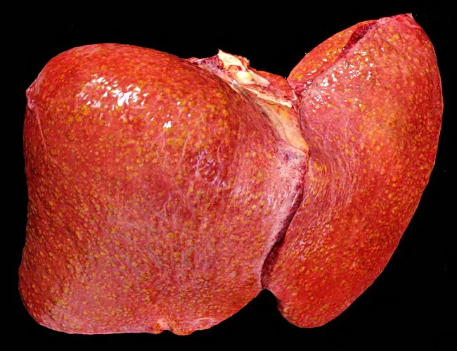
Gross pathology photograph of a human liver showing cirrhotic nodularity with coarse surface, reflecting end-stage hepatic disease. The image depicts hepatocellular nodules separated by fibrous septa, typical of chronic liver injury. In Wilson disease, impaired copper transport due to ATP7B mutation leads to copper deposition in hepatocytes, contributing to inflammation, steatosis in early disease, and progression to chronic hepatitis and cirrhosis. The macroscopic features include enlarged, nodular liver with a firm texture. While histology is not shown, cirrhosis features such as bridging fibrosis and regenerative nodules would be expected. This image serves as an educational reference for macroscopic evaluation of cirrhosis within copper storage disorders and complements histopathology with architectural assessment. Clinically significant for teaching end-stage liver disease, differential diagnosis with viral hepatitis and alcohol-related liver disease, and correlation with systemic copper accumulation; useful in pathology, hepatology, medical education, and radiology training for recognizing advanced liver damage in Wilson disease. This description highlights the relationship between macroscopic appearance and underlying pathophysiology: copper-induced hepatocellular injury triggers stellate cell activation, extracellular matrix deposition, nodularity, and eventual loss of normal lobular architecture. In practice, gross pathology complements biochemical copper quantification, hepatic copper staining (rhodanine) and ATP7B genotyping in diagnosis. The image can also aid educational case libraries and digital pathology databases for differential diagnosis training.
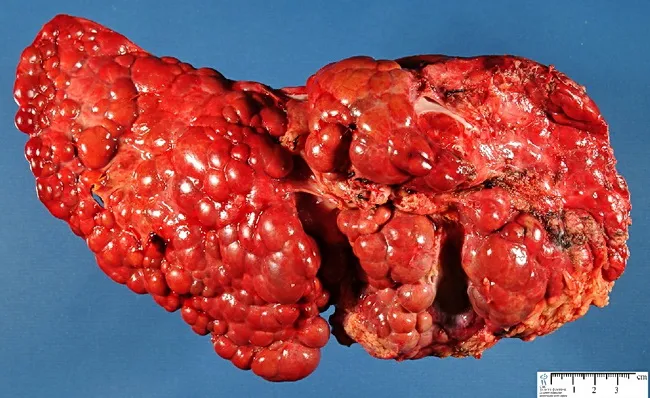
Gross pathology photograph of the liver showing advanced cirrhosis with coarse, nodular surface. The specimen demonstrates macronodular cirrhosis, in which regenerative nodules exceed 3 mm in diameter, a histologic and macroscopic hallmark of fibrosis and architectural disarray associated with chronic tyrosinemia type I. The context is hereditary infantile tyrosinemia due to fumarylacetoacetate hydrolase deficiency, a metabolic liver disease that progresses to cirrhotic remodeling and risk of hepatocellular carcinoma if untreated. The hepatic nodularity and firm consistency reflect extensive parenchymal scarring with bridging fibrous septa linking nodules. Color is a deep reddish-brown, with variable areas of congested or mottled parenchyma; surface nodularity may obscure normal lobular anatomy. This image provides a macroscopic correlate to the cellular and architectural changes described in histology, including regenerative nodules embedded in dense fibrous tissue. Clinically, such findings support severe liver disease in tyrosinemia and justify monitoring for portal hypertension and hepatocellular carcinoma risk. In educational and research contexts, this photograph is valuable for teaching gross hepatic morphology, cirrhotic evolution in metabolic disorders, and the distinction between macronodular vs micronodular cirrhosis. Potential uses include medical education, pathology slide correlation, and diagnostic radiology-pathology correlation studies. These features reinforce differential considerations for cirrhosis etiologies in pediatrics worldwide today.
portal hypertension portosystemic collateral varices caput medusae
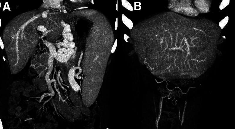
This diagnostic image displays two coronal views (A and B) from a Computed Tomography Portal Venography (CTV), illustrating advanced sequelae of portal hypertension secondary to liver cirrhosis. Panel A highlights extensive collateral circulation, featuring tortuous, markedly dilated esophageal and gastric varices. These appear as irregular, winding vascular clusters in the upper gastrointestinal region. Panel B demonstrates the recanalization of the umbilical vein, seen as a distinct vessel extending from the liver toward the umbilical region. Additionally, this panel reveals caput medusae, characterized by prominent superficial abdominal wall varices. The images collectively demonstrate the pathophysiological redistribution of portal blood flow through portosystemic shunts. Key anatomical structures visible include the cirrhotic liver, an enlarged spleen (splenomegaly), and multiple dilated venous networks, providing high educational value for understanding the clinical manifestations of portal venous congestion and decompensated cirrhosis.
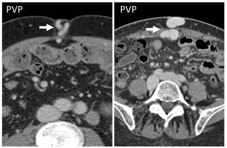
Diagnostic axial CT scans of the abdomen in the portal venous phase (PVP) from two different patients with liver cirrhosis. The images demonstrate the classic CT appearance of 'caput medusae,' characterized by prominent, tortuous, contrast-enhanced collateral vessels (indicated by white arrows) within the subcutaneous fat of the anterior abdominal wall at the level of the umbilicus. These findings result from the recanalization of the umbilical vein as a portosystemic shunt in the setting of portal hypertension. The left image shows a more slender, tortuous morphology of the umbilical collateral, while the right image displays larger, more rounded, hyperattenuating vascular cross-sections in the periumbilical region. Both images show typical abdominal anatomy, including the subcutaneous and mesenteric fat, bowel loops, abdominal wall musculature, and the lumbar vertebrae posteriorly. This visual finding is a crucial diagnostic indicator of advanced liver disease and significant portal venous pressure elevation.
LIVER UNIT - CIRRHOSIS & PORTAL HYPERTENSION
General Medicine Long Notes | BDS 3rd Year | University Exam Pattern (India)
QUESTION 1: CIRRHOSIS
Define Cirrhosis. Discuss Etiology, Pathogenesis, Clinical Features & Complications.
DEFINITION
- Replacement of normal liver parenchyma by fibrous tissue
- Formation of regenerative nodules
- Distortion of the normal hepatic architecture leading to portal hypertension and progressive loss of liver function
- Sabiston Textbook of Surgery: "Cirrhosis is characterized by chronic liver inflammation that replaces normal liver parenchyma with diffuse fibrosis."
- Textbook of Family Medicine: "The term cirrhosis refers to a progressive diffuse, fibrosing, and nodular condition that disrupts the entire normal architecture of the liver."
ETIOLOGY
- Most common cause in developed countries (USA: ~48% of cirrhosis deaths)
- Requires 10-15 years of chronic heavy drinking
- Risk: >80 g/day in males; >60 g/day in females
- Only 10-20% of heavy drinkers develop cirrhosis (genetic susceptibility)
- Leading cause in India, SE Asia, Sub-Saharan Africa
- ~20-30% of chronic HBV develop cirrhosis over decades
- Major cause in developing countries
- 70-85% of HCV infections become chronic; 5-30% progress to cirrhosis
- Leading indication for liver transplantation in Western countries
- Drugs: Methotrexate, isoniazid, methyldopa, amiodarone, nitrofurantoin
- Autoimmune Hepatitis (AIH): Anti-smooth muscle antibody (ASMA) positive; young women
- Fastest growing cause globally
- Associated with obesity, type 2 DM, metabolic syndrome, dyslipidemia
- NAFLD → Non-Alcoholic Steatohepatitis (NASH) → cirrhosis
- Wilson's disease (copper accumulation)
- Haemochromatosis (iron overload)
- Alpha-1 antitrypsin deficiency
- Glycogen storage diseases
- Primary Biliary Cholangitis (PBC) - anti-mitochondrial antibody (AMA) positive; middle-aged women
- Primary Sclerosing Cholangitis (PSC) - associated with IBD
- Biliary obstruction (secondary biliary cirrhosis)
- Cardiac cirrhosis (chronic right heart failure - "nutmeg liver")
- Budd-Chiari syndrome (hepatic vein occlusion)
- Cryptogenic (cause unknown ~10%)
PATHOGENESIS
- Normally, HSCs (Ito cells) are quiescent, storing Vitamin A in lipid droplets in the perisinusoidal space of Disse
- Injury signals (reactive oxygen species, inflammatory cytokines: TGF-β1, PDGF, TNF-α) from damaged hepatocytes, Kupffer cells, and sinusoidal endothelial cells activate HSCs
- Activated HSCs transform into myofibroblast-like cells
- Activated HSCs produce excessive Type I and III collagen (major fibrous component of scar tissue)
- They also produce Matrix Metalloproteinase Inhibitors (TIMPs) that prevent collagen breakdown
- Result: Net fibrosis accumulation in the liver
- Fibrosis begins in portal tracts → portal fibrosis → bridges form between portal tracts (bridging fibrosis) → eventually surrounds nodules (cirrhosis)
- The surviving hepatocytes undergo regenerative hyperplasia, forming clusters of hepatocytes (nodules) encircled by fibrous tissue
- Micronodular cirrhosis: Nodules <3mm (uniform size) - typically alcoholic, early stage
- Macronodular cirrhosis: Nodules >3mm (variable size) - typical of viral hepatitis; higher HCC risk
- Normal lobular and acinar architecture is replaced by fibrous septa and nodules
- Sinusoidal blood flow is obstructed → increased intrahepatic resistance → portal hypertension
- Dynamic component: HSCs contract in response to vasoconstricting substances (endothelin, angiotensin II) and produce insufficient nitric oxide (NO) in sinusoids → further vasoconstriction
- Hepatocyte mass is reduced → decreased synthetic function
- Portal hypertension → splanchnic arterial vasodilation (mediated by NO, endocannabinoids) → reduced systemic vascular resistance → effective arterial hypovolemia → activation of RAAS, SNS, ADH → sodium and water retention → ascites and edema (Sleisenger & Fordtran's)
CLINICAL FEATURES
- Often discovered incidentally on routine blood tests or imaging
- Mild non-specific symptoms: fatigue, weakness, anorexia, vague abdominal discomfort
- Mild hepatomegaly (firm, non-tender, irregular surface)
SYMPTOMS:
- Fatigue and weakness (most common)
- Anorexia, weight loss, muscle wasting
- Nausea, abdominal distension
- Jaundice (yellow discoloration of skin and eyes)
- Pruritus (bile salt deposition)
- Dark urine, pale stools
- Ankle/leg swelling
- Hematemesis (variceal bleeding)
- Confusion, behavioral changes (encephalopathy)
- Loss of libido, menstrual irregularities (in women)
SIGNS (organized by system):
- Jaundice (scleral icterus, skin yellowing)
- Muscle wasting, cachexia
- Fetor hepaticus (musty, sweet smell - dimethyl sulfide exhaled due to portosystemic shunting)
- Spider naevi (telangiectasias): >5 are significant; upper body (face, chest, arms), arms; appear as central arteriole with radiating vessels; blanch on pressure; caused by hyperestrogenism
- Palmar erythema: Erythema of thenar and hypothenar eminences ("liver palms"); also due to hyperestrogenism
- Leukonychia: White nails (hypoalbuminemia)
- Terry's nails: White nails with distal brown band
- Clubbing
- Purpura and bruising: Coagulopathy + thrombocytopenia
- Pigmentation (haemochromatosis)
- Xanthelasma (PBC)
- Gynaecomastia (males) - bilateral breast enlargement
- Testicular atrophy (males)
- Loss of body hair (axillary and pubic hair)
- Menstrual irregularities (females)
- Hepatomegaly: Firm, irregular, non-tender (in early cirrhosis); small, shrunken liver in advanced cirrhosis (fibrosis and hepatocyte loss)
- Splenomegaly: Portal hypertension → splenic venous congestion
- Ascites: Shifting dullness, fluid thrill (if massive), protruberant abdomen, eversion of umbilicus
- Caput medusae: Dilated tortuous periumbilical veins radiating from umbilicus - pathognomonic of portal hypertension
- Asterixis (hepatic flap): Flapping tremor of outstretched hands - hepatic encephalopathy; irregular, arrhythmic tremor with brief lapses of sustained posture
- Confusion, drowsiness, altered consciousness
- Anemia (multiple causes: GI blood loss, hypersplenism, folate deficiency)
- Bruising, petechiae
INVESTIGATIONS
- ALT, AST: May be elevated (active hepatitis) or normal/low (burnt-out cirrhosis)
- AST:ALT ratio >2:1 suggests alcoholic liver disease
- Serum Bilirubin: Elevated in decompensated disease
- Serum Albumin: Low (reduced synthetic function - important prognostic marker)
- PT/INR: Prolonged (reduced clotting factor synthesis) - important prognostic marker
- Platelet count: Low (hypersplenism - trapped in enlarged spleen; the most reliable single lab test for portal hypertension and large varices)
- Serum sodium: Low (dilutional hyponatremia - poor prognosis)
- Serum creatinine/urea: Elevated in hepatorenal syndrome
- GGT: Elevated, especially in alcoholic disease
- Serum globulins: Elevated (immune dysregulation)
| Parameter | 1 point | 2 points | 3 points |
|---|---|---|---|
| Serum Bilirubin (mg/dL) | <2 | 2-3 | >3 |
| Serum Albumin (g/dL) | >3.5 | 2.8-3.5 | <2.8 |
| PT (seconds prolonged) | <4 | 4-6 | >6 |
| Ascites | None | Mild/controlled | Moderate/severe/refractory |
| Encephalopathy grade | None | Grade 1-2 | Grade 3-4 |
- Class A (5-6): Compensated; 1-year survival ~100%
- Class B (7-9): Significant compromise; 1-year survival ~80%
- Class C (10-15): Decompensated; 1-year survival ~45%; transplant evaluation needed
- MELD = 3.78(Ln bilirubin) + 11.2(Ln INR) + 9.57(Ln creatinine) + 6.43
- Used for transplant listing and organ allocation; MELD >15 = transplant consideration
- MELD-Na (includes serum sodium) - currently used for allocation in India and USA
- Nodular, shrunken liver with coarse echotexture
- Splenomegaly, ascites
- Portal vein diameter >13 mm (portal hypertension)
- Enlarged portal and splenic veins
- Loss of hepatic vein pulsatility
- Screen for HCC (every 6 months)
- Better characterization of liver nodules
- Varices visualization
- HCC detection (arterial enhancement + portal washout)
- Non-invasive assessment of liver stiffness (proxy for fibrosis)
- Values: F0 (<7.9 kPa) to F4 cirrhosis (>12.5 kPa)
- Alternative to liver biopsy for fibrosis staging
- All cirrhotic patients at diagnosis: screening for esophageal/gastric varices
- Repeat every 1-3 years if no varices; annually if small varices
- Gold standard for confirming diagnosis, determining etiology, grading/staging
- Histology: Fibrous septa, regenerative nodules, distorted architecture
- Matrix stains (Masson's trichrome or Sirius Red) to quantify fibrosis
- Risk: Bleeding (coagulopathy + thrombocytopenia in cirrhosis)
- Viral markers: HBsAg, anti-HCV, HBV/HCV DNA
- Autoimmune: ANA, ASMA, AMA, anti-LKM1
- Iron studies: Serum ferritin, transferrin saturation (hemochromatosis)
- Serum ceruloplasmin, 24-hour urine copper (Wilson's)
- Alpha-1 antitrypsin level and phenotype
- AFP (alpha-fetoprotein) - HCC screening
COMPLICATIONS OF CIRRHOSIS
- Portal hypertension → Esophageal/gastric varices, ascites, splenomegaly
- Spontaneous Bacterial Peritonitis (SBP)
- Hepatic encephalopathy
- Hepatorenal syndrome
- Hepatocellular carcinoma (HCC)
- Coagulopathy and bleeding
- Hepatopulmonary syndrome
- Portopulmonary hypertension
- Malnutrition and muscle wasting
QUESTION 2: PORTAL HYPERTENSION
Write in Detail about Portal Hypertension
DEFINITION
ANATOMY OF PORTAL CIRCULATION
ETIOLOGY / CLASSIFICATION
1. PRE-SINUSOIDAL (pre-hepatic or pre-sinusoidal intrahepatic):
- Portal vein thrombosis (most common pre-hepatic cause; associated with hypercoagulable states, malignancy, sepsis)
- Splenic vein thrombosis
- Increased portal flow: Arteriovenous fistula, tropical splenomegaly, massive splenomegaly
- Schistosomiasis (most common cause of portal hypertension worldwide - granulomatous obstruction of portal tracts)
- Primary Biliary Cholangitis (early)
- Myeloproliferative disorders (infiltration)
2. SINUSOIDAL (intrahepatic, at the level of sinusoids):
- Cirrhosis - most common cause in India and globally (all types)
- Alcoholic hepatitis
- Acute hepatitis
- NAFLD
3. POST-SINUSOIDAL:
- Budd-Chiari syndrome (hepatic vein occlusion): Abrupt onset; hepatomegaly, ascites, abdominal pain; associated with myeloproliferative disorders (polycythemia vera), prothrombotic states, OCPs
- Veno-occlusive disease (sinusoidal obstruction syndrome): After bone marrow transplantation, chemotherapy
- Cardiac disease (most common post-hepatic cause): Congestive heart failure, constrictive pericarditis, tricuspid regurgitation → "cardiac cirrhosis" (nutmeg liver)
PATHOPHYSIOLOGY
Portal pressure = Portal blood flow × Portal resistance
- Fibrosis and regenerative nodules distort sinusoidal architecture
- Mechanical compression of sinusoids and small portal veins
- Collagen deposition in the space of Disse → "capillarization" of sinusoids (loss of sinusoidal fenestrations)
- Activated hepatic stellate cells contract in response to vasoconstrictors (endothelin-1, angiotensin II)
- Reduced intrahepatic NO production (impaired eNOS activity in sinusoidal endothelial cells) → net intrahepatic vasoconstriction
- This dynamic component is reversible - target for beta-blockers and NO donors
- Portal hypertension triggers release of vasodilators (NO, prostacyclin, glucagon, endocannabinoids) in the splanchnic circulation
- Splanchnic arterial vasodilation → increased splanchnic blood flow → increased portal flow
- Effective arterial blood volume decreases → baroreceptors stimulated → RAAS + SNS activation → increased cardiac output (hyperdynamic circulation)
- Net result: High-output state with low peripheral resistance - "hyperdynamic circulation"
PORTOSYSTEMIC COLLATERALS
| Site | Vessels | Clinical Result |
|---|---|---|
| Lower esophagus / gastric fundus | Left gastric vein (portal) ↔ Azygos vein (systemic) | Esophageal/gastric varices - most dangerous |
| Umbilicus | Paraumbilical veins (portal) ↔ Epigastric veins (systemic) | Caput medusae - periumbilical dilated veins |
| Rectum | Superior rectal vein (portal) ↔ Middle/inferior rectal veins (systemic) | Haemorrhoids (anorectal varices) |
| Retroperitoneum | Splenic vein ↔ Retroperitoneal veins | Splenorenal collaterals |
CLINICAL FEATURES OF PORTAL HYPERTENSION
- Splenomegaly: Splenic venous congestion → spleen enlarges; hypersplenism (pancytopenia)
- Esophageal varices: Portosystemic collaterals at gastroesophageal junction; risk of life-threatening hemorrhage
- Gastric varices: Fundal varices; harder to treat than esophageal
- Caput medusae: Periumbilical vein dilation; visible on abdominal examination
- Haemorrhoids (anorectal varices)
- Ascites: Sodium and water retention from splanchnic vasodilation → underfilling → RAAS activation
- Hepatic encephalopathy (ammonia bypasses liver via collaterals)
- Hepatorenal syndrome (renal hypoperfusion)
- Portopulmonary hypertension
- Hepatopulmonary syndrome
DIAGNOSIS OF PORTAL HYPERTENSION
- Gold standard
- Catheter inserted via jugular vein into hepatic vein
- HVPG = Wedged hepatic venous pressure (WHVP) - Free hepatic venous pressure (FHVP)
- Normal: <5 mmHg; Portal hypertension: >5 mmHg; Clinical significance: >10-12 mmHg
- Used in research and for assessing response to treatment
- Portal vein diameter >13 mm
- Reversed (hepatofugal) portal flow
- Splenomegaly
- Collateral vessels (paraumbilical vein, coronary vein)
- Ascites
- Varices, collateral vessels, splenomegaly, ascites
- Liver morphology (nodular, shrunken)
- Direct visualization of esophageal/gastric varices
- Grading of esophageal varices (Baveno criteria):
- Small (<5 mm) vs Large (>5 mm)
- High-risk features: Large size, red wale marks, blue colour
MANAGEMENT OF PORTAL HYPERTENSION
- Non-selective beta-blockers (NSBB): Propranolol 20-40 mg BD or Carvedilol 6.25 mg OD (titrated to HR 55-60 bpm or maximum tolerated dose)
- Carvedilol is now preferred over propranolol (more effective portal pressure reduction - blocks both beta1, beta2, and alpha1)
- NSBB reduce cardiac output (beta-1 block) and splanchnic vasoconstriction (beta-2 block) → reduces portal flow and pressure
- NSBB OR Endoscopic Variceal Ligation (EVL) - both equally effective
- EVL preferred if NSBB not tolerated
- IV access, fluid resuscitation (cautious - avoid over-expansion)
- Vasoactive drugs (start as soon as variceal bleed suspected, before endoscopy):
- Terlipressin (synthetic vasopressin analogue) - drug of choice; 2 mg IV every 4 hours x 5 days; reduces splanchnic blood flow
- Octreotide/Somatostatin - reduces splanchnic vasodilation; 50 mcg IV bolus then 50 mcg/hr infusion
- Antibiotics (prophylactic - reduces infection risk, SBP, mortality): IV Cefotaxime or oral Norfloxacin x 7 days
- Urgent endoscopy within 12 hours: EVL (band ligation) is first-line; endoscopic injection sclerotherapy (EIS) if EVL not possible
- Balloon tamponade (Sengstaken-Blakemore tube): Temporary measure if endoscopy unavailable/fails; inflates gastric and esophageal balloons to mechanically compress varices
- TIPS (Trans-jugular Intrahepatic Portosystemic Shunt): For refractory bleeding (failure of 2 endoscopic attempts); stent placed between hepatic and portal vein (radiological); bypasses portal resistance; reduces portal pressure markedly
- Blood products: Packed red cells (target Hb 7-8 g/dL; avoid overload); platelets if <50,000; FFP; vitamin K
- Avoid NSAIDs, beta-blockers in acute bleed
- Combination of EVL + NSBB is superior to either alone
- EVL every 2-4 weeks until varices obliterated
- NSBB continued long-term
- TIPS for patients who fail combination therapy
QUESTION 3: COMPLICATIONS OF CIRRHOSIS
Ascites, Varices & Hepatic Encephalopathy
A. ASCITES
Definition
Pathogenesis
- Portal hypertension → splanchnic arterial vasodilation (mediated by NO, prostacyclin)
- Splanchnic vasodilation → effective arterial underfilling (despite increased total blood volume)
- Baroreceptors sense reduced effective volume → activate:
- RAAS: Angiotensin II → aldosterone → Na+/H2O retention
- Sympathetic nervous system: Na+ retention, renal vasoconstriction
- ADH/vasopressin: Free water retention → dilutional hyponatremia
- Na+/H2O retention → increased capillary filtration (Starling forces)
- Hypoalbuminemia: Reduced oncotic pressure (insufficient hepatic albumin synthesis) → fluid leaks into peritoneal cavity
- Result: Fluid accumulates faster than lymphatics can drain → ascites
Clinical Features
- Increasing abdominal girth (most common complaint)
- Ankle edema
- Dyspnea (if massive ascites - diaphragm pushed up)
- Umbilical eversion
- On examination:
- Distended abdomen
- Shifting dullness: Dull to percussion in flanks, shifts with patient position (detects >500 mL)
- Fluid thrill: Detected when massive ascites (>3L) present; place one hand on flank, flick opposite flank; vibration transmitted through fluid
- Visible dilated veins (portal hypertension)
- "Puddle sign" (detects as little as 120 mL) - patient on all fours, percuss umbilical area
Diagnosis
- Ultrasound: Most sensitive; detects as little as 100 mL
- Diagnostic paracentesis (ascitic fluid analysis):
- Appearance: Straw-colored (transudate), turbid/cloudy (SBP), bloody (HCC, TB)
- SAAG (Serum-Ascites Albumin Gradient) = Serum albumin - Ascites albumin
- SAAG ≥1.1 g/dL = portal hypertension-related ascites (cirrhosis, cardiac, Budd-Chiari)
- SAAG <1.1 g/dL = non-portal hypertension causes (malignancy, TB, nephrotic syndrome, pancreatitis)
- Total protein <25 g/L (transudate) vs >25 g/L (exudate)
- Cell count: WBC >500/mm³ or PMN >250/mm³ = SBP
- Cytology: Malignant cells
- Culture and sensitivity (for SBP)
- Amylase (elevated in pancreatic ascites)
Grading of Ascites (International Ascites Club):
- Grade 1: Mild ascites only detectable on ultrasound
- Grade 2: Moderate ascites; symmetrical abdominal distension
- Grade 3: Large ascites with marked abdominal distension
Management
- Sodium restriction: <2 g/day (88 mEq/day) - fundamental; reduce dietary salt
- Fluid restriction: Only if hyponatremia (Na <125 mEq/L); otherwise not routinely needed
- Avoid NSAIDs (cause sodium retention and renal impairment)
- Avoid ACE inhibitors/ARBs in decompensated cirrhosis (cause hypotension and AKI)
- Spironolactone (first-line): Aldosterone antagonist (K+-sparing); 100 mg/day, increase by 100 mg every 3-5 days up to 400 mg/day; onset: 3-4 days; treats the underlying pathophysiology (hyperaldosteronism)
- Furosemide (add-on): Loop diuretic; 40 mg/day (with spironolactone in ratio 100:40); accelerates diuresis
- Target: Weight loss of 0.5 kg/day (with peripheral edema: max 1 kg/day)
- Monitor: Serum electrolytes (K+, Na+), renal function (creatinine)
- Avoid diuretics if: Serum Na <120 mEq/L; creatinine >3 mg/dL; hepatic encephalopathy
- For refractory ascites (resistant to diuretics) or grade 3/tense ascites
- Remove ≥5 L per session
- Mandatory albumin infusion: 8 g/L of ascites removed (to prevent paracentesis-induced circulatory dysfunction - PICD, which can trigger HRS)
- Example: Remove 8L → infuse 64g albumin (8 × 8g) IV
- For refractory ascites (not controlled by LVP + diuretics)
- Reduces portal pressure → reduces ascites formation
- Complication: Worsening hepatic encephalopathy (20% risk)
- Contraindicated: MELD >18, heart failure, advanced liver disease
- Definitive treatment
- All patients with ascites should be evaluated for transplantation (5-year survival with ascites: 30-40%)
B. ESOPHAGEAL VARICES (Variceal Hemorrhage)
Definition
Epidemiology
- Present in 50% of patients at time of cirrhosis diagnosis
- Risk of first bleed: ~25-35% over lifetime
- Mortality per bleeding episode: 10-15%
- Re-bleeding risk: 60-70% within 2 years without prophylaxis
Pathogenesis
- Large size (>5mm)
- Red wale marks (longitudinal red streaks on varix surface)
- Advanced Child-Pugh class (C)
Clinical Features
- Sudden, massive hematemesis (bright red blood or "coffee ground" vomiting)
- Melena (dark tarry stools) - from blood passing through GI tract
- Hematochezia (fresh blood per rectum) if massive bleeding
- Hypovolemic shock: Tachycardia, hypotension, cold clammy skin, pale mucosae
- Rapid onset of hepatic encephalopathy (blood in GI tract → ammonia load)
- Urgent OGD (oesophago-gastro-duodenoscopy): Diagnostic + therapeutic; within 12 hours
- FBC, coagulation (PT/INR), LFTs, group and crossmatch, U&E
- Chest X-ray (aspiration)
- ECG
Management
- Large-bore IV access (2 wide-bore cannulae)
- IV fluid resuscitation (crystalloids initially, then packed red cells)
- Target Hb 7-8 g/dL (NOT higher - over-transfusion worsens portal pressure)
- Correct coagulopathy: Vitamin K IV, FFP (for INR >1.5), platelets (if <50,000)
- Avoid NSAIDs, sedatives, nephrotoxic drugs
- Airway protection: If encephalopathy, consider elective intubation before endoscopy
- Terlipressin (1-2 mg IV every 4-6 hrs x 5 days) - drug of choice; reduces splanchnic blood flow by vasoconstriction; also reduces mortality
- Octreotide (50 mcg IV bolus + 50 mcg/hr infusion x 5 days) - somatostatin analogue; reduces glucagon → splanchnic vasoconstriction
- Continue for 5 days (reduces re-bleeding risk)
- Reduces SBP, bacteremia, re-bleeding, and mortality
- IV Ceftriaxone 1g daily (preferred) or Ciprofloxacin 400 mg BD x 7 days
- Mandatory in all patients with cirrhosis and variceal bleed
- Endoscopic Variceal Ligation (EVL) / Band Ligation: Treatment of choice
- Rubber bands placed around varices at endoscopy → strangulate and obliterate varices
- More effective and fewer complications than sclerotherapy
- Endoscopic Injection Sclerotherapy (EIS): If EVL not available
- Sclerosant (polidocanol, sodium tetradecyl sulphate) injected into or around varices
- Thromboses the varix
- Complications: Ulceration, stricture, perforation, mediastinitis
- Temporary measure (only for 24 hours max) when endoscopy fails or not available
- Inflatable gastric balloon (250-300 mL) + esophageal balloon (inflated to 25-35 mmHg)
- Mechanically compresses varices
- Risk: Aspiration, esophageal rupture, pressure necrosis
- Bridge to definitive therapy only
- Indicated: Failure to control bleeding after 2 endoscopic attempts
- Radiological procedure: Stent placed between hepatic vein and portal vein branch → decompresses portal system
- Also used as pre-emptive ("early") TIPS in high-risk patients (HVPG >20, Child C)
- Complications: Hepatic encephalopathy (20%), stent stenosis
- Combination therapy: Non-selective beta-blockers (carvedilol/propranolol) + EVL repeated every 2-4 weeks until varices eradicated
- TIPS or surgical shunts for refractory cases
C. HEPATIC ENCEPHALOPATHY (HE)
Definition
Pathogenesis (Ammonia toxicity theory - most accepted)
- Ammonia (NH3) production: Gut bacteria (gram-negative aerobic bacilli) degrade dietary proteins and urea → produce ammonia
- Reduced hepatic clearance: Cirrhotic liver fails to convert ammonia to urea (impaired urea cycle)
- Portosystemic shunting: Portal blood (rich in NH3) bypasses liver via collaterals → directly enters systemic circulation
- Blood-brain barrier crossing: NH3 crosses BBB into astrocytes
- Astrocyte swelling: Astrocytes convert NH3 → Glutamine (osmotic load) → astrocyte swelling → cerebral edema
- Neurotransmitter imbalance: Glutamine accumulation disrupts synaptic transmission; GABA-ergic tone increases (hence benzodiazepine-like effect); manganese and false neurotransmitters accumulate
- Result: Neuropsychiatric dysfunction, ranging from minimal cognitive impairment to coma
- Benzodiazepine-like substances (endogenous)
- Manganese deposition in basal ganglia (explains Parkinsonism)
- Inflammatory cytokines (worsened by infection, inflammation)
- Zinc deficiency (needed for urea cycle)
PRECIPITATING FACTORS (Must be identified and treated)
- GI Bleeding (blood in gut → ammonia load - most common)
- Alcohol binge / Alcoholic hepatitis
- Sedatives/opiates/benzodiazepines (avoid!)
- Protein excess in diet
- High nitrogen load
- Infection (SBP, UTI, pneumonia - sepsis worsens HE)
- Diuretic excess (hypokalemia and alkalosis enhance NH3 toxicity)
- Electrolyte imbalance (hyponatremia, hypokalemia)
- Surgery / constipation / dehydration
CLINICAL FEATURES - West Haven Grading System
| Grade | Clinical Features |
|---|---|
| Grade 0 (Minimal/Covert HE) | No obvious clinical features; only detected by psychometric tests (Number Connection Test) |
| Grade I (Mild) | Mild confusion, shortened attention span, altered sleep rhythm (day-night reversal), impaired arithmetic; slight personality change, irritability |
| Grade II (Moderate) | Asterixis (flapping tremor) - most characteristic sign; lethargy, drowsiness; disorientation in time; slurred speech; inappropriate behavior |
| Grade III (Severe) | Gross disorientation in time and place; stuporous (arousable); hyperreflexia; Babinski sign; incoherence; extreme agitation |
| Grade IV (Coma) | Unresponsive; no response to painful stimuli; may/may not have decerebrate posturing |
- Asterixis (hepatic flap): Pathognomonic of grade II HE
- Ask patient to hold arms outstretched with dorsiflexed wrists
- Brief, irregular lapses in sustained posture → flapping tremor
- Due to disrupted efferent motor signals from diencephalon
- Fetor hepaticus: Musty, sweet smell of breath (dimethyl sulfide)
- Jaundice, altered sleep-wake cycle, cognitive changes
- Serum ammonia: Elevated (but does NOT correlate perfectly with grade)
- EEG: Triphasic waves (grade II-III); generalized slowing
- Number Connection Test / Psychometric Hepatic Encephalopathy Score (PHES): For minimal HE
- CT brain: To exclude structural causes (bleed, stroke, subdural hematoma - must rule out before attributing confusion to HE)
- MRI brain: T1 hyperintensity in basal ganglia (manganese deposition) in chronic portosystemic shunting
Management
- GI bleed → stop bleeding, clear blood from GI tract
- Infection → appropriate antibiotics (cultures first)
- Constipation → lactulose, bowel clearance
- Hypokalemia → IV potassium replacement
- Sedatives → stop and reverse (flumazenil for BZD - limited benefit)
- Dehydration → careful fluid repletion
- Non-absorbable synthetic disaccharide (galactose + fructose)
- Mechanism:
- Acidifies the colon (pH ↓) → converts NH3 (absorbed) → NH4+ (non-absorbed) → trapped in colon
- Osmotic cathartic effect → increases bowel motility → purges ammonia-rich stool
- Alters colonic bacterial flora (reduces NH3-producing gram-negative bacilli)
- Dose:
- Acute: 20-30 g (30-45 mL) orally every 1-2 hours until bowel movement
- Maintenance: 20-30 g orally every 8-12 hours, titrated to 2-3 soft stools/day
- Coma/unable to swallow: Lactulose enema (300 mL in 700 mL water; retain 30 minutes)
- Side effects: Bloating, flatulence, abdominal cramps, diarrhea (with overdose)
- Non-absorbable antibiotic (targets GI tract)
- Suppresses intestinal flora (gram-negative aerobic bacilli) → reduces ammonia and toxin production
- Used in combination with lactulose (not as monotherapy per current guidelines)
- Dose: 550 mg orally twice daily
- Excellent tolerability; minimal systemic absorption
- Especially effective for prevention of recurrent HE
- Expensive (cost barrier in India)
- Do NOT restrict protein (outdated concept - worsens malnutrition/sarcopenia)
- Adequate nutrition: 1.2-1.5 g protein/kg/day (preferably vegetable proteins and branched-chain amino acids)
- Branched-chain amino acids (BCAA: valine, leucine, isoleucine): Reduce plasma aromatic amino acids (which compete with BCAA for BBB transport); supplement in malnourished patients
- Zinc supplementation: Zinc is a cofactor of urea cycle enzymes; deficiency worsens HE; Zinc sulphate 220 mg BD
- Neomycin (alternative antibiotic): Older agent; risk of nephrotoxicity and ototoxicity with long-term use; not preferred
- L-Ornithine L-Aspartate (LOLA): Stimulates urea cycle and glutamine synthesis; reduces NH3; used in some guidelines
- Avoid sedatives, opioids, benzodiazepines (aggravate HE); if sedation essential, use low-dose haloperidol
- Flumazenil (BZD antagonist): May transiently improve HE (limited evidence)
- TIPS reduction or closure if TIPS-related HE
- Positioning: Elevate head of bed 30°
- Airway protection: Intubate if grade III-IV (aspiration risk)
- Monitor and treat hypoglycemia
- Prevent falls and injuries
- Treat cerebral edema if fulminant hepatic failure
QUESTION 4: MANAGEMENT OF CIRRHOSIS
Detailed Management of Cirrhosis
GOALS OF MANAGEMENT
- Treat the underlying cause (where possible)
- Prevent progression to decompensation
- Screen for and manage complications
- Liver transplantation for end-stage disease
A. TREATMENT OF UNDERLYING CAUSE
| Cause | Specific Treatment |
|---|---|
| Chronic Hepatitis B | Tenofovir (TDF/TAF) or Entecavir - lifelong if cirrhosis present; suppresses HBV DNA; prevents further fibrosis and can partially reverse fibrosis |
| Chronic Hepatitis C | Direct-acting antivirals (Sofosbuvir + Velpatasvir x 12 weeks); SVR achieved in >95%; prevents progression; may reverse fibrosis |
| Alcohol | Complete abstinence from alcohol - mandatory; all complications improve or stabilize; partial reversal of early fibrosis possible |
| NASH/NAFLD | Weight loss (5-10% body weight improves histology); Mediterranean diet; exercise; treat DM, hypertension, dyslipidemia; no approved drug yet (semaglutide/resmetirom showing promise) |
| Autoimmune Hepatitis | Prednisolone 40-60 mg/day (induction) + Azathioprine 1-2 mg/kg/day (maintenance); long-term |
| Wilson's disease | D-Penicillamine (copper chelation) or Trientine; low-copper diet |
| Haemochromatosis | Therapeutic phlebotomy (remove 400-500 mL blood weekly until ferritin <50 ng/mL); deferoxamine if phlebotomy not possible |
| PBC | Ursodeoxycholic acid (UDCA) 13-15 mg/kg/day; slows progression; Obeticholic acid (add-on) |
| Cardiac cirrhosis | Treat underlying cardiac disease (HF management, TIPS for tricuspid disease) |
B. PREVENTION OF DECOMPENSATION
- Absolute alcohol abstinence (even in non-alcoholic cirrhosis)
- Dietary advice: Adequate nutrition; 1.2-1.5 g protein/kg/day; sodium restriction (<2g/day if ascites); small frequent meals; late evening snack (prevents overnight catabolism)
- Regular exercise: Maintains muscle mass (sarcopenia worsens prognosis)
- Vaccinations: All cirrhotic patients should receive:
- Hepatitis A vaccine (if not immune)
- Hepatitis B vaccine (if not immune)
- Influenza (annual)
- Pneumococcal (every 5 years)
- COVID-19 vaccine
- Avoid hepatotoxins:
- NSAIDs (cause renal impairment, GI bleeding, Na retention)
- ACE inhibitors/ARBs in decompensated disease
- High-dose paracetamol (>2 g/day)
- Herbal medicines (many are hepatotoxic)
- Avoid sedatives and opioids (precipitate encephalopathy)
- Non-selective beta-blockers (carvedilol/propranolol): For clinically significant portal hypertension (HVPG >10-12); prevent first decompensation episode (PRE-DESCI trial)
- Statins (simvastatin): Reduce intrahepatic resistance (increase NO); emerging evidence for benefit in compensated cirrhosis; not yet standard of care
- Rifaximin: For prevention of recurrent hepatic encephalopathy (550 mg BD)
- Norfloxacin 400 mg OD: Primary prophylaxis for SBP in high-risk patients (ascites protein <1.5 g/dL + Child C or MELD >18)
C. SURVEILLANCE / SCREENING
| Complication | Screening Method | Frequency |
|---|---|---|
| HCC | Ultrasound + AFP | Every 6 months |
| Esophageal varices | Upper GI endoscopy | At diagnosis; repeat per grade |
| Osteoporosis | DEXA scan (especially PBC, alcohol) | At diagnosis; every 2 years |
| Malnutrition | Nutritional assessment (MUAC, 6-minute walk) | Regular |
D. MANAGEMENT OF SPECIFIC COMPLICATIONS
| Complication | First-line Treatment | Second-line / Refractory |
|---|---|---|
| Ascites | Salt restriction + Spironolactone ± Furosemide | LVP + albumin; TIPS; liver transplant |
| SBP | IV Cefotaxime + IV Albumin | Norfloxacin prophylaxis after episode |
| Variceal bleed (acute) | Terlipressin + EVL + antibiotics | TIPS; balloon tamponade |
| Variceal prophylaxis | NSBB (carvedilol) + EVL | TIPS; surgical shunt |
| Hepatic encephalopathy | Identify precipitant + Lactulose | Rifaximin; dietary protein management |
| Hepatorenal syndrome | Terlipressin + Albumin; discontinue diuretics | Liver transplantation |
| HCC | Resection / ablation / TACE / Sorafenib (depending on BCLC stage) | Liver transplantation |
| Coagulopathy | Vitamin K IV; FFP (if bleeding); avoid over-correction | Tranexamic acid |
E. LIVER TRANSPLANTATION
- MELD ≥15 (or MELD-Na ≥11)
- Child-Pugh score Class C
- Decompensation: Refractory ascites, recurrent variceal bleed, SBP, hepatorenal syndrome
- Hepatopulmonary syndrome
- HCC within Milan criteria (single nodule ≤5 cm, or ≤3 nodules all ≤3 cm, no vascular invasion or extrahepatic spread)
- Quality of life severely impaired
- Active alcohol/drug use (6-month abstinence usually required)
- Active malignancy outside HCC Milan criteria
- Active infection (systemic sepsis)
- Advanced cardiopulmonary disease
- Non-compliance
- 1-year patient survival: >90%
- 5-year patient survival: >80%
- Recurrence of original disease possible (HCV: eliminated with DAAs; HBV: HBIG + antivirals post-transplant)
F. GENERAL SUPPORTIVE CARE
- Pruritus: Cholestyramine (bile acid sequestrant); rifampicin; naltrexone; UDCA; antihistamines (limited benefit)
- Leg cramps: Quinine; taurine; vitamin E; zinc supplements
- Muscle wasting / Sarcopenia: High protein diet; BCAA supplementation; exercise; testosterone (in selected males)
- Bone disease: Calcium 1-1.5 g/day; Vitamin D 400-800 IU/day; bisphosphonates (if T-score <-2.5); screen for osteoporosis
- Anaemia: Iron (if deficient); B12/folate; erythropoietin (if renal anaemia); treat bleeding source
SUMMARY - HIGH-YIELD EXAM POINTS
| Topic | Key Point |
|---|---|
| Cirrhosis definition | Diffuse fibrosis + regenerative nodules + architectural distortion |
| Most common cause globally | Alcohol + Chronic viral hepatitis (HBV, HCV) |
| Most common cause in India | Alcohol + Chronic HBV |
| Pathognomonic histological feature | Fibrous septa encircling regenerative nodules |
| Compensated vs decompensated | Decompensation = ascites / variceal bleed / encephalopathy / jaundice |
| Portal hypertension normal pressure | 5-7 mmHg (normal); >10-12 mmHg = clinically significant |
| Portal hypertension measurement | HVPG (Hepatic Venous Pressure Gradient) |
| Portal hypertension type in cirrhosis | Sinusoidal |
| Most common cause of portal HTN worldwide | Cirrhosis (sinusoidal) |
| Non-cirrhotic portal HTN | Schistosomiasis (pre-sinusoidal); portal vein thrombosis (pre-hepatic) |
| Portosystemic collateral sites | Esophagus, umbilicus, rectum, retroperitoneum |
| Caput medusae | Periumbilical veins - portal HTN sign |
| Best single lab test for portal HTN | Platelet count (low = significant portal HTN) |
| Ascites diagnosis - best test | SAAG ≥1.1 g/dL = portal hypertension |
| Ascites treatment - first line | Salt restriction + Spironolactone |
| LVP - mandatory add-on | Albumin 8g/L of fluid removed |
| Variceal bleed - vasoactive drug | Terlipressin (drug of choice) |
| Variceal bleed - endoscopic treatment | EVL (Endoscopic Variceal Ligation) |
| Balloon tamponade device | Sengstaken-Blakemore tube |
| TIPS indication | Refractory ascites / refractory variceal bleed |
| Hepatic encephalopathy - pathogenesis | Ammonia (NH3) crosses BBB → astrocyte swelling |
| HE - most characteristic sign | Asterixis (hepatic flap) - grade II |
| HE grade with coma | Grade IV |
| HE first-line treatment | Lactulose (20-30g TDS, titrate to 2-3 stools/day) |
| HE second-line | Rifaximin 550 mg BD + Lactulose |
| Most common HE precipitant | GI bleeding |
| Child-Pugh Class A/B/C | A = compensated (100% 1-yr survival); C = severe (45%) |
| MELD score components | INR + Bilirubin + Creatinine |
| MELD for transplant listing | MELD ≥15 |
| HCC surveillance | 6-monthly USG + AFP |
| Liver transplant - Milan criteria for HCC | Single nodule ≤5 cm OR ≤3 nodules all ≤3 cm |
| Definitive treatment for cirrhosis | Liver transplantation |
* Define jaundice. Classify and describe causes & clinical features. * Differentiate hemolytic, hepatocellular, obstructive jaundice. * Investigations in jaundice.
* Define jaundice. Classify and describe causes & clinical features. * Differentiate hemolytic, hepatocellular, obstructive jaundice. * Investigations in jaundice.
jaundice scleral icterus yellow skin bilirubin
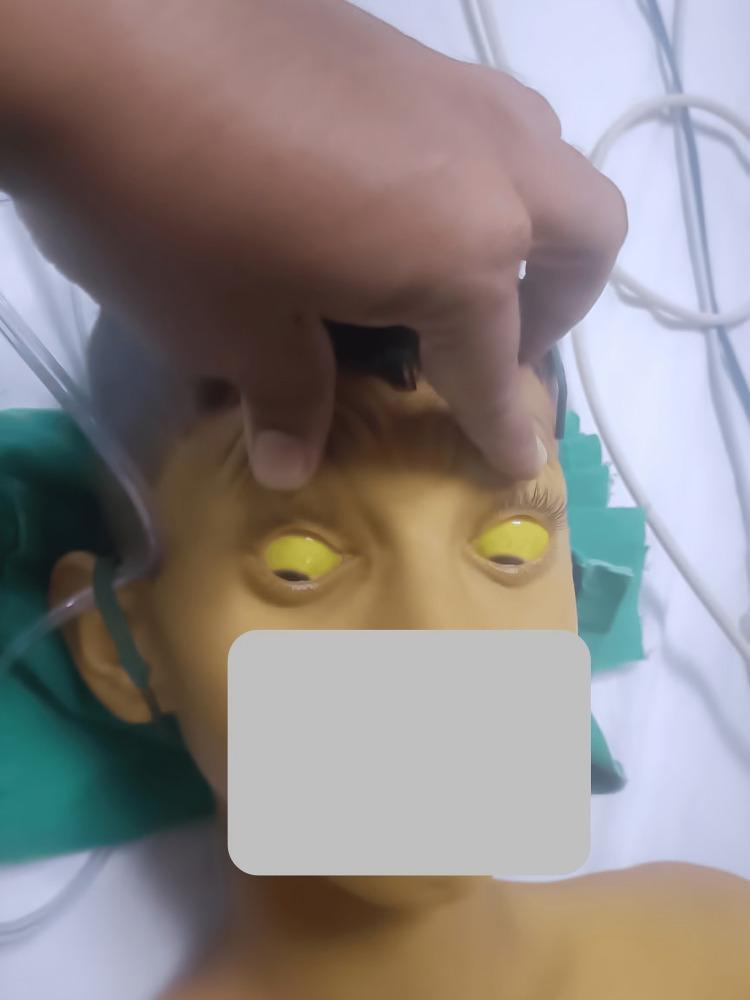
This clinical photograph displays a pediatric patient demonstrating severe systemic jaundice. The primary diagnostic feature is profound scleral icterus, characterized by a bright yellow discoloration of the entire visible sclera in both eyes. The upper eyelids are manually elevated to fully reveal the extent of the icterus. Additionally, the patient's facial skin, forehead, and upper chest exhibit a marked yellowish hue (jaundice), consistent with hyperbilirubinemia. The patient appears to be in a clinical setting, as evidenced by medical tubing and linens visible in the background. This image is an educational example of the physical manifestations of liver dysfunction or hemolytic processes, such as hepatitis, Wilsonian crisis, or acute toxicity, which can lead to significant accumulation of bilirubin in mucosal surfaces and skin.
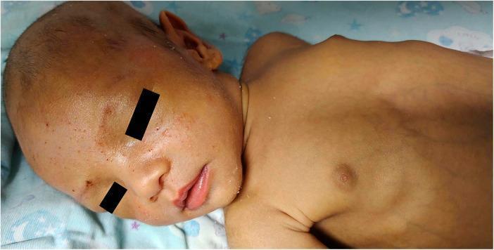
Clinical photograph of a 53-day-old infant demonstrating severe generalized jaundice. The infant's skin exhibits a profound yellow-to-bronze discoloration across the face, trunk, and upper extremities. Scleral icterus is visible despite black bars masking the eyes for privacy. The facial skin shows mild textural irregularities and scattered superficial erythematous papules. The torso reveals prominent superficial venous patterning and a visible umbilical hernia, which are common clinical findings in pediatric cholestasis. This image represents the clinical manifestation of conjugated hyperbilirubinemia, specifically in the context of Dubin-Johnson syndrome (associated with ABCC2 gene mutations), illustrating the significant accumulation of bilirubin pigments in the skin and mucous membranes during early infancy.
JAUNDICE - General Medicine Long Notes
BDS 3rd Year | University Exam Pattern (India)
QUESTION 1: DEFINE JAUNDICE. CLASSIFY AND DESCRIBE CAUSES & CLINICAL FEATURES.
DEFINITION
- Normal serum bilirubin: ≤1 mg/dL
- Clinical jaundice becomes visible when serum bilirubin exceeds 2-3 mg/dL
- Jaundice is NOT a disease - it is a symptom of an underlying disorder
- The word "icterus" is used specifically for yellow discoloration of the sclerae
NORMAL BILIRUBIN METABOLISM (Essential for understanding jaundice)
- Red blood cells (RBCs) have a lifespan of ~120 days
- Senescent RBCs are phagocytosed by macrophages in the spleen, liver (Kupffer cells), and bone marrow
- Haemoglobin is broken down: Hb → Globin (amino acids recycled) + Haem
- Haem is acted upon by haem oxygenase → Biliverdin (green pigment) → reduced by biliverdin reductase → Bilirubin (red-orange pigment)
- This bilirubin is Unconjugated Bilirubin (UCB) = "Indirect Bilirubin" (by van den Bergh test)
- UCB is water-insoluble, lipid-soluble, and toxic (especially to the brain - kernicterus in neonates)
- UCB is poorly soluble in plasma → tightly bound to albumin for transport to the liver
- UCB-albumin complex cannot be filtered by the kidney → no bilirubinuria in prehepatic jaundice
- UCB dissociates from albumin at hepatocyte sinusoidal membrane
- Enters hepatocyte via facilitated transport (OATP transporters)
- Binds to intracellular protein ligandin (Y protein)
- Enzyme: Bilirubin UDP-Glucuronosyl Transferase (bilirubin UGT) in hepatocyte endoplasmic reticulum
- UCB + 2 molecules of UDP-Glucuronic acid → Bilirubin Diglucuronide = Conjugated Bilirubin (CB) = "Direct Bilirubin"
- CB is water-soluble, non-toxic, can be filtered by kidneys → bilirubinuria in hepatic/obstructive jaundice
- CB is actively transported (energy-dependent, rate-limiting) across the canalicular membrane into bile canaliculi by MRP2 transporter
- Defect here → Dubin-Johnson Syndrome (conjugated hyperbilirubinemia)
- CB enters intestine via bile
- Gut bacteria deconjugate and reduce CB → Urobilinogen (colorless)
- Most urobilinogen → oxidized by bacteria → Stercobilin → gives feces its characteristic brown color
- Some urobilinogen reabsorbed (enterohepatic circulation):
- Re-excreted by liver into bile (hepatic recycling)
- Some enters systemic circulation → kidneys → oxidized to Urobilin → excreted in urine → gives urine its characteristic yellow color
CLASSIFICATION OF JAUNDICE
Classification Based on Site of Defect:
- Excess bilirubin production overwhelming hepatic capacity
- Type: Unconjugated hyperbilirubinemia
- Failure of hepatocytes to take up, conjugate, or secrete bilirubin
- Type: Mixed (both unconjugated + conjugated hyperbilirubinemia)
- Obstruction to outflow of bile from liver to duodenum
- Type: Conjugated hyperbilirubinemia
Classification Based on Solubility:
| Type | Serum Bilirubin elevated | Van den Bergh Reaction |
|---|---|---|
| Unconjugated hyperbilirubinemia | Indirect (unconjugated) | Indirect positive (Prompt in methanol; delayed in direct) |
| Conjugated hyperbilirubinemia | Direct (conjugated) | Direct positive (immediate with diazo reagent in aqueous medium) |
| Mixed | Both | Both positive |
CAUSES OF JAUNDICE
I. PRE-HEPATIC (Haemolytic / Prehepatic) Jaundice
- Hereditary spherocytosis (membrane defect)
- Glucose-6-phosphate dehydrogenase (G6PD) deficiency (enzyme defect)
- Pyruvate kinase deficiency
- Sickle cell anaemia (Hb S)
- Thalassaemia (alpha and beta)
- Autoimmune haemolytic anaemia (warm/cold antibody)
- Malaria (Plasmodium falciparum - major cause in India)
- Mismatched blood transfusion (transfusion reaction)
- Pernicious anaemia (megaloblastic)
- Thalassaemia major (intramedullary destruction)
- After major trauma, surgery, or internal bleeding
- RBCs broken down locally → bilirubin load
- Immature liver (insufficient bilirubin UGT)
- High rate of RBC breakdown (fetal Hb replacement)
- Appears day 2-3 of life; resolves by day 10-14
II. HEPATIC (Hepatocellular) Jaundice
- Viral hepatitis (most common cause in India) - HAV, HBV, HCV, HEV, HCV
- Infectious mononucleosis (EBV)
- Leptospirosis (Weil's disease - also cholestatic component)
- Malaria (also haemolytic)
- Typhoid fever (rare)
- Yellow fever
- Bacterial sepsis (especially gram-negative)
- Alcohol (alcoholic hepatitis, alcoholic cirrhosis)
- Drugs: Paracetamol (overdose), isoniazid, rifampicin, halothane, statins, oral contraceptives
- Chemical toxins: Carbon tetrachloride, phosphorus, industrial solvents
- Mushroom poisoning (Amanita phalloides)
- Autoimmune hepatitis
- Chronic hepatitis B and C
- Primary biliary cholangitis (PBC) - also cholestatic
- Non-alcoholic steatohepatitis (NASH)
- Wilson's disease (copper accumulation)
- Haemochromatosis (iron overload)
- Alpha-1 antitrypsin deficiency
- Gilbert's syndrome: Reduced bilirubin UGT (~30% of normal); benign; mild unconjugated hyperbilirubinemia precipitated by fasting, stress, illness; most common hereditary cause of jaundice
- Crigler-Najjar syndrome:
- Type I: Complete absence of bilirubin UGT; severe; kernicterus; fatal without liver transplant
- Type II: Partial deficiency; responds to phenobarbitone
- Dubin-Johnson syndrome: Defect in canalicular transport of CB; conjugated hyperbilirubinemia; benign; black liver (melanin-like pigment); urinary coproporphyrin pattern abnormal
- Rotor syndrome: Similar to DJS; no black pigment
III. POST-HEPATIC (Obstructive / Cholestatic) Jaundice
- Drug-induced cholestasis (oral contraceptive pills, chlorpromazine, erythromycin estolate, anabolic steroids)
- Intrahepatic cholestasis of pregnancy (ICP)
- Primary Biliary Cholangitis (PBC) - autoimmune destruction of intrahepatic bile ducts
- Primary Sclerosing Cholangitis (PSC) - fibroinflammatory stricturing of bile ducts
- Viral hepatitis (cholestatic variant)
- Choledocholithiasis (common bile duct stone) - most common cause of surgical obstructive jaundice in India
- Bile duct worms (Ascaris)
- Cholangiocarcinoma (bile duct carcinoma / Klatskin tumor at hilum)
- Primary Sclerosing Cholangitis (PSC)
- Benign bile duct stricture (post-cholecystectomy, post-traumatic)
- Carcinoma head of pancreas (most common malignant cause; classic painless jaundice in elderly)
- Periampullary carcinoma (at ampulla of Vater)
- Chronic pancreatitis (fibrosis compresses CBD)
- Enlarged lymph nodes at porta hepatis (lymphoma, metastasis)
CLINICAL FEATURES
I. General Features Common to All Types of Jaundice
- Yellow discoloration of skin and eyes
- Dark urine (tea/cola-colored) - bilirubinuria (in conjugated hyperbilirubinemia)
- Pale/clay-colored stools (acholic stools) - in obstructive jaundice (no stercobilin reaching gut)
- Pruritus (itching) - due to bile salt deposition in skin; more severe in obstructive jaundice
- Anorexia, nausea, malaise
- Dark-colored tears and sweat (severe jaundice)
- Scleral icterus (earliest and most reliable sign - sclerae have high elastin content with high affinity for bilirubin)
- Yellow skin (generalised, especially palms, soles)
- Hepatomegaly / splenomegaly (depending on cause)
II. Specific Features by Type
PRE-HEPATIC (Haemolytic) Jaundice:
- Mild jaundice (bilirubin rarely exceeds 5 mg/dL - liver capacity not overwhelmed)
- Lemon-yellow tinge (mild, pale yellow)
- Stools are dark (increased stercobilin - more CB reaching gut)
- Urine: Normal or slightly dark (increased urobilinogen, no bilirubinuria)
- Splenomegaly (site of RBC destruction + sequestration)
- Features of anaemia: Pallor, fatigue, palpitations, exertional dyspnea
- No pruritus (no cholestasis)
- No hepatomegaly (unless haemolysis involves liver)
HEPATIC (Hepatocellular) Jaundice:
- Moderate to severe jaundice (orange-yellow)
- Features of liver cell failure:
- Anorexia, nausea, vomiting
- Right hypochondrial pain (liver capsule distension)
- Fever (viral hepatitis, alcoholic hepatitis)
- Malaise and profound fatigue
- Tender hepatomegaly (in acute hepatitis)
- Splenomegaly (in chronic liver disease/cirrhosis)
- Urine: Dark (bilirubinuria) + increased urobilinogen
- Stools: Normal or slightly pale
- Mild pruritus
- Signs of chronic liver disease (if cirrhotic): Spider naevi, palmar erythema, gynaecomastia, ascites, caput medusae
- Raised ALT/AST (hepatocellular damage)
POST-HEPATIC (Obstructive) Jaundice:
- Severe, progressive jaundice (deep green-yellow/"biliverdin jaundice" in prolonged cases)
- Severe pruritus (bile salts in skin; characteristic - patient scratches vigorously; scratch marks visible)
- Pale/clay/putty-colored stools (acholic) - no stercobilin reaching gut
- Dark urine (bilirubinuria - conjugated bilirubin filtered by kidneys)
- Steatorrhoea - fat malabsorption (no bile for fat emulsification)
- Vitamin K deficiency - fat-soluble vitamins not absorbed → coagulopathy (prolonged PT)
- Vitamin D deficiency - osteomalacia with prolonged cholestasis
- No fever (unless complicated by cholangitis/infection in bile duct)
- Abdominal pain: Colicky (CBD stone - sudden onset, biliary colic); painless + progressive (carcinoma head of pancreas - classic)
- Courvoisier's sign (Courvoisier's Law): Palpable, non-tender, distended gallbladder + painless progressive jaundice = malignant obstruction (carcinoma head of pancreas or periampullary carcinoma)
- Exception: This sign is ABSENT if obstruction is from CBD stone (stone usually implies previous cholecystitis → GB fibrosis → cannot distend)
- Weight loss (suggests malignancy)
- Xanthomata/xanthelasma (prolonged cholestasis → hypercholesterolemia)
- Hepatomegaly (smooth, non-tender in malignant obstruction)
QUESTION 2: DIFFERENTIATE HAEMOLYTIC, HEPATOCELLULAR & OBSTRUCTIVE JAUNDICE
COMPREHENSIVE DIFFERENTIATION TABLE
| Feature | HAEMOLYTIC (Pre-hepatic) | HEPATOCELLULAR (Hepatic) | OBSTRUCTIVE (Post-hepatic) |
|---|---|---|---|
| Other names | Prehepatic jaundice | Medical jaundice | Surgical / cholestatic jaundice |
| Site of defect | RES (spleen, marrow) - excessive RBC destruction | Liver cells (hepatocytes) | Bile ducts (intra or extrahepatic) |
| Mechanism | Excess bilirubin production | Failure of uptake/conjugation/secretion | Obstruction of bile flow |
| Common causes | Haemolytic anaemias, malaria, G6PD deficiency, sickle cell | Viral hepatitis, alcoholic hepatitis, cirrhosis, drugs | CBD stone, carcinoma head of pancreas, cholangiocarcinoma |
| Color of jaundice | Mild, lemon-yellow | Moderate, orange-yellow | Deep, green-yellow (prolonged: biliverdin) |
| Severity | Mild (rarely >5 mg/dL) | Moderate to severe | Severe and progressive |
| Onset | Variable | Subacute (hepatitis) or insidious | Gradual (malignancy) or sudden (stone) |
| Pruritus | Absent | Mild/absent | Severe (bile salts in skin) |
| Abdominal pain | Absent (or LUQ if spleen) | RUQ discomfort | Colicky (stone); Painless (malignancy) |
| Fever | Present if haemolytic crisis | Common (viral hepatitis, alcoholic) | Usually absent (unless cholangitis) |
| URINE COLOR | Normal or slightly dark | Dark (bilirubinuria + urobilinogen ↑) | Very dark (bilirubinuria) |
| Urine bilirubin | Absent (UCB not filtered by kidneys) | Present | Present |
| Urine urobilinogen | Increased | Increased | Absent/very low (no bile reaching gut) |
| STOOL COLOR | Dark (increased stercobilin) | Normal or slightly pale | Pale/clay/acholic (no stercobilin) |
| Steatorrhoea | Absent | Absent | Present (no bile for fat digestion) |
| Splenomegaly | Present (prominent - RBC destruction) | May be present (portal hypertension) | Absent (unless portal cause) |
| Hepatomegaly | Absent | Present (tender in acute hepatitis) | Present (smooth in malignancy) |
| Gallbladder | Not palpable | Not palpable | Palpable (Courvoisier's sign in malignancy) |
| BILIRUBIN TYPE | Unconjugated (Indirect) ↑↑ | Both (Conjugated + Unconjugated) ↑ | Conjugated (Direct) ↑↑ |
| Van den Berg test | Indirect positive (delayed) | Both positive | Direct positive (prompt) |
| Total bilirubin | Mildly elevated (<5 mg/dL) | Moderately elevated | Markedly elevated (can be very high) |
| ALT/AST (SGOT/SGPT) | Normal | Markedly elevated (thousands) | Mildly elevated (2-5x) |
| Alkaline Phosphatase (ALP) | Normal | Mildly elevated | Markedly elevated (5-10x) |
| GGT | Normal | Elevated (alcoholic hepatitis) | Markedly elevated |
| Serum Albumin | Normal | Low (if chronic liver disease) | Normal (initially) |
| Prothrombin Time (PT) | Normal | Prolonged (liver synthetic failure) | Prolonged (responds to Vitamin K IV) |
| Response to Vitamin K | No effect | No improvement (hepatocytes damaged) | PT corrects (absorption issue, not liver) |
| CBC | Anaemia present; reticulocytosis; abnormal RBC morphology | Normal or mild anaemia | Normal (anaemia if malignancy) |
| Reticulocyte count | Elevated (compensatory erythropoiesis) | Normal | Normal |
| Blood film | Sickle cells, spherocytes, target cells (cause-dependent) | Normal | Normal |
| LDH | Elevated (RBC breakdown) | Elevated (hepatocellular damage) | Normal |
| Haptoglobin | Low/absent (bound to free Hb from haemolysis) | Normal | Normal |
| Serum cholesterol | Normal | Low (in severe liver failure) | Elevated (bile acid retention) |
| Coombs test | Positive (in immune haemolytic anaemia) | Negative | Negative |
| Ultrasound abdomen | Normal liver; splenomegaly | Hepatomegaly; cirrhotic liver | Dilated bile ducts (diagnostic); gallstones |
| Xanthomata | Absent | Absent | Present (prolonged cholestasis) |
| Weight loss | Not prominent | Moderate (chronic liver disease) | Prominent (malignancy) |
KEY DISTINGUISHING RULES (Exam High-yield):
- Haemolytic jaundice → no bilirubinuria (UCB bound to albumin, not filtered)
- Hepatocellular & Obstructive → bilirubinuria (CB is water-soluble, passes into urine)
- "Black water fever" test: Shake urine → yellow froth = bilirubinuria
- Haemolytic: Elevated (excess CB formed → excess urobilinogen produced)
- Hepatocellular: Elevated (liver cannot recycle urobilinogen from enterohepatic circulation)
- Obstructive: Absent (no bile reaching gut → no urobilinogen formed)
- Haemolytic: Dark brown (excess stercobilin)
- Obstructive: Pale/clay/acholic (no bile/stercobilin reaching gut)
- Hepatocellular: Variable (may be pale in cholestatic hepatitis)
- Obstructive jaundice: Prolonged PT CORRECTS with Vitamin K IV (fat-soluble vitamin, not absorbed → give IV)
- Hepatocellular: Prolonged PT does NOT respond significantly to Vitamin K (hepatocytes damaged, cannot synthesize factors)
- Obstructive jaundice: ALP markedly elevated (>3x upper normal) + mildly elevated transaminases
- Hepatocellular: ALT/AST markedly elevated + mildly elevated ALP
- "ALP up: think obstruction; ALT up: think hepatocytes"
- Palpable gallbladder + painless jaundice = malignant obstruction (carcinoma head of pancreas)
- Stone jaundice = gallbladder NOT palpable (fibrosis from previous cholecystitis)
QUESTION 3: INVESTIGATIONS IN JAUNDICE
APPROACH TO INVESTIGATION
- Confirm hyperbilirubinemia
- Determine whether it is pre-hepatic, hepatic, or post-hepatic
- Identify the underlying cause
- Assess severity and complications
A. URINE EXAMINATION
| Test | Haemolytic | Hepatocellular | Obstructive |
|---|---|---|---|
| Bilirubin (dipstick) | Absent | Present | Present |
| Urobilinogen | Markedly increased | Increased | Absent |
| Colour | Slightly dark | Dark (amber) | Very dark (cola/tea) |
| Froth test | White froth | Yellow froth | Yellow froth |
- Bilirubinuria (bile in urine): Detected by Fouchet test or Gmelin test or urine dipstick
- "Foamy urine" with yellow froth suggests bilirubinuria (conjugated bilirubin)
- Normal: White froth
- Urobilinogen: Detected by Ehrlich's aldehyde test
- Increased in haemolytic and hepatocellular jaundice
- Absent in complete obstructive jaundice
B. BLOOD TESTS
1. Serum Bilirubin (Total, Direct/Conjugated, Indirect/Unconjugated)
- Direct bilirubin (conjugated): Reacts immediately with diazo reagent in aqueous medium
- Indirect bilirubin (unconjugated): Reacts only after adding methanol (accelerator)
- Total bilirubin = Direct + Indirect
| Normal | Haemolytic | Hepatocellular | Obstructive | |
|---|---|---|---|---|
| Total bilirubin | <1 mg/dL | ↑ (2-5 mg/dL) | ↑↑ (variable) | ↑↑↑ (may be very high) |
| Indirect (UCB) | 0.2-0.8 mg/dL | ↑↑↑ | ↑ | Normal/mildly↑ |
| Direct (CB) | 0-0.3 mg/dL | Normal | ↑ | ↑↑↑ |
| D:T ratio | <20% | <20% | >30% | >50% |
2. Liver Function Tests (LFTs)
- Indicate hepatocellular damage
- ALT (SGPT) is more specific for liver (also in kidney, heart)
- AST (SGOT) is less specific (also in heart, muscle)
- ALT > AST: Viral hepatitis, NASH
- AST > ALT (>2:1): Alcoholic liver disease (alcohol depletes pyridoxine needed for ALT synthesis)
- Normal: ALT <40 IU/L; AST <40 IU/L
- In viral hepatitis: Can rise to 500-5000 IU/L
- In obstructive jaundice: Only mildly elevated (<3x normal)
- Found in bile duct epithelium, bone, intestine, placenta
- Elevated in cholestasis (obstructive jaundice, PBC, PSC)
- Liver-specific ALP confirmed by concurrent GGT elevation
- ALP >3x normal = obstructive etiology until proven otherwise
- Markedly elevated ALP with mildly raised bilirubin: Consider bone disease (isoenzyme fractionation needed)
- More specific for liver than ALP
- Elevated in: Alcoholic liver disease (markedly elevated), cholestasis, drug-induced hepatitis
- Not elevated in bone disease (helps differentiate from ALP due to bone origin)
- A very sensitive (but non-specific) indicator of alcohol abuse
- Synthesized by hepatocytes
- Low albumin = chronic liver disease / liver synthetic failure
- Normal in acute viral hepatitis, haemolytic jaundice, obstructive jaundice (initially)
- Low albumin + prolonged PT = poor prognosis in liver disease
- Clotting factors II, VII, IX, X synthesized by liver (Vitamin K-dependent)
- Prolonged PT in: Both hepatocellular and obstructive jaundice
- Differentiating test: Vitamin K injection
- Obstructive: PT corrects after 24-48 hours of Vitamin K (absorption problem; parenteral K works)
- Hepatocellular: PT does NOT correct (hepatocytes cannot use Vitamin K - cells damaged)
- Prolonged PT that does not respond to Vitamin K = severe hepatocellular failure
- Total protein low: Liver failure
- Globulins elevated: Autoimmune hepatitis (IgG), PBC (IgM), alcoholic liver disease
3. Complete Blood Count (CBC) with Peripheral Smear
- Anaemia + reticulocytosis + abnormal RBC forms → haemolytic jaundice
- Spherocytes → hereditary spherocytosis or autoimmune haemolysis
- Sickle cells → sickle cell anaemia
- Target cells → thalassaemia, liver disease
- Bite cells/blister cells → G6PD deficiency
- Leukocytosis → viral hepatitis (early leucopenia with lymphocytosis), bacterial cholangitis, alcoholic hepatitis
- Thrombocytopenia → liver cirrhosis (hypersplenism), severe viral hepatitis
- Macrocytosis → alcoholic liver disease, B12/folate deficiency
4. Reticulocyte Count
- Elevated (>2%) in haemolytic jaundice - compensatory marrow response
- Normal in hepatocellular and obstructive jaundice
5. Specific Tests for Haemolysis
- Coombs test (DAT): Positive in immune haemolytic anaemia (warm autoimmune, drug-induced)
- Serum LDH: Elevated (RBC breakdown)
- Serum haptoglobin: Low/absent (binds free Hb from lysed RBCs)
- Free haemoglobin in plasma (haemoglobinaemia)
- G6PD enzyme assay: Deficiency
- Osmotic fragility test: Hereditary spherocytosis
- Hb electrophoresis: Sickle cell, thalassaemia
- Malaria smear (thick and thin smear) + malaria antigen test
6. Serum Cholesterol and Bile Acids
- Elevated in obstructive jaundice (bile cannot be excreted → cholesterol accumulates)
- Causes xanthomata and xanthelasma in prolonged cholestasis
C. LIVER DISEASE-SPECIFIC MARKERS
| Condition | Test |
|---|---|
| Viral hepatitis A | Anti-HAV IgM (acute), Anti-HAV IgG (past/immune) |
| Viral hepatitis B | HBsAg (active), IgM Anti-HBc (acute), HBeAg (replication), HBV DNA, Anti-HBs (recovery/immunity) |
| Viral hepatitis C | Anti-HCV, HCV RNA (PCR) |
| Autoimmune hepatitis | ANA, anti-smooth muscle antibody (ASMA), anti-LKM1; serum IgG elevated |
| Primary Biliary Cholangitis | Anti-mitochondrial antibody (AMA - M2 subtype); serum IgM elevated |
| Primary Sclerosing Cholangitis | pANCA; AMA negative; elevated ALP, GGT |
| Wilson's disease | Low serum ceruloplasmin (<20 mg/dL), elevated 24-hr urine copper, Kayser-Fleischer rings (slit-lamp) |
| Haemochromatosis | Elevated serum ferritin, elevated transferrin saturation (>45%), HFE gene mutation (C282Y) |
| Alpha-1 antitrypsin deficiency | Low serum alpha-1 antitrypsin level; Pi phenotype ZZ |
| Carcinoma head of pancreas | CA 19-9 (tumor marker - not diagnostic alone), CEA; CT findings |
| Hepatocellular carcinoma | Alpha-fetoprotein (AFP) - markedly elevated; imaging |
D. IMAGING STUDIES
1. Ultrasound Abdomen (First-line Imaging - done in all patients)
- Completely non-invasive, no radiation, cheap, widely available
- Dilated bile ducts (CBD >8mm without cholecystectomy, >10mm post-cholecystectomy) = obstructive jaundice
- Gallstones in gallbladder or CBD
- CBD stone - echogenic focus in CBD with posterior acoustic shadowing
- Pancreatic mass (carcinoma head of pancreas)
- Hepatomegaly (hepatitis, fatty liver, congestion)
- Cirrhotic liver (nodular, shrunken, coarse echogenicity)
- Splenomegaly (portal hypertension, haemolytic anaemia)
- Ascites
- Liver metastases or HCC
- Cannot visualize CBD stones in 50% cases (gas in duodenum)
- Operator-dependent
- Cannot evaluate functional biliary anatomy
2. CT Abdomen (CT Scan with Contrast)
- Better characterization of pancreatic and biliary pathology
- Detects: Pancreatic carcinoma, cholangiocarcinoma, lymph nodes, liver metastases, HCC
- CT cholangiography for biliary tree visualization
- Assesses resectability of malignant lesions (vascular involvement)
3. MRI / MRCP (Magnetic Resonance Cholangiopancreatography)
- Gold standard non-invasive investigation for biliary and pancreatic duct visualization
- MRCP: Non-invasive "road map" of biliary tree; detects CBD stones, strictures, cholangiocarcinoma
- No radiation, no contrast injection needed for biliary visualization
- Cannot be used for therapeutic intervention (unlike ERCP)
- Preferred over ERCP as first-line for diagnostic purposes
4. ERCP (Endoscopic Retrograde Cholangiopancreatography)
- Gold standard for extrahepatic obstructive jaundice (both diagnostic AND therapeutic)
- Endoscope advanced to ampulla → contrast injected into CBD and pancreatic duct
- Diagnostic: Shows site and nature of obstruction (stone, stricture, tumor)
- Therapeutic: Can relieve obstruction in the same procedure:
- Sphincterotomy: Cuts sphincter of Oddi to allow stone passage
- Stone extraction (balloon catheter, basket retrieval)
- Stenting: For malignant strictures (provides biliary drainage)
- Tissue biopsy / brush cytology of strictures
- Complications: Pancreatitis (2-3%), bleeding, perforation, cholangitis
- Use: CBD stones, suspected cholangiocarcinoma, pancreatic carcinoma
5. PTC (Percutaneous Transhepatic Cholangiography)
- Needle through skin into dilated intrahepatic bile duct → contrast injection
- Used when ERCP fails or not accessible
- Can also be used for biliary drainage (PTBD - percutaneous transhepatic biliary drainage)
6. Endoscopic Ultrasound (EUS)
- Highly sensitive for small CBD stones, small pancreatic tumors, ampullary tumors
- Can perform fine-needle aspiration (FNA) of pancreatic masses
E. LIVER BIOPSY
- Diagnosis and staging of chronic hepatitis (B, C, autoimmune)
- Grading fibrosis in chronic liver disease
- Diagnosis of suspected NASH, drug-induced liver injury
- Unexplained hepatomegaly or elevated transaminases
- Diagnosis of cholestatic liver disease (PBC, PSC)
- Staging of HCC / liver metastases
- Contraindicated: Coagulopathy (PT >3 seconds prolonged, platelets <60,000), obstructive jaundice (risk of bile peritonitis)
- Viral hepatitis: Lobular inflammation, hepatocyte necrosis, viral inclusions
- Alcoholic hepatitis: Mallory-Denk bodies (alcoholic hyaline), steatosis, neutrophil infiltration
- PBC: Granulomatous bile duct destruction
- Haemochromatosis: Iron (haemosiderin) deposits (Perls' stain)
- Wilson's disease: Copper (rhodanine stain)
F. SPECIAL TESTS
- Serum amylase and lipase: Pancreatitis, carcinoma head of pancreas
- CA 19-9: Pancreatic carcinoma marker (elevated; not specific)
- CEA: Colorectal liver metastases
- AFP: HCC
- Thyroid function tests: Hypothyroidism (mild jaundice, cholestasis)
APPROACH ALGORITHM FOR JAUNDICE (Exam Flow Chart)
JAUNDICE
    ↓
Clinical history + examination
    ↓
Urine: bilirubin present?
    ↓
NO                           YES
Pre-hepatic (UCB)          Hepatic or Obstructive
(Haemolytic)               (CB)
    ↓                           ↓
CBC, reticulocyte count,    LFTs (ALT/ALP ratio)
haemolysis screen               ↓
                        ALT >> ALP:          ALP >> ALT:
                        HEPATOCELLULAR        OBSTRUCTIVE
                            ↓                    ↓
                        Viral markers        Ultrasound
                        Autoimmune panel     (dilated ducts?)
                        Liver biopsy              ↓
                                          MRCP / ERCP
                                          CT scan
SUMMARY - HIGH-YIELD EXAM POINTS
| Topic | Key Fact |
|---|---|
| Definition | Yellow discoloration due to hyperbilirubinemia; visible at serum bilirubin >2-3 mg/dL |
| Normal bilirubin | ≤1 mg/dL; mostly unconjugated (>95%) |
| First visible sign | Scleral icterus (sclerae have high elastin affinity for bilirubin) |
| Prehepatic (haemolytic) type | Unconjugated ↑; no bilirubinuria; dark stools; no pruritus |
| Hepatocellular type | Mixed ↑; bilirubinuria; ALT >> ALP; Vitamin K doesn't correct PT |
| Obstructive type | Conjugated ↑; bilirubinuria; pale stools; severe pruritus; ALP >> ALT |
| Courvoisier's sign | Palpable GB + painless jaundice = malignant obstruction |
| Courvoisier's law exception | Stone = no palpable GB (GB fibrosed from previous cholecystitis) |
| Pale stools | Obstructive jaundice ONLY (no stercobilin reaching gut) |
| Absent urine urobilinogen | Complete obstructive jaundice (no bile → no urobilinogen formed) |
| Vitamin K response | Corrects PT in obstructive; NOT in hepatocellular |
| Best initial imaging | Ultrasound abdomen (dilated ducts = obstruction) |
| Non-invasive biliary imaging | MRCP (gold standard for diagnosis) |
| Therapeutic biliary procedure | ERCP (diagnose + treat in one procedure) |
| Marker of haemolysis | Low haptoglobin + elevated LDH + reticulocytosis + Coombs test |
| Gilbert's syndrome | Benign, unconjugated; mild jaundice with fasting/stress; bilirubin UGT deficiency |
| Dubin-Johnson | Benign, conjugated; defect in canalicular transport; black liver |
| Best liver synthetic function tests | Serum albumin + Prothrombin time (INR) |
| CBD stone vs pancreatic Ca jaundice | Stone: Colicky pain, fever (Charcot's triad); Ca: Painless, weight loss, palpable GB |