
Preservation of viscera in suspected poisoning cases
Reading File
This is a forensic medicine / forensic pathology topic. Let me search the medical library for this.
Finding Sources
Finding Sources
Excellent - I found the dedicated section. Let me read both the main section and Parikh's coverage in parallel.
Reading File
Reading File
Let me read more of the main section which was truncated, and also get the Parikh's despatch section:
Reading File
Reading File
Let me get the rest of the main textbook content which was truncated (the additional viscera section and dispatch/labeling details):
Reading File
Reading File
I now have comprehensive content from both major forensic medicine textbooks. Let me compile the full answer.
Preservation of Viscera in Suspected Poisoning Cases
The Essentials of Forensic Medicine and Toxicology, 36th ed. (2026) | Parikh's Textbook of Medical Jurisprudence, Forensic Medicine and Toxicology
When to Preserve Viscera
Viscera must be preserved in the following circumstances:
- Death is suspected to be due to poisoning (by police or doctor)
- Deceased was intoxicated or known to use drugs
- Cause of death not found even after full autopsy
- Unusual smell, color, or unidentifiable material detected in stomach contents
- Anaphylactic deaths
- Deaths due to burns
- Advanced decomposition
- Accidental death involving a driver of a vehicle or machine operator
- When the investigating officer requests it
- When medical officer suspects poison by smell or other findings during an injury autopsy
Routine Viscera - Must Be Preserved in All Suspected Cases
| Organ/Fluid | Quantity | Rationale |
|---|---|---|
| Stomach + contents | Entire (wall if empty) | Most poisons taken orally; highest initial concentration here |
| Upper small intestine + contents | ~30 cm length | Ongoing absorption, quantifies ingestion timeline |
| Liver | 200-300 g | Major detoxicating organ; concentrates poisons even when blood levels are low |
| Kidney | Half of each kidney | Organ of excretion; large amounts present in urine |
| Blood | 30 mL (minimum 10 mL) | Systemic levels; from peripheral vein |
| Urine | 30 mL | Evidence of absorption and excretion |
Note: GI tract contents must be kept in separate bottles from solid viscera, because the relative amounts of poison in stomach vs. intestine vs. solid organs give information about time since ingestion.
Containers
- Use 1-litre capacity, clean, wide-mouthed, white glass bottles with glass stoppers
- Rubber inserts under caps must NOT be used - they extract certain poisons (chloroform, phenols)
- Clean glass containers with sulphuric acid-chromate solution, rinse with distilled water, and dry
- Polyethylene bags/containers can be used, but volatile poisons may diffuse through plastic
- For lungs or tissues to be analyzed for volatile substances, use nylon bags (impermeable to such substances)
- Blood must be collected in screw-capped bottles of ~30 mL
Preservatives
(A) Saturated Common Salt (NaCl) Solution
- Used in all cases of suspected poisoning
- Exceptions - NOT used in poisoning by:
- Corrosive acids (except phenol/carbolic acid)
- Corrosive alkalis
- Corrosive sublimate (mercuric chloride)
- Aconite
(B) Rectified Spirit (Alcohol)
- Used when saturated salt is contraindicated (corrosive acid cases, except carbolic acid)
- NOT used in suspected poisoning by:
- Alcohol, kerosene
- Chloroform, ether
- Chloral hydrate
- Formic acid, formaldehyde, acetic acid
- Phenol
- Phosphorus (diminishes phosphorescence)
- Paraldehyde (organic acids and paraldehyde are soluble in alcohol)
(C) Blood Preservatives (special rules)
- Routine: 10 mg/mL sodium or potassium fluoride (inhibits glycolysis, enzyme enolase, and bacterial growth) + 3 mg potassium oxalate (anti-coagulant) per 10 mL blood
- Fluoride should also be added to urine, CSF, and vitreous humor if alcohol estimation is required, and also for cocaine, cyanide, and CO analysis
- Oxalic acid / ethylene glycol / fluoride poisoning: Use 30 mg sodium citrate instead of potassium oxalate
- CO and CN poisoning: Use airtight caps; add a layer of liquid paraffin (1-2 cm) over blood to prevent exposure to atmospheric oxygen
- Do NOT use heparin or EDTA - they interfere with detection of methanol
(D) Urine Preservatives
- 1 mL concentrated HCl, or 100 mg thymol, or 100 mg sodium fluoride per 10 mL urine
- Alternatively: equal quantity of saturated NaCl or rectified spirit; or toluene (preferred)
(E) When Preservative is NOT Necessary
- Viscera can be analyzed within 24 hours
- Sample can be kept in a refrigerator or ice box
- Bones, hair, nails
- Lungs for detecting inhaled poisons
Important: Viscera must NOT be preserved in formaldehyde - extraction of poison (especially non-volatile organic compounds) becomes very difficult.
Additional Viscera in Specific Poisonings
| Organ/Tissue | Quantity | Poisons to Detect |
|---|---|---|
| Heart | As required | Strychnine, digitalis, yellow oleander |
| Brain (cerebrum/cerebellum) | 100 g | Alkaloids, organophosphates, opiates, CO, cyanide, strychnine, barbiturates, anesthetics, volatile organic poisons |
| Spinal cord | Entire length | Strychnine, gelsemium |
| Gallbladder/Bile | Bile via puncturing gall bladder in situ | Morphine, cocaine, methadone, barbiturates, major tranquilizers |
| Vitreous humor | As available | Alcohol, chloroform, cocaine, morphine, TCAs, urea, creatinine, sugar, electrolytes |
| CSF | As available | Alcohol |
| Lung (one lung) | Full lung | Gaseous poisons, HCN, alcohol, chloroform - tie trachea, collect bronchial air in nylon bag (heat-sealed) |
| Fatty tissue (abdominal wall / perinephric region) | 10 g | Pesticides, insecticides |
| Skin (with underlying fat/muscle) | 10 cm radius around injection/absorption site | Insulin, morphine, heroin, cocaine, corrosive poisons |
| Bone (femur shaft) | 10 cm | Chronic/subacute poisoning by arsenic, antimony, thallium, radium |
| Hair | 20-30 strands with roots (head hair) | Chronic heavy metal poisoning (arsenic, etc.) |
| Nails | All nails (removed entirely) | Heavy metals |
| Muscle (thigh) | 50-100 g (3x3 cm) | Used when internal organs are badly putrefied |
| Uterus + appendages + upper vagina | Entire | Criminal abortion cases; foreign bodies sent separately |
Instructions for Packing and Dispatch
- Separate bottles: Stomach + small intestine (one bottle); liver + kidney (second bottle); blood separately; urine separately; brain in its own bottle
- Processing: Stomach and intestines are opened before preservation. Liver and kidney are cut into 0.5-1 cm pieces or minced
- Quantity of preservative should be equal to or more than the viscera in bulk
- Fill only two-thirds of bottle capacity - to avoid bursting from gases of decomposition
- Sealing: Stoppers well-fitted, covered with cloth, tied with tape/string, ends sealed; seal as soon as possible to prevent loss of volatile substances
- Labelling - each bottle must carry:
- Name, age, sex of victim
- Autopsy number
- Police station and crime number
- Organs contained
- Date and place of autopsy
- Preservative used
- Signature of the doctor
- Infection warning (if contents are infectious, e.g., HIV, HBV, TB, anthrax)
- Preservative sample: 25 mL rectified spirit or 25 g NaCl separately bottled and sent - to exclude the preservative itself as a contaminant
- Secured transport: Sealed bottles placed in a locked, sealed box with personal/departmental seal; sample seal and signature in a separate sealed envelope
- Documentation: Copies of inquest report, PM report, and Magistrate's authorization sent to FSL along with viscera
- Authorization: Viscera not analyzed without written authorization from a Magistrate, or a police officer not below the rank of Deputy Superintendent
- Chain of custody: Viscera box handed to police constable with receipt; constable delivers it personally to FSL and obtains a receipt
Why Each Organ is Preserved - Rationale
- Stomach/GI: Most poisons are ingested; highest initial concentration; helps estimate quantity and time of ingestion
- Liver: Concentrates absorbed poisons even when blood levels have declined; major metabolic organ
- Kidney: Organ of excretion; contains large amounts of poison being cleared
- Blood: Reflects systemic levels; must be from peripheral vein (central blood levels may show postmortem redistribution)
- Urine: Proof of absorption and excretion; cannot be added with evil intent as it comes from the body
- Muscle: Drug levels in muscle more accurately reflect blood levels than liver or kidney
Postmortem note: After death, cellular barriers of mucous and serous membranes break down, so substances in the GI tract can migrate to thoracic and abdominal organs - causing a false rise in apparent blood levels. This is why separate bottling of GI contents vs. solid viscera is essential.
The Essentials of Forensic Medicine and Toxicology, 36th Edition (2026), pp. 131-132 | Parikh's Textbook of Medical Jurisprudence, Forensic Medicine and Toxicology, pp. 140
Numbered bottle for viscera preservation from fmt reddy
The numbered bottle system for viscera preservation is a classic exam topic from FMT Reddy. Let me pull the exact details from the textbook.
Searching sources...
Finding Sources
Finding Sources
Finding Sources
Finding Sources
Finding Sources
The library has "The Essentials of Forensic Medicine and Toxicology 36th edition" - this IS the Reddy textbook (K.S. Narayana Reddy). Let me search it specifically for the numbered bottle system.
Finding Sources
Finding Sources
Finding Sources
The numbered bottle content was in the block I already read. Let me also check P.C. Dikshit (another FMT textbook in the library) and re-read the Reddy section more carefully for any numbered bottle table I may have missed.
Finding Sources
The "numbered bottle" system (Bottle No. 1, 2, 3...) is a classic way this topic is presented in Reddy. The content I already retrieved from the Reddy textbook (block2, lines 2422-2435) contains the dispatch instructions. Let me now also read the Dikshit section which may have the explicit numbered format, and also re-read the Reddy section in the answer block (block6) for any numbered table:
Reading File
Reading File
I now have all the information needed. The "numbered bottle" system as described in FMT Reddy (The Essentials of Forensic Medicine and Toxicology, by K.S. Narayana Reddy) does not use an explicitly numbered "Bottle No. 1, 2, 3..." table format in the digital text - rather, the same content is described in a sequential, paragraph-based dispatch instruction format. The numbered system is a popular way teachers and notes present this content. Here is the complete, accurate breakdown directly from Reddy's textbook:
Numbered Bottle System for Viscera Preservation
As described in FMT Reddy (The Essentials of Forensic Medicine and Toxicology, 36th Ed.)
Bottle No. 1
Contents: Stomach + its contents + upper 30 cm of small intestine + its contents
- Stomach and intestines are opened before being placed in the bottle
- Preservative: Saturated common salt (NaCl) solution
Bottle No. 2
Contents: Liver (200-300 g) + half of each kidney
- Cut into pieces of 0.5-1 cm thickness, or minced in a grinder
- Preservative: Saturated common salt (NaCl) solution
Bottle No. 3
Contents: Blood - 30 mL (minimum 10 mL), collected from a peripheral vein
- Preservative: 10 mg/mL sodium fluoride (inhibits glycolysis, enzyme enolase, bacterial growth) + 3 mg potassium oxalate (anticoagulant) per 10 mL blood
- Use screw-capped bottle (~30 mL)
Bottle No. 4
Contents: Urine - 30 mL
- Preservative: 1 mL concentrated HCl, or 100 mg thymol, or 100 mg sodium fluoride per 10 mL; or equal quantity saturated NaCl or rectified spirit; Toluene is preferred
Bottle No. 5 (Control/Blank)
Contents: Sample of the preservative used (25 mL rectified spirit or 25 g NaCl)
- Sent separately to exclude the possibility of any poison present in the preservative itself as a contaminant
Additional Bottles (Poison-Specific)
| Bottle | Contents | Poisons |
|---|---|---|
| Brain (100 g) | Cerebrum or cerebellum | Alkaloids, organophosphates, opiates, CO, cyanide, strychnine, barbiturates, anesthetics, volatile organics |
| Heart | Full heart | Strychnine, digitalis, yellow oleander |
| Spinal cord | Entire length | Strychnine, gelsemium |
| Lung (one full lung) | In nylon bag, heat-sealed; tie trachea and collect bronchial air | Gaseous poisons, HCN, alcohol, chloroform |
| Bile/Gallbladder | Puncture gall bladder in situ | Morphine, cocaine, methadone, barbiturates, major tranquilizers |
| Vitreous humor | As available | Alcohol, cocaine, morphine, TCAs, chloroform |
| CSF | As available | Alcohol |
| Fatty tissue (abdominal wall) | 10 g | Pesticides, insecticides |
| Bone (femur shaft, 10 cm) | - | Chronic arsenic, antimony, thallium, radium |
| Hair (20-30 strands with roots) | - | Chronic heavy metals |
| Nails (all, removed entirely) | - | Heavy metals |
| Skin (10 cm radius + fat + muscle around injection site) | Control from opposite side | Insulin, morphine, heroin, cocaine, corrosive poisons |
| Muscle (thigh, 50-100 g) | Used if organs are putrefied | Reflects blood levels accurately |
| Uterus + appendages + upper vagina | Separately: any foreign bodies | Criminal abortion |
Key Rules for All Bottles (Reddy)
- Preservative quantity must equal or exceed the bulk of viscera
- Fill only two-thirds of bottle capacity - space for decomposition gases
- Stopper: Well-fitted, covered with cloth, tied with tape, ends sealed; seal immediately to prevent loss of volatile substances
- Label must contain: name, age, sex; autopsy number; police station; crime number; organs contained; date and place of autopsy; preservative used; doctor's signature; infection warning if applicable
- Do NOT use formaldehyde - makes extraction of poisons (especially non-volatile organics) very difficult
- Do NOT use rubber inserts under caps - extract chloroform and phenols
- Sealed bottles placed in a locked, sealed box with personal/departmental seal; sample seal + signature in a separate sealed envelope
- Copies of inquest report, PM report, and Magistrate's authorization sent to FSL with the viscera
- Analysis is done only with authorization from a Magistrate or a police officer not below the rank of Deputy Superintendent
- Viscera box handed to police constable with receipt; constable delivers to FSL personally and obtains a receipt
Preservative Exceptions - Quick Summary
| Preservative | NOT used when |
|---|---|
| Saturated NaCl | Corrosive acids (except phenol), alkalis, corrosive sublimate, aconite |
| Rectified spirit | Alcohol, kerosene, chloroform, ether, chloral hydrate, formic acid, formaldehyde, acetic acid, phenol, phosphorus, paraldehyde |
| Potassium oxalate (in blood) | Oxalic acid / ethylene glycol poisoning - use sodium citrate instead |
| Sodium fluoride (in blood) | Fluoride poisoning - use sodium nitrite instead |
- The Essentials of Forensic Medicine and Toxicology, 36th Edition (Reddy), pp. 131-132
Tardieu spots
Tardieu spots are subpleural/subpericardial petechial hemorrhages classically seen in asphyxial deaths. Let me pull the full details from the forensic medicine textbooks.
Searching sources...
Finding Sources
Finding Sources
Excellent - found content in multiple textbooks including images. Let me read all the key sections in parallel.
Reading File
Reading File
Reading File
Reading File
Let me also read the Parikh section on the petechiae and the Reddy main asphyxia section for the definition/description of Tardieu spots:
Reading File
Finding Sources
Now let me fetch the images from Reddy:
Loading Image
Loading Image
I now have comprehensive content from all major forensic medicine textbooks plus images. Here is the complete answer:
Tardieu Spots
Definition and Eponym
Tardieu spots are petechial hemorrhages (pinpoint or slightly larger hemorrhages) found on the serous surfaces of organs - classically the visceral pleura and epicardium - and other areas of the body in deaths due to asphyxia.
Named after Auguste Ambroise Tardieu, a French Police Surgeon (médecin légiste) who first described and reported them in 1866.
Mechanism of Formation
Tardieu spots develop through two main pathophysiological processes:
- Increased venous stasis - obstruction to venous return causes congestion, raising intracapillary/venous pressure to the point of rupturing small vessels
- Increased capillary permeability due to hypoxia - anoxia enlarges pores between endothelial cells, allowing blood (plasma and cells) to transudate into tissues
The combined result is rupture of unsupported capillaries at sites of peak pressure and minimal tissue support.
These signs develop when mechanical obstruction to breathing is maintained for about 30 seconds. (Parikh's)
Appearance
- Shape: Usually round, well-defined
- Color: Dark red (may appear purple)
- Size: Usually pinhead size (but vary - can be scanty or so numerous as to resemble a measles rash involving the skin of the face and eyelids)
- When spots aggregate, they can form larger purpura (patches of purplish discoloration)
- Better visible in fair-skinned persons and in fresh bodies
- Tend to disappear as putrefaction sets in
Classic Locations
Tardieu spots are found where capillaries are least supported - i.e., where there is little surrounding connective tissue:
| Location | Notes |
|---|---|
| Visceral/subpleural surface of lungs | Most prominent; especially in interlobar fissures and around the hilum - "true" Tardieu spots |
| Epicardium/pericardium | Subpericardial petechiae - very classic |
| Conjunctivae | Highly sensitive sign; easily visible |
| Face and eyelids | Skin of face; especially prominent above level of neck compression |
| Epiglottis | Serous surface |
| Meninges | Petechiae in white matter of brain; larger patches in subarachnoid space |
| Brain | White matter; superficial vessels rupture from acute venous engorgement |
| Thymus | Especially numerous in infants and children |
In carotid artery obstruction: facial, cerebral, and orbital petechiae are LESS prominent than in pure venous obstruction (because arterial supply is also cut off, reducing overall blood flow and pressure). Sudden complete carotid obstruction can cause facial pallor.
In strangling: spots are more pronounced above the level of constriction of the neck.
In traumatic asphyxia: spots are more pronounced in the chest.
Subpleural Tardieu Spots (Visceral Pleura)
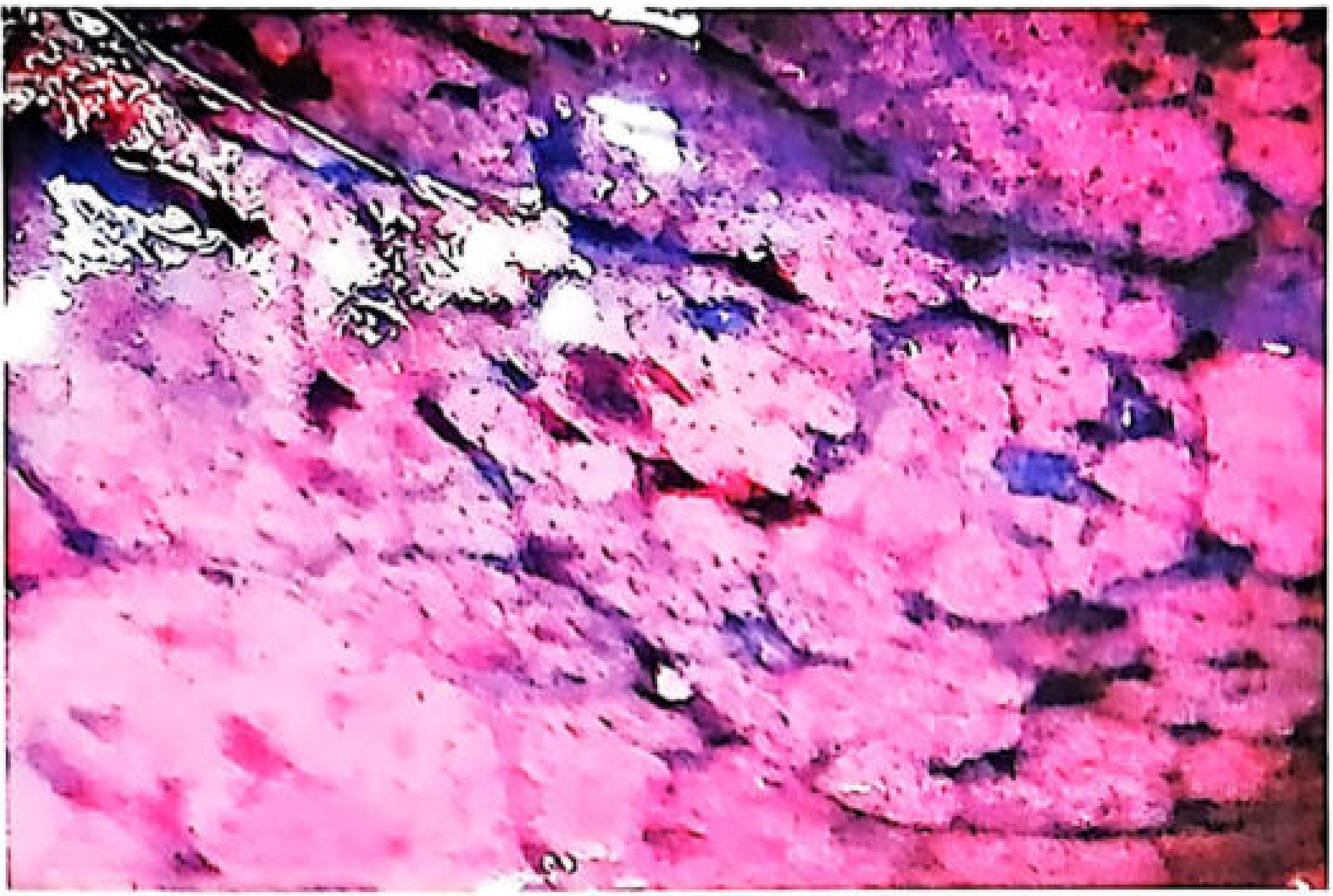
True Tardieu spots on the visceral pleural surface - The Essentials of Forensic Medicine and Toxicology, 36th Ed. (Reddy)
Tardieu Spots on Skin (Dependent Areas / Hanging)
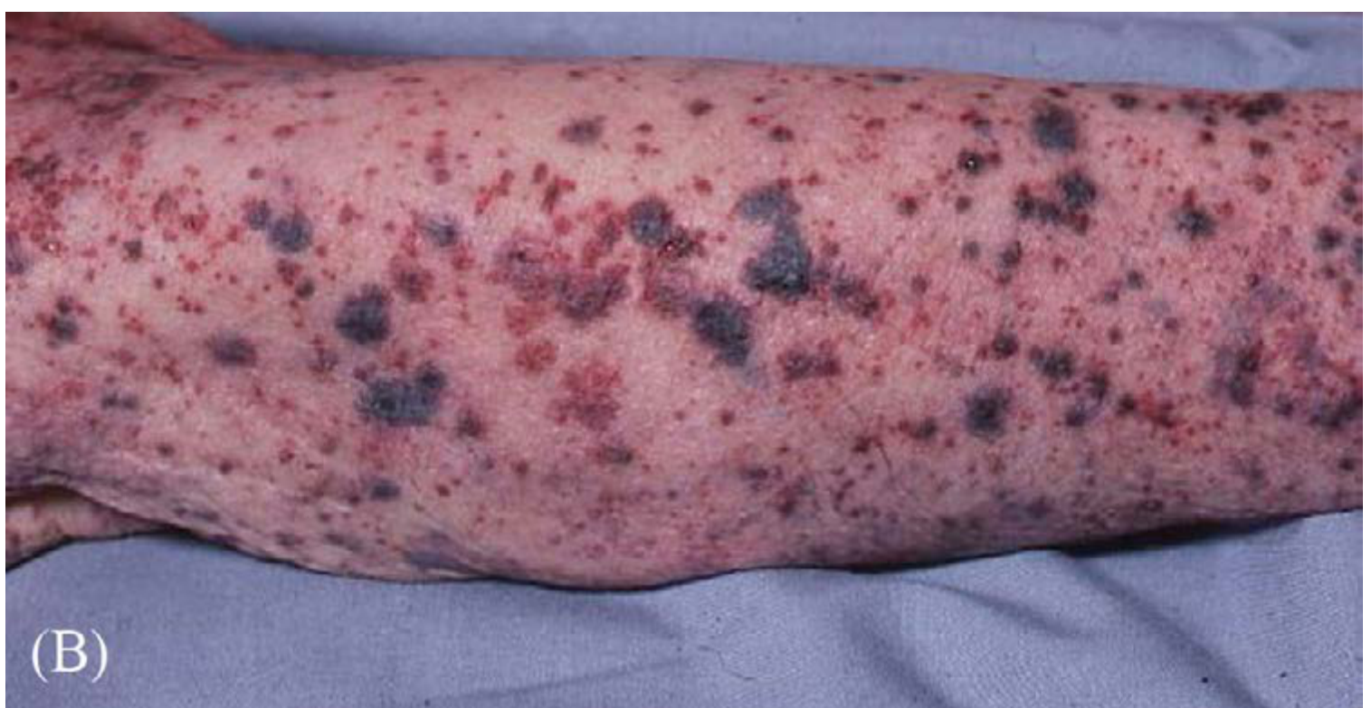
Tardieu spots on arm hanging over a bed - DiMaio's Forensic Pathology, 3rd Ed.
True vs. False (Postmortem) Tardieu Spots
This is a critical distinction in forensic practice:
| Feature | True (Antemortem) Tardieu Spots | False (Postmortem) Petechiae |
|---|---|---|
| Origin | Antemortem - due to asphyxial hypoxia + pressure | Postmortem - gravitational settling of blood rupturing capillaries |
| Location | Serous surfaces (visceral pleura, epicardium), conjunctiva, face | Skin of dependent areas - hands, legs; NOT on serous surfaces of heart/lungs |
| Appearance | Small, round, circumscribed, dark-red | Larger, less circumscribed |
| Mechanism | Hypoxia + raised intracapillary pressure | Gravitational blood pooling in dependent areas with capillary over-distension |
| Timing | Develop within ~30 seconds of obstruction | Usually take 18-24 hours; in hanging limbs may appear as early as 2-4 h |
| Significance | Strong indicator of mechanical asphyxia | NOT indicative of asphyxia |
Gordon and Mansfield (and later Shapiro, 1955) questioned the antemortem origin of many of these spots. Zaini and Knight (1981) confirmed that only one-third of subpleural hemorrhages are true petechiae and two-thirds are false - due to intrapleural venous channels, air blisters, thickened pleural plaques, and dust pigments. They are difficult to distinguish by naked eye. (Dikshit)
Conditions Where Tardieu Spots are Seen
- All forms of mechanical asphyxia - hanging, strangulation, smothering, drowning, traumatic asphyxia
- Hanging - Rosen's Emergency Medicine specifically notes them as "helpful tipoffs to a hanging mechanism"
- Terminal stages of asphyxia in any cause of death
- Coronary deaths (differential)
- Diseases with hemorrhagic tendency (differential) - in disease, spots are smaller, more widely distributed, and more numerous
Important Notes
- Absence of Tardieu spots does NOT exclude death from asphyxia - they are rarely prominent in drowning
- Numerous petechiae can be produced as a common autopsy artifact during reflection of scalp flaps in any type of death - of no significance
- Petechiae seen in the reflected scalp are due to tearing of small vessels during autopsy, NOT asphyxia
- Skin behind the ears is a good site to look for petechiae after blood has drained from the tissues
- When asphyxial signs including Tardieu spots are found, a close look in good light is necessary
The Essentials of Forensic Medicine and Toxicology, 36th Ed. (Reddy), p. 144 | Parikh's Textbook of Medical Jurisprudence, pp. 199-200 | P.C. Dikshit Textbook of Forensic Medicine and Toxicology, p. 290 | DiMaio's Forensic Pathology, 3rd Ed., p. 33
How do you differentiate it from measles rash
Good question - this is actually a clinical/forensic trap. The previous Reddy text itself used the phrase "numerous like measles rash" to describe Tardieu spots on the face and eyelids. Let me check the textbooks for any explicit differentiation.
Searching sources...
Finding Sources
Finding Sources
Finding Sources
The textbooks don't have an explicit head-to-head comparison of Tardieu spots vs. measles rash - this is because the forensic texts only use "measles rash" as a simile to describe how numerous and widespread Tardieu spots can appear on the face and eyelids in severe asphyxia. They are not saying Tardieu spots look identical to measles - it's a comparison of density/distribution. Let me answer directly from what the textbooks say about each:
Clarifying the Comparison
The phrase in Reddy - "numerous like measles rash involving the skin of the face and eyelids" - is a descriptive simile, not a diagnostic confusion. It means: in severe asphyxia, Tardieu spots on the face can be so densely packed that they superficially resemble the widespread spotted rash of measles. It is NOT saying the two are clinically mistaken for each other in living patients.
That said, here is how you distinguish them:
Tardieu Spots vs. Measles Rash - Differentiation
| Feature | Tardieu Spots | Measles Rash (Rubeola) |
|---|---|---|
| Nature | Petechial hemorrhages - blood outside vessels | Maculopapular exanthem - inflammatory skin lesion |
| Color | Dark red / purple (non-blanching on pressure) | Erythematous red (blanches on pressure initially) |
| Feel | Flat, do NOT blanch on pressure (glass test negative for blanching) | Maculopapular - slightly raised, blanchable initially |
| Distribution | Face, eyelids, conjunctivae; also serous surfaces internally | Starts on forehead/hairline/behind ears → descends to trunk → extremities (cephalocaudal spread) |
| Conjunctival involvement | Petechiae ON the conjunctiva | Conjunctivitis (redness/discharge) - NOT petechiae |
| Internal findings | Present on visceral pleura, epicardium at autopsy | Absent internally (no serous surface petechiae) |
| Associated signs | Context of asphyxial death - ligature mark, cyanosis, congestion | Koplik's spots on buccal mucosa (pathognomonic), prodrome of 3 Cs (cough, coryza, conjunctivitis), fever |
| History/context | Found at autopsy in suspected asphyxial death | Living patient with febrile illness, upper respiratory prodrome |
| Coalescence | Individual spots that may aggregate into purpura but remain distinct | Lesions coalesce into confluent semiconfluent patches |
| Scalp/ears | Petechiae behind ears possible (from pressure/venous obstruction) | Rash prominent behind ears and hairline at onset |
| Timing | Develop within seconds to minutes of obstruction | Rash appears day 3-5 of illness, after prodrome |
Key Point for Forensic Context
In forensic autopsy, the differentiation is straightforward because:
- You are examining a dead body - measles is a disease of the living
- Tardieu spots are petechiae (non-blanching, subepithelial hemorrhages) - confirmed by pressing a glass slide against them: they do not blanch
- Internal serous surfaces (subpleural, subpericardial, subconjunctival) show the same petechiae - measles rash has no internal equivalent
- The histology shows extravasated RBCs in the skin/serous surface (Tardieu), vs. inflammatory infiltrate + epidermal changes (measles)
The measles analogy in Reddy is purely about visual density of spots on the face - it does not imply diagnostic confusion between the two.
The Essentials of Forensic Medicine and Toxicology, 36th Ed. (Reddy) | Parikh's Textbook of Medical Jurisprudence | Sherris & Ryan's Medical Microbiology, 8th Ed.
What are the other signs of mechanical asphyxia besides Tardieu spots?
Searching sources...
Finding Sources
Finding Sources
Reading File
Reading File
Reading File
Let me get the rest of the Dikshit classical signs section (fluidity of blood, oedema of lungs, right heart dilatation details):
Reading File
Reading File
I have all the content needed. Here is the complete answer:
Signs of Mechanical Asphyxia (Other Than Tardieu Spots)
The classical signs of asphyxia are traditionally listed as a group - Tardieu spots are just one of them. All these signs are non-specific and can occur in deaths from other causes. They carry diagnostic weight only when found together in the right context.
Cardinal Signs (The Classical 4/5)
1. Cyanosis
- Bluish-purple discoloration of skin, lips, tongue, and mucous membranes
- Due to diminished oxygen tension in blood and a rise in reduced haemoglobin
- Becomes apparent only when at least 5 g% of reduced haemoglobin is present
- Seen prominently in the face, lips, fingernails, and organs with rich venous/capillary beds (lungs, meninges, liver, spleen, kidneys)
- In neck compression: cyanosis almost invariably follows congestion of the face because venous blood accumulates and contains much reduced haemoglobin
- Limitations:
- Terminal cyanosis is common in many forms of death; not specific to asphyxia
- After 24 hours postmortem, cyanosis may be entirely due to postmortem change
- Absence of cyanosis does not exclude asphyxia
2. Congestion of Internal Organs
- Due to obstructed venous return and capillo-venous distension
- Hypoxia causes capillary dilatation → stasis → pooling of dark blood
- The vicious cycle: Asphyxia → capillary dilatation → stasis → reduced venous return to heart → reduced pulmonary blood flow → further asphyxia
- Systemic and pulmonary congestion + dilatation of right side of heart are signs of asphyxial death
- Congestion immediately above a ligature mark on the neck is of great diagnostic significance
- Face, lips, and tongue become swollen and reddened when the neck is compressed
- Limitations: Right ventricular dilatation and pulmonary congestion are common to many forms of death; not specific. Blood redistribution by gravity and rigor mortis also confounds interpretation.
3. Fluidity of Blood (Liquid, Unclotted Blood)
- Blood remains fluid and dark at autopsy; clots do not form normally
- Historically thought to be pathognomonic of asphyxia
- Now known to be due to fibrinolytic enzymes (fibrinolysin) present in the cadaver - amount depends on rapidity of death, not on the nature of death
- High agonal catecholamine levels in rapid deaths may drive fibrinolysis
- Left side of heart is empty; right side contains dark fluid blood (Reddy)
- Limitation: NOT characteristic of asphyxia specifically - can occur in any rapid death
4. Petechial Haemorrhages (Tardieu Spots)
(Already covered in your previous question)
Additional Signs Described in Detailed Classifications
5. Pulmonary Oedema
- Some degree of pulmonary oedema is common but not invariable in asphyxial deaths
- Mechanism: either due to heart failure or increased capillary permeability (debated)
- In anoxic deaths, oedema of more than minor degree indicates death was not very rapid
- Lungs should be weighed at autopsy to quantify oedema
- Limitation: Common in many modes of death; little specific value
6. Pulmonary / Submucosal Haemorrhages
- Large submucosal haemorrhages in the pharynx, especially over the dorsum of the cricoid cartilage (where the cricoid is pressed against the anterior surface of the spine)
- Due to: (a) direct trauma, and (b) rupture of the large thin-walled submucous venous plexus at this site from severe venous congestion
- Also found in a variety of deaths unconnected with trauma
7. Increased Capillary Permeability
- Results from anoxia + stasis; pores between endothelial cells enlarge
- Blood (plasma and cells) transudes into tissues and tissue spaces, leading to:
- Gelatinous moistening of organs
- Development of excess fluid in serous sacs (pleura, pericardium)
- Oedema of mediastinal tissues and lungs (can develop rapidly)
- Petechial haemorrhages (Tardieu spots) - from further capillary rupture
Summary Table
| Sign | Mechanism | Specificity |
|---|---|---|
| Cyanosis | ↑ reduced Hb (≥5 g%) | Non-specific; common terminal sign |
| Visceral congestion | Obstructed venous return + capillo-venous dilatation | Non-specific |
| Fluidity of blood | Fibrinolysis; rapid death | Non-specific; not exclusive to asphyxia |
| Tardieu spots | ↑ venous pressure + hypoxic capillary fragility | Relatively more specific when on serous surfaces |
| Pulmonary oedema | Heart failure / ↑ capillary permeability | Non-specific |
| Pulmonary/submucosal haemorrhage | Venous congestion; trauma to cricoid | Somewhat suggestive |
| ↑ Capillary permeability | Hypoxia + stasis | Underlying mechanism of several above signs |
Important Caveat (DiMaio)
"The classical signs of asphyxia are visceral congestion, petechiae, cyanosis and fluidity of blood. These are nonspecific, however, and can occur in deaths from other causes."
No single sign alone confirms mechanical asphyxia. Diagnosis requires integration of all autopsy findings with scene investigation and circumstances of death.
The Essentials of Forensic Medicine and Toxicology, 36th Ed. (Reddy), p. 334 | P.C. Dikshit Textbook of Forensic Medicine and Toxicology, pp. 289-290 | Parikh's Textbook of Medical Jurisprudence, pp. 199-200 | DiMaio's Forensic Pathology, 3rd Ed., p. 248
Asphyxia long question for fmt mbbs exam from ksn reddy 36 th edition
Searching sources...
Finding Sources
Two major asphyxia sections found. Let me read both in parallel - the main ASPHYXIA chapter (block2) and the comprehensive block4 version:
Reading File
Reading File
Reading File
Reading File
Reading File
Reading File
Reading File
I now have the complete Reddy asphyxia chapter content. Let me also get the internal finding details I need from the block2 section between lines 3650-3720:
Reading File
Here is the complete long-question answer on Asphyxia, structured exactly as expected in an MBBS FMT exam, sourced directly from KSN Reddy 36th Edition:
ASPHYXIA
KSN Reddy - The Essentials of Forensic Medicine and Toxicology, 36th Edition
Definition
Asphyxia is a condition caused by interference with respiration, or due to lack of oxygen in respired air, due to which the organs and tissues are deprived of oxygen (together with failure to eliminate CO₂), causing unconsciousness or death.
The term asphyxia indicates a mode of dying, rather than a cause of death.
Physiological Basis (Important Facts)
- Normal arterial oxygen saturation: 90-100 mmHg in young/middle-aged; 60-85 mmHg in persons above 60 years. In severe to fatal asphyxia, it falls to 20-40 mmHg
- Brain weight is only 1.4% of body weight but uses 20% of total oxygen. Nervous tissue is the first to be affected
- In total ischemia of brain:
- Cerebral cortex: cessation of function begins after 8-15 seconds
- Brainstem ganglia: after 25-30 seconds
- Irreparable damage - cortex: ~3 minutes; basal ganglia: 6-7 minutes; vagal center: 9-10 minutes
- Thumb rule: Breathing stops within 20 seconds of cardiac arrest; heart stops within 20 minutes of stopping breathing
- If the heart continues for several minutes after breathing stops, lung weight may increase to 450-500 g
Classification / Types of Asphyxia
(1) Mechanical Asphyxia
Air passages are blocked mechanically:
- (a) Closure of external respiratory orifices - smothering (hand/cloth/mud over nose & mouth)
- (b) Closure of air passages by external pressure on neck - hanging, strangulation, throttling
- (c) Closure by foreign bodies in larynx/pharynx - choking (café coronary)
- (d) Air passages filled with fluid - drowning
- (e) External compression of chest/abdomen interfering with respiratory movements - traumatic asphyxia
(2) Pathological
- Entry of oxygen prevented by disease: bronchitis, acute oedema of glottis, laryngeal spasm, tumours, abscess
- Paralysis of respiratory muscles: acute poliomyelitis
(3) Toxic
- (a) Reduced oxygen-carrying capacity of Hb - CO poisoning
- (b) Enzymatic processes blocked - cyanide poisoning (cytochrome oxidase inhibition)
- (c) Respiratory center paralyzed - opium, barbiturates, strychnine
- (d) Respiratory muscles paralyzed - gelsemium
(4) Environmental
- Insufficiency of oxygen in inspired air (enclosed places, disused refrigerator, trunk)
- Irrespirable gases in atmosphere (sewer gas, CO, CO₂)
- Exposure to high altitude
(5) Traumatic
- Pulmonary thromboembolism from femoral vein thrombosis
- Pulmonary fat embolism from fracture of long bones
- Pulmonary air embolism from incised wound of internal jugular vein
- Bilateral pneumothorax from chest/lung injuries
(6) Postural Asphyxia
Unconscious/stuporous person (from alcohol, drugs, or disease) lying with upper half of body lower than the remainder
(7) Iatrogenic
Mainly associated with anaesthesia
Pathology of Mechanical Asphyxia
Compression of the neck leads to:
- Obstruction of venous return from the brain → venous congestion
- Obstruction of lymphatic drainage
- Partial or complete obstruction of carotid arteries → cerebral ischaemia
- Pressure on carotid sinus → reflex cardiac arrest
Vicious cycle of asphyxia (Fig. 6.1):
Asphyxia → capillary dilatation → stasis → reduced venous return to heart → reduced pulmonary blood flow → deficient oxygenation → further asphyxia
Gordon's Classification of Anoxia:
- Anoxic anoxia - no oxygen in air breathed (e.g., high altitude)
- Stagnant anoxia - decreased blood flow, vascular blockage
- Anaemic anoxia - reduced O₂-carrying capacity (anaemia, CO poisoning, methaemoglobinaemia)
- Histotoxic anoxia - cells fail to use oxygen (cyanide - blocks cytochrome oxidase)
Symptoms / Stages of Mechanical Asphyxia
Stage 1 - Stage of Dyspnoea
- Excess CO₂ stimulates respiratory centre
- Respiratory rate and amplitude increase; BP and pulse rate increase
- Slight cyanosis
Stage 2 - Stage of Convulsions
- Breathing effort is mostly expiratory
- Face is deeply congested, BP raised, pulse fast
- Neck veins become swollen
- Convulsions occur, then victim becomes insensible and reflexes are abolished
Stage 3 - Stage of Exhaustion
- Respiratory centre is paralyzed
- Complete insensibility; reflexes lost; pupils widely dilated
- Breathing is gasping, mostly inspiratory with long intervals between gasps
- BP falls, muscles relax, respiration ceases → death
- Heart may continue to beat for some minutes after respiration has ceased
- Total duration: 3 to 5 minutes
Postmortem Appearances in Asphyxial Deaths
A. External Findings
Face and general appearance:
- Postmortem hypostasis is well developed
- Face may be pale (slow asphyxia) or distorted, congested, cyanosed, purple, swollen, and oedematous
- Ears and fingernails are bluish
- Eyes are prominent, conjunctivae congested, pupils dilated
- Tongue is protruded in most cases
- Frothy and bloody mucus escapes from mouth and nostrils
- Postmortem discharge of semen from meatus is common (not specific to asphyxia)
B. Internal Findings
The Classical 4 Cardinal Signs (All Non-specific):
1. Cyanosis
- Bluish-purple discoloration due to diminished oxygen tension + rise in reduced haemoglobin
- Becomes apparent only when ≥ 5 g/100 mL of reduced haemoglobin is present
- Seen in skin (especially hypostatic areas), lips, ears, tip of nose, fingernails, cheeks; internally in lungs, liver, spleen, kidneys, meninges
- Methhaemoglobin and sulphhaemoglobin also cause cyanosis
- Limitation: Non-specific; common terminal sign; after 24 hours may be entirely postmortem
2. Petechial Haemorrhages - Tardieu Spots
- Most marked where capillary congestion is most prominent - above the level of obstruction in strangulation
- Size: 0.1 to 2 mm (larger = ecchymoses)
- Seen in: skin, sclerae, conjunctivae, outer and inner surfaces of eyelids, oral mucosa
- Internally: visceral pleura (interlobar fissures, around hilum), pericardium, brain (white matter), subarachnoid space, thymus (especially in infants/children)
- When on the visceral pleura = "true Tardieu spots"
- Limitation: Non-specific; can appear postmortem; seen in heart disease, meningococcal septicaemia, blood dyscrasias, coronary thrombosis, severe increase in intrathoracic pressure
3. Congestion of Internal Organs
- Due to obstructed venous return + capillo-venous distension
- Hypoxia → capillary dilatation → stasis → pooling of dark blood
- Lungs, liver, spleen, kidneys, meninges - all congested
- Systemic and pulmonary congestion + right ventricular dilatation are signs of asphyxial death
- Limitation: Non-specific; common to many forms of death
4. Fluidity of Blood
- Blood remains fluid, dark, and unclotted at autopsy
- Due to release of fibrinolysin (fibrinolytic enzymes) from endothelium - a defence mechanism to keep blood flowing during stagnation; depends on rapidity of death, not nature of death
- Left chambers of heart are empty; right side contains dark fluid blood
- Obsolete cardiac findings note (Reddy): Dilatation of the right heart chambers and fluidity of blood are now considered obsolete signs that should be disregarded - seen in any congestive death
Lungs at autopsy:
- Right lung weight ~300 g = compatible with sudden rhythm disturbance
- Heavier lungs (450-500 g or more) = indicates cessation of respiration with heart continuing to beat for several minutes
Histological Signs of Asphyxia in Tissues
- Partial disruption of alveolar septa with distinctive haemorrhage within the alveoli and intra-alveolar oedema fluid
- Brick-red discoloration of nerve cells in the cerebral cortex (stained neurological sections). Pallor and vacuolar degeneration of Purkinje cells in the cerebellum
- Vacuolar degeneration of liver cells in prolonged suffocation
Asphyxial Stigmata (Important Concept)
- Asphyxia is NOT a pathological entity and cannot be clearly recognized from morbid anatomical findings alone
- The triad of:
- Cyanosis
- Facial, palpebral, bulbar, subpleural and subepicardial petechiae
- Visceral congestion
- ...are all due to raised venous pressure - merely consistent with, but not diagnostic of asphyxia from anoxic anoxia
- Reliable local indications of fatal obstructing trauma must be demonstrated to establish mechanical asphyxia
- In putrefaction, mechanical asphyxia is indicated by petechiae under eyelids, conjunctivae, sclerae, and facial skin
Variations in Intensity of Asphyxial Signs
- If heart failure occurs before respiratory failure during asphyxia, asphyxial signs may be less marked
- In many cases of foetal hypoxia, asphyxial signs are absent (e.g., sudden blockage of trachea by food, plastic bag over head, entering a space devoid of oxygen)
- Vomiting occurs due to medullary suboxia → air passages may be filled with inhaled vomit at the end of asphyxia. About 20-25% of all individuals aspirate food agonally regardless of cause of death
Delayed Deaths Following Asphyxia
- Higher cortical centres suffer first from hypoxic injury → then basal ganglia → then vital centres
- Delayed deaths follow periods of unconsciousness from anoxic cerebral damage and subsequent hypostatic pneumonia
- Other lethal sequelae: massive subcutaneous and mediastinal emphysema from tracheal/laryngeal lacerations; oedematous swelling of pharyngeal tissues/aryepiglottic folds occluding airway
- When death occurs hours, days, or weeks after the asphyxial episode, the proximate cause of death is the traumatic incident
Medicolegal Importance
- Asphyxia may be suicidal, homicidal, or accidental
- Most common form - hanging (almost always suicidal)
- Most common homicidal form - strangulation
- Cause of death must be expressed within a broad framework as "consistent with asphyxia" when findings are inconclusive
- All four classical signs (cyanosis, Tardieu spots, congestion, fluidity of blood) are non-specific - diagnosis requires integration with scene findings and circumstances
The Essentials of Forensic Medicine and Toxicology, 36th Edition (KSN Reddy), pp. 143-146, 334
Hydrostatic test
Searching sources...
Finding Sources
Finding Sources
Finding Sources
Reading File
Reading File
Finding Sources
Reading File
Finding Sources
Now let me get the Reddy (KSN) specific content on the hydrostatic test:
Finding Sources
Finding Sources
Finding Sources
Finding Sources
Reading File
Finding Sources
Reading File
I now have comprehensive content from all three major forensic medicine textbooks. Here is the complete answer:
Hydrostatic Test (Lung Float Test)
Definition and Eponym
The Hydrostatic Test (also called the Lung Float Test or Raygat's Test) is a medicolegal test performed at autopsy on a newborn to determine whether the infant was born alive (live-born) or was stillborn, based on changes in the specific gravity of the lungs following establishment of respiration.
- First described by: Scheyer in 1683
- Also known as: Raygat's test
Note: Do not confuse with Fodere's test / Static test (based on weight of lung relative to body weight) or Plouquet's test - these are different, related tests.
Basis / Principle
The test depends on the fundamental fact that:
- Before respiration: The lungs are unexpanded, collapsed, and liver-like
- Specific gravity = 1040 to 1050 (heavier than water) → SINK
- After respiration: Alveoli are filled with air, lungs are light and spongy
- Specific gravity falls to ~940 (lighter than water) → FLOAT
"Even with compression, if the alveoli are aerated at all, they will still contain some residual air after squeezing." (Dikshit)
The lung volume increases with breathing. A lung containing air is lighter than water and therefore floats. The test determines buoyancy of the lungs.
Procedure (Step-by-Step)
Step 1 - Remove the "Pluck":
- Lungs are removed at autopsy with the bronchi, trachea, and larynx intact as one unit (the "pluck")
Step 2 - Test the whole pluck:
- The entire pluck is placed in a container of water
- If it floats → positive (preliminary)
Step 3 - Test individual lobes:
- Each lung is then separated and placed in water
- Floating → positive for that lobe
Step 4 - Cut into fragments:
- Each lung is cut into 15-20 small fragments
- Each fragment placed in water and tested
- This excludes irregular/partial aeration
Step 5 - Squeeze test:
- Floating fragments are squeezed between thumb and index finger under the water surface
- Air bubbles escape; if still floats → confirms residual air
- Fragments are then wrapped in cloth and squeezed with a weight
- If pieces STILL float after all this compression → positive test → residual air present → respiration has been established
Control:
- Before performing the test, a piece of liver is dropped into water as a control
- If the liver also floats (due to putrefaction), the test result is unreliable and of no use
Interpretation of Results
| Result | Interpretation |
|---|---|
| All portions of the pluck and all fragments float (after squeezing), body not putrefied | Complete respiration has taken place - child was live-born |
| Only some fragments float | Partial respiration has taken place |
| All pieces of lungs sink | Lungs unexpanded - infant has not breathed - possibly stillborn |
Differences in Lung Before and After Respiration
| Feature | Before Respiration (Stillborn) | After Respiration (Live-born) |
|---|---|---|
| Chest shape | Flat; circumference 1-2 cm less than abdomen at umbilical level | Expands; arch-shaped / drum-shaped |
| Diaphragm position | At level of 4th rib | At level of 6th-7th rib |
| Lung filling | Fills 75% of pleural cavity volume | Fills 75% of pleural cavity volume |
| Margins | Sharp | Rounded |
| Consistency | Dense, firm, non-crepitant, liver-like | Soft, spongy, elastic, crepitant |
| Colour | Uniformly reddish-brown, bluish or deep violet | Mottled/marbled; mosaic appearance on surface (polygonal areas) |
| On pressing | Little froth-less blood comes out | Frothy blood comes out |
| Weight | 30-40 g | 60-70 g |
| Hydrostatic test | Sinks | Floats |
| Specific gravity | ~1040-1050 | ~940-950 |
Related Tests
Fodere's Test / Static Test (Weight Test)
- Based on increased blood flow to lungs after respiration is established
- Before respiration: Lung weight = 1/70th of body weight
- After respiration: Lung weight = 1/35th of body weight (weight doubles)
Plouquet's Test
- Variant of static test; also uses ratio of lung weight to body weight
Fallacies of the Hydrostatic Test
A. Expanded (Respired) Lungs May SINK (False Negative)
- Diseases such as:
- Acute pulmonary oedema
- Pneumonia
- Congenital syphilis
- Atelectasis (non-expansion) due to:
- Feeble respiration (air not reaching alveoli)
- Complete absorption of air from lungs by blood (if circulation continued after respiration stopped, or in asphyxia)
- More air expelled during expiration due to recoil of lung tissue
- Obstruction by alveolar duct membrane
- Drowning - when birth occurs in a toilet bowl or bucket of water, the infant may die of drowning; lungs are waterlogged and may sink
B. Unexpanded (Non-respired) Lungs May FLOAT (False Positive)
- Putrefactive gases - foetal lungs are resistant to putrefaction (due to less blood); soft greenish gas bubbles of different sizes form, which shift under pressure and sink - distinguishing feature is that putrefactive bubbles shift position on pressure and sink when squeezed
- Artificial inflation - mouth-to-mouth resuscitation or blowing air via tube inflates the lungs partially (stomach also contains air in these cases)
- Alcoholic fixation
When Hydrostatic Test is NOT Necessary
The test need not be performed if:
- Foetus is < 180 days (not viable)
- Foetus is a monster (fetal anomaly incompatible with life)
- Foetus is macerated or mummified
- Umbilical cord is separated or cicatrized (shows the child lived independently)
- Stomach contains milk (proof of survival and feeding)
- Whole of the thoracic contents float (already obvious)
Applications / Significance of the Test
- If body is not putrefied and all portions float after squeezing → complete respiration established
- If some fragments float → partial respiration occurred
- If all pieces sink → lungs unexpanded; infant has not breathed
In putrefied bodies: floating lungs cannot be confidently attributed to respiration; sinking portions do not exclude prior expansion (putrefaction may have destroyed lung tissue, expelling respired air along with putrefactive gases).
Medicolegal Value and Limitations (Parikh)
The hydrostatic test is now considered of little to no value in forensic work for the following reasons:
- Even the slightest degree of decomposition invalidates interpretation
- Resuscitation attempts (mouth-to-mouth) make evaluation impossible
- The lungs of a stillborn may float while the lungs of a live-born (even those who lived for days) may sink - this paradox has been documented
- Foetal trial breathing - some expansion of air sacs occurs in the foetus towards the end of pregnancy as amniotic fluid moves in and out of the bronchial tree; thus an undoubted stillbirth may sometimes reveal extensive alveolar expansion (Meixner found apparently respired alveoli in lung sections from a dead infant taken from the uterus of a dead mother)
- Shapiro states it is not possible to distinguish histologically the lungs of stillborn infants from those of live-born
"The best way to seek proof of respiration is to look at, feel, and listen to the lungs." (Parikh)
Summary
| Hydrostatic Test | |
|---|---|
| Also called | Raygat's test; Lung float test |
| Described by | Scheyer (1683) |
| Principle | Specific gravity change after aeration (1040-1050 → 940) |
| Positive = floats | Respiration established → live born |
| Negative = sinks | No respiration → stillborn (possibly) |
| Overall forensic value | Unreliable; considerable fallacies; not used alone |
The Essentials of Forensic Medicine and Toxicology, 36th Ed. (Reddy) | P.C. Dikshit Textbook of Forensic Medicine and Toxicology, p. 328 | Parikh's Textbook of Medical Jurisprudence, p. 470
Gettlers test
Searching sources...
Finding Sources
Finding Sources
Found in three textbooks. Let me read all three in parallel:
Reading File
Reading File
Reading File
Excellent - comprehensive content from all three textbooks. Here is the complete answer:
Gettler's Test (Gettler Chloride Test)
Definition
Gettler's test is a chemical/laboratory test used in the medicolegal diagnosis of drowning. It is based on the difference in chloride concentration between the blood from the right and left sides of the heart, which arises because water absorbed from the lungs dilutes or concentrates the blood reaching the left heart depending on the type of water (fresh vs. salt water) in which drowning occurred.
First published by: Gettler in 1921
Normal Chloride Level
- Normally, chloride content is almost equal in the right and left chambers of the heart
- Normal value: approximately 600 mg per 100 mL
- A difference of ≥ 25 mg/100 mL between the two sides is considered significant and indicates drowning
Physiological Basis
When a person drowns and aspirates water into the lungs, the water is absorbed across the alveolar membrane into the pulmonary circulation. The nature of what happens depends on the tonicity of the drowning medium relative to blood:
Fresh Water Drowning
- Freshwater is hypotonic relative to blood
- Water is absorbed from the alveoli into the pulmonary circulation (osmosis)
- Blood reaching the left heart is diluted → chloride content is lower on the left than the right
- Blood gets diluted by as much as 72% in 3 minutes (Reddy)
- Chloride in left heart blood: up to 50% lower than normal
Salt Water Drowning
- Salt water is hypertonic relative to blood
- Water is drawn from the pulmonary circulation into the alveolar spaces (reverse osmosis)
- Blood reaching the left heart is concentrated (haemoconcentration) → chloride content is higher on the left than the right
- Volume of fluid drawn out can be up to 42% (Reddy)
- Chloride in left heart blood: increases by 30 to 40%
Interpretation
| Condition | Left Heart Chloride | Right Heart Chloride | Result |
|---|---|---|---|
| Normal (no drowning) | ~600 mg/100 mL | ~600 mg/100 mL | Equal |
| Fresh water drowning | LOW (↓ up to 50%) | Normal/higher | Left < Right |
| Salt water drowning | HIGH (↑ 30-40%) | Normal/lower | Left > Right |
| Significant difference | ≥ 25 mg/100 mL difference | → Indicates drowning |
Historical Development
| Year | Researcher | Contribution |
|---|---|---|
| 1902 | Carrara | Established disproportionate dilution of left heart blood in freshwater vs. saltwater based on specific gravity and freezing point |
| 1903 | Placzela | Emphasized diagnostic value of specific gravity method |
| 1921 | Gettler | Published the chloride test - difference of 25 mg/100 mL between sides indicates drowning |
| 1944 | Mortiz | Suggested magnesium as more reliable than chloride, particularly for saltwater drowning |
| 1955 | Freimuth et al. | Using specific gravity of heart plasma: negative differences may occur in both drowning and non-drowning; positive values usually indicate death by means other than drowning |
Other Related Blood Tests in Drowning
Plasma Specific Gravity (Reddy)
- In drowning: specific gravity of plasma from left side < right side
- In non-drowning: the reverse (left > right)
Magnesium Test (Mortiz, 1944)
- In saltwater drowning: magnesium content of the blood on the left side > right side by more than 1.25 mg/1000 mL
- More reliable than chloride for saltwater drowning
Serum Strontium (Abdallah et al., 1985; Azparren et al., 1994)
- Raised serum strontium levels in drowning deaths
- Reciprocally related to the volume of water aspirated
- Can differentiate true drowning from postmortem immersion
- In sea water drowning: difference in strontium concentration between left and right heart blood is always > 75 μg/L
Limitations / Fallacies of Gettler's Test
The test is considered of doubtful/unreliable value (Reddy) and of no help in diagnosing drowning (DiMaio) for the following reasons:
- Postmortem diffusion - after death, chloride can diffuse between cardiac chambers, equalising levels and erasing the difference
- Putrefaction - decomposition alters blood chemistry, making interpretation impossible
- Negative results are better interpreted than positive results - because the deceased might have consumed the same water while alive (Reddy)
- Non-specific - similar chloride shifts can occur in other conditions
- Freimuth's finding - positive specific gravity difference between sides usually indicates death by causes other than drowning
- Considerable doubt about reliability of all chemical tests exists across the literature (Dikshit)
Medicolegal Significance
- The test, when positive (showing significant left-right chloride difference), supports a diagnosis of drowning
- The type of drowning (fresh vs. salt water) can be inferred from the direction of the difference
- Negative test does not exclude drowning - it may simply mean the test was unreliable in that case
- It must always be corroborated with:
- Clinical/scene findings
- Diatom test
- Autopsy findings (frothy fluid in airways, waterlogged lungs, washerwoman's hands, cutis anserina, etc.)
- Plasma specific gravity
- Strontium levels
Summary Table
| Feature | Gettler's Test |
|---|---|
| Based on | Chloride difference between right and left heart blood |
| Described by | Gettler, 1921 |
| Significant difference | ≥ 25 mg/100 mL |
| Fresh water | Left chloride lower than right |
| Salt water | Left chloride higher than right |
| Current status | Doubtful/unreliable - not used alone |
The Essentials of Forensic Medicine and Toxicology, 36th Ed. (Reddy) | P.C. Dikshit Textbook of Forensic Medicine and Toxicology, p. 313 | DiMaio's Forensic Pathology, 3rd Ed., p. 395
Disaster cycle from park
Your credits are exhausted. You can purchase a top-up or subscribe to a plan to continue using Orris. If you're already subscribed, your daily credits will reset tomorrow.
This is a shared conversation. Sign in to Orris to start your own chat.