
Anatomy of Nose with detailed explanation in simple and understandable way
Reading File
Now let me search the medical library for nose anatomy content, in parallel with relevant heading and paragraph searches.
Finding Sources
Finding Sources
Searching the Web
anatomy of the nose nasal cavity detailed explanation
I have multiple great sources. Let me read the key textbook sections in parallel - the Cummings Otolaryngology anatomy section, Bailey & Love, and Miller's Anesthesia for comprehensive coverage.
Reading File
Reading File
Reading File
Excellent! I have rich textbook content. Let me fetch the key anatomy diagrams and also read the KJ Lee block for nasal cavity detail:
Loading Image
Loading Image
Loading Image
Reading File
I now have excellent textbook content and high-quality diagrams. Let me also fetch the blood supply image and read more of the KJ Lee section on nasal cavity (blood supply, innervation):
Reading File
Finding Sources
I now have comprehensive material from multiple authoritative textbooks plus diagrams. Let me compile everything into a clear, detailed, easy-to-understand explanation.
Anatomy of the Nose - A Detailed, Simple Explanation
The nose is the first part of the respiratory tract. It does far more than just smell - it warms, moistens, and filters every breath you take. Here is a complete walkthrough of its anatomy from outside to inside.
1. External Nose (The Part You Can See)
The outer nose is a pyramid-shaped structure made of bone in the upper third and cartilage in the lower two-thirds.
Key Landmarks
| Landmark | What it Is |
|---|---|
| Nasion | The root of the nose where it meets the forehead (corresponds to the nasofrontal suture) |
| Radix | The nasal root region, centered at the nasion |
| Dorsum | The "bridge" of the nose - the top line running from nasion to tip |
| Rhinion | The bony-cartilaginous junction point along the nasal bridge |
| Tip (Apex) | The pointed front end of the nose |
| Alae | The rounded, wing-shaped sides of the nostrils |
| Columella | The vertical strip of tissue separating the two nostrils |
| Nares (Nostrils) | The two external openings through which air enters |
Skeleton of the External Nose
Nasal Skeleton Diagram (Bailey & Love's Surgery):
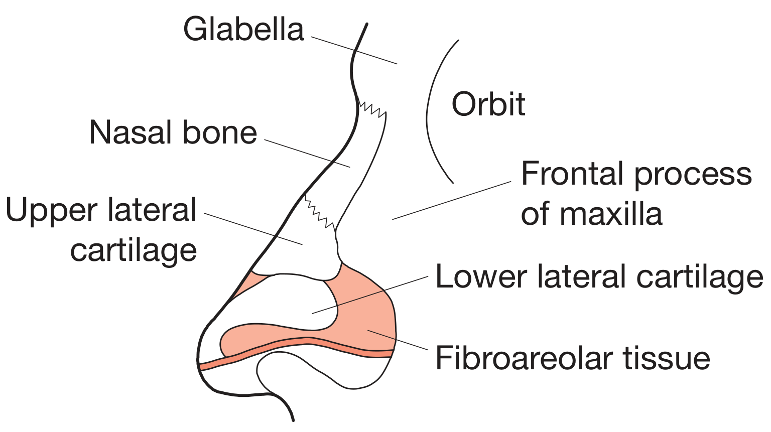
The skeleton has two layers:
a) Bony Framework (upper nose)
- Two nasal bones - these are the small rectangular bones you feel at the top of your nose when you press it. Together with the frontal processes of the maxilla (cheek bones), they form the firm upper nose.
b) Cartilaginous Framework (lower nose)
- Upper Lateral Cartilages (ULC) - flat, triangular cartilages that form the middle third of the nose. They attach to the nasal bones above (this junction is called the "keystone area" - very important surgically because destroying it can cause the nose to collapse).
- Lower Lateral Cartilages (LLC) - paired, C-shaped cartilages that give the nose tip its shape. Each has:
- A medial crus (inner limb, forms the columella)
- A lateral crus (outer limb, forms the nostril rim)
- Sesamoid cartilages - small, accessory cartilages lateral to the LLCs, providing additional support to the nostril walls.
Layers of the Nose Skin (Outside In)
A useful mnemonic: "Subcutaneous Fat DeeP"
- S - Superficial fatty layer (attached to dermis)
- F - Fibromuscular layer (the nasal SMAS - similar to the face's SMAS)
- D - Deep fatty layer (contains the neurovascular supply)
- P - Periosteum / perichondrium (directly over bone/cartilage)
The optimal surgical dissection plane is between the deep fatty layer and periosteum because it is relatively avascular and heals with minimal scarring.
Nasal Muscles
The muscles of the nose act on the nostrils and nose shape:
- Elevators (shorten nose, dilate nostrils): Procerus, levator labii superioris alaeque nasi
- Depressors (lengthen nose, dilate nostrils): Depressor septi, alar nasalis
- Compressors (lengthen nose, constrict nostrils): Transverse nasalis, compressor narium minor
2. The Nasal Septum (The Dividing Wall)
The septum divides the nose into right and left sides. Think of it as the central "partition wall" of the nose.
Nasal Septum Diagram:
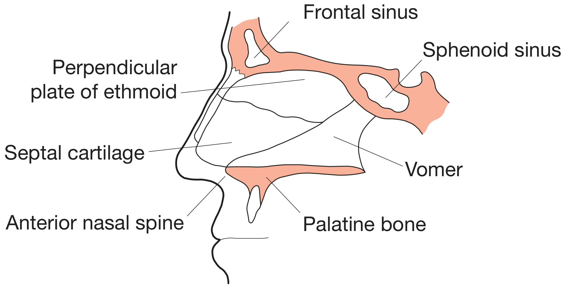
It is made of three parts:
| Part | Location | Material |
|---|---|---|
| Quadrangular (Septal) Cartilage | Anterior (front) | Hyaline cartilage |
| Perpendicular Plate of Ethmoid | Superior (upper-back) | Bone |
| Vomer | Inferior-posterior (lower-back) | Bone |
Minor bony contributions also come from the crest of the maxilla, palatine bone, sphenoid crest, and nasal spine of the frontal bone.
The septum is covered on both sides by a mucous membrane called the mucoperichondrium (over cartilage) or mucoperiosteum (over bone).
Membranous (Mobile) Septum
The very front of the septum has no cartilage - it is just a soft tissue membrane connecting the columella to the caudal (lower) edge of the septal cartilage. This is where surgeons make incisions during septoplasty.
Keystone Area
Where the nasal bones, upper lateral cartilages, and septal cartilage all converge. This is a load-bearing junction - disrupting it without proper reconstruction causes the nose bridge to collapse.
3. Nasal Cavity (The Internal Space)
The nasal cavity is the internal hollow space that extends from the nostrils (anterior nares) all the way back to the nasopharynx (posterior nares/choanae). It is about 6-7 cm long.
It is bounded:
- Above (roof): Cribriform plate of ethmoid, nasal bones
- Below (floor): Hard palate (maxilla and palatine bones)
- Medially: Nasal septum
- Laterally: Turbinates and paranasal sinus openings
Regions of the Nasal Cavity
- Vestibule - just inside the nostril, lined by skin and contains vibrissae (nasal hairs) that filter large particles
- Respiratory region - the main middle area, lined by pseudostratified ciliated columnar epithelium with goblet cells (produces mucus)
- Olfactory region - the uppermost area near the roof, contains special olfactory neurons for smell
4. Lateral Wall - The Turbinates (Conchae)
This is one of the most important parts of nasal anatomy.
Turbinates (Right Lateral Wall):
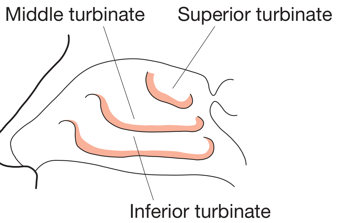
The lateral wall of the nasal cavity has three curved, shelf-like bony projections called turbinates (conchae):
| Turbinate | Origin | Function |
|---|---|---|
| Inferior turbinate | Separate bone (maxilloturbinal) | Largest; main air conditioner - warms and humidifies air |
| Middle turbinate | Part of ethmoid bone | Key surgical landmark; covers the ostiomeatal complex |
| Superior turbinate | Part of ethmoid bone | Smallest; near olfactory region |
A rare supreme turbinate exists in some people above the superior turbinate.
Meatuses (Channels Under Each Turbinate)
Below each turbinate is a passage called a meatus. These are important because various ducts drain into them:
| Meatus | Location | What Drains Into It |
|---|---|---|
| Inferior meatus | Below inferior turbinate | Nasolacrimal duct (tears from eyes!) |
| Middle meatus | Below middle turbinate | Frontal, maxillary, and anterior ethmoid sinuses (via the ostiomeatal complex) |
| Superior meatus | Below superior turbinate | Posterior ethmoid sinuses |
| Sphenoethmoidal recess | Above superior turbinate | Sphenoid sinus |
Think of the meatuses as "drains" - the sinuses need somewhere to empty their mucus, and these channels are it.
5. Paranasal Sinuses (Air-Filled Cavities Around the Nose)
There are four paired sinuses, all named after the bone they sit in:
| Sinus | Location | Drains Into |
|---|---|---|
| Frontal sinus | Behind forehead | Middle meatus |
| Maxillary sinus | In cheekbones (largest sinus) | Middle meatus |
| Ethmoid sinuses | Between eyes; divided into anterior and posterior groups | Anterior → middle meatus; Posterior → superior meatus |
| Sphenoid sinus | Behind nose, deep in skull | Sphenoethmoidal recess |
Functions of sinuses:
- Lighten the skull
- Warm and humidify air
- Add resonance to voice
- Produce mucus to keep the nasal cavity moist
6. Blood Supply
The nose has a rich blood supply from both the internal and external carotid arteries:
External Carotid Artery supplies most of the nose via:
- Maxillary artery → Sphenopalatine artery (main supply to the nasal cavity)
- Facial artery → Superior labial artery (anteroinferior septum)
- Greater palatine artery (anteroinferior septum via the incisive canal)
Internal Carotid Artery supplies via:
- Ophthalmic artery → Anterior ethmoidal artery (upper nasal cavity and septum)
- Ophthalmic artery → Posterior ethmoidal artery (upper posterior cavity)
Kiesselbach's Plexus (Little's Area)
All these arteries meet and form a rich anastomotic network on the anterior nasal septum called Kiesselbach's plexus (or Little's area). This is why most nosebleeds (epistaxis) happen right here - the anastomotic network is very superficial and prone to bleeding with minor trauma.
Clinical tip: About 90% of nosebleeds come from Kiesselbach's plexus on the anterior septum.
Venous drainage goes via the ophthalmic and facial veins and the pterygoid/pharyngeal plexuses. The connection to the ophthalmic vein (which drains to the cavernous sinus) is clinically important - nasal infections can potentially spread intracranially via this route.
7. Nerve Supply
The nose receives both sensory and special sensory (smell) innervation:
Special Sense - Smell (Olfaction)
- Olfactory nerve (CN I) - special sensory nerve fibres in the olfactory epithelium (roof of nasal cavity) pass through the cribriform plate of the ethmoid to reach the olfactory bulb in the brain.
General Sensation
- Ophthalmic branch of trigeminal nerve (V1) - innervates the upper nose via the anterior and posterior ethmoidal nerves
- Maxillary branch of trigeminal nerve (V2) - innervates the lower and posterior nasal cavity via the sphenopalatine (pterygopalatine ganglion) branches and greater/lesser palatine nerves
- Nasociliary nerve - anterior upper nasal cavity
Autonomic Supply
- Parasympathetic (from facial nerve via vidian nerve): causes nasal secretion and vasodilatation (why your nose runs when you cry or in cold weather)
- Sympathetic (from superior cervical ganglion via vidian nerve): causes vasoconstriction and reduces secretions (how decongestant sprays work)
8. Nasal Valves (Functional Bottlenecks)
The nasal valve is the narrowest part of the nasal airway and contributes the most to nasal resistance:
- Internal nasal valve - the angle between the upper lateral cartilage and the nasal septum (should be about 10-15 degrees). This is the main airflow bottleneck.
- External nasal valve - the entrance of the nostril, bounded by the columella, nasal floor, and alar rim.
A collapsed or narrow nasal valve is a common cause of nasal obstruction.
9. Functions of the Nose - Putting It All Together
| Function | How It's Done |
|---|---|
| Air filtration | Nasal hairs (vestibule) trap large particles; mucus traps smaller particles |
| Warming | Turbinates increase surface area; rich blood supply warms cold air |
| Humidification | Mucous membrane secretions add moisture to dry air |
| Smell (olfaction) | Olfactory epithelium in the roof detects odor molecules |
| Mucociliary clearance | Cilia beat in coordinated waves, moving mucus (with trapped particles) backward to the throat |
| Resonance | Nasal cavity + paranasal sinuses amplify and shape voice sound |
| Immune defense | IgA antibodies in nasal secretions; lymphoid tissue traps pathogens |
Quick Summary Diagram - Mental Map
External Nose
  ├── Bony part (upper 1/3): Two nasal bones
  └── Cartilaginous part (lower 2/3): ULC + LLC + sesamoids

Nasal Septum (central dividing wall)
  ├── Front: Quadrangular/septal cartilage
  ├── Upper-back: Perpendicular plate of ethmoid
  └── Lower-back: Vomer

Nasal Cavity (internal space)
  ├── Roof: Cribriform plate → olfactory nerve passes through
  ├── Floor: Hard palate
  ├── Medial wall: Septum
  └── Lateral wall:
        ├── Inferior turbinate → inferior meatus (nasolacrimal duct)
        ├── Middle turbinate → middle meatus (frontal, maxillary, ant. ethmoid sinuses)
        └── Superior turbinate → superior meatus (post. ethmoid)
                              → sphenoethmoidal recess (sphenoid sinus)

Blood Supply
  ├── Sphenopalatine artery (main)
  ├── Anterior & posterior ethmoidal arteries
  └── All meet at Kiesselbach's plexus (Little's area) → most nosebleeds here

Nerve Supply
  ├── CN I (olfactory) → smell
  ├── CN V1 (ophthalmic) → upper nose sensation
  └── CN V2 (maxillary) → lower nose sensation
Sources:
- Bailey and Love's Short Practice of Surgery, 28th Edition - Basic Anatomy of the Nose and Paranasal Sinuses
- Cummings Otolaryngology Head and Neck Surgery - Anatomy and Embryology of the Nose
- K.J. Lee's Essential Otolaryngology - Nasal Cavity
- Miller's Anesthesia, 10th Edition - Synopsis of Ear, Nose, and Throat Anatomy
This is a shared conversation. Sign in to Orris to start your own chat.