
MacEwen’s triangle 2. Foramen magnum By vishram singh
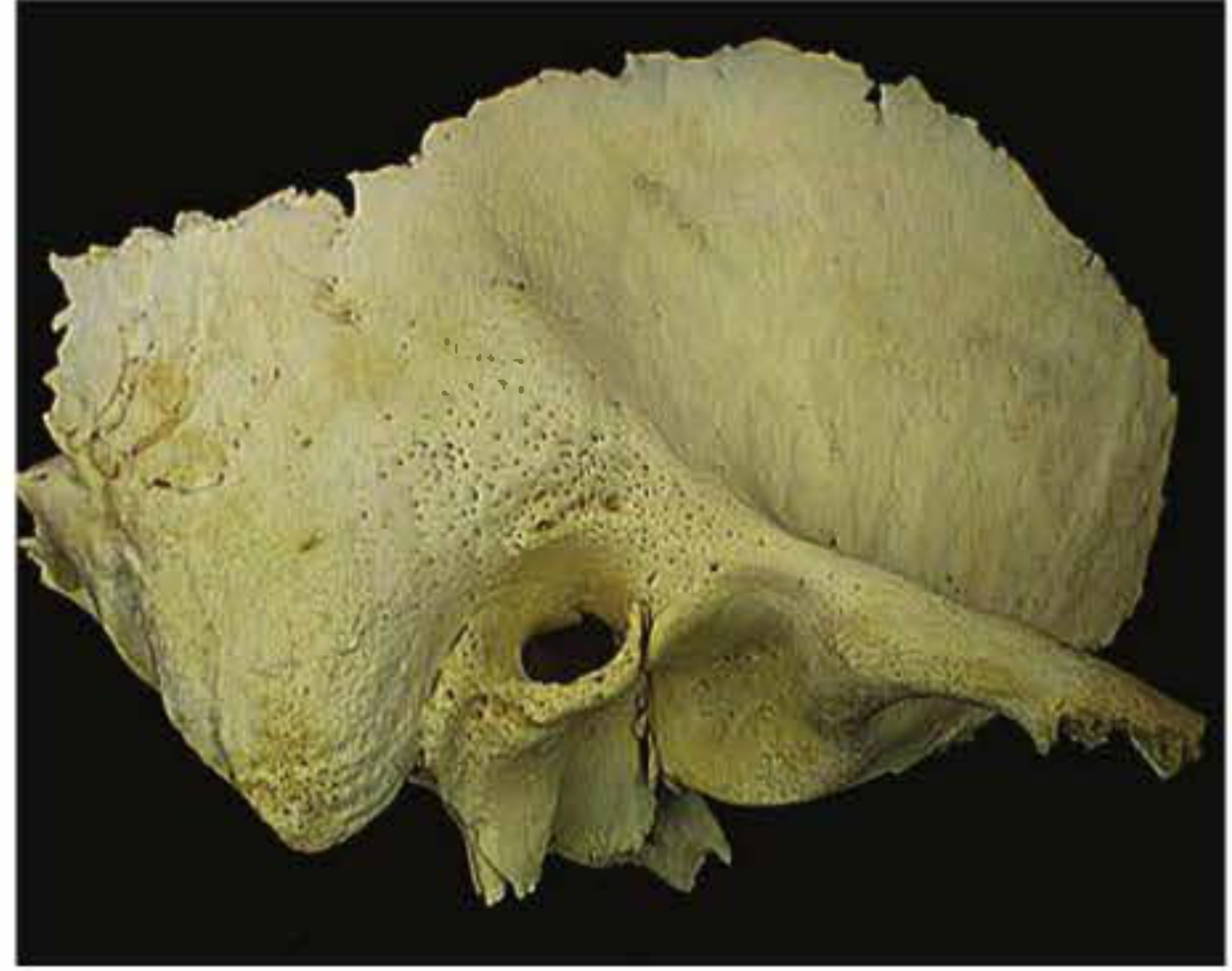
1. MacEwen's Triangle (Suprameatal Triangle / Fossa Mastoidea)
Definition
Boundaries
| Boundary | Structure |
|---|---|
| Superior | Temporal line (linea temporalis) - the ridge that also marks the floor of the middle cranial fossa |
| Posterior | A tangent drawn along the posterior wall of the external auditory canal |
| Anteroinferior | Posterosuperior rim of the external auditory canal (spine of Henle forms a nearby reference point) |
Key Features
- Overlies the mastoid antrum laterally - the mastoid antrum lies approximately 12-15 mm deep to this triangle in adults
- Cribrose (cribriform) area: the triangle is perforated by multiple small vessels, giving it a sieve-like (cribrose) appearance - hence the alternative name
- The spine of Henle (suprameatal spine) is a small bony prominence at the posterosuperior rim of the canal; it lies within or just anterior to the triangle and acts as a useful landmark
Surgical Importance (Vishram Singh emphasis)
- Surface marking for the mastoid antrum: The triangle serves as a guide for cortical mastoidectomy - the surgeon drills through this area to reach the antrum
- Mastoiditis and subperiosteal abscess: The abscess from mastoiditis points and discharges through this triangle
- Access for mastoid exploration: Used in the Schwartze mastoidectomy (cortical mastoidectomy) to gain access to the antrum and mastoid air cells
- Depth to antrum: In an adult, the antrum lies about 12-15 mm deep to the triangle; in a newborn, it is essentially at the surface (only 2-4 mm deep)
2. Foramen Magnum
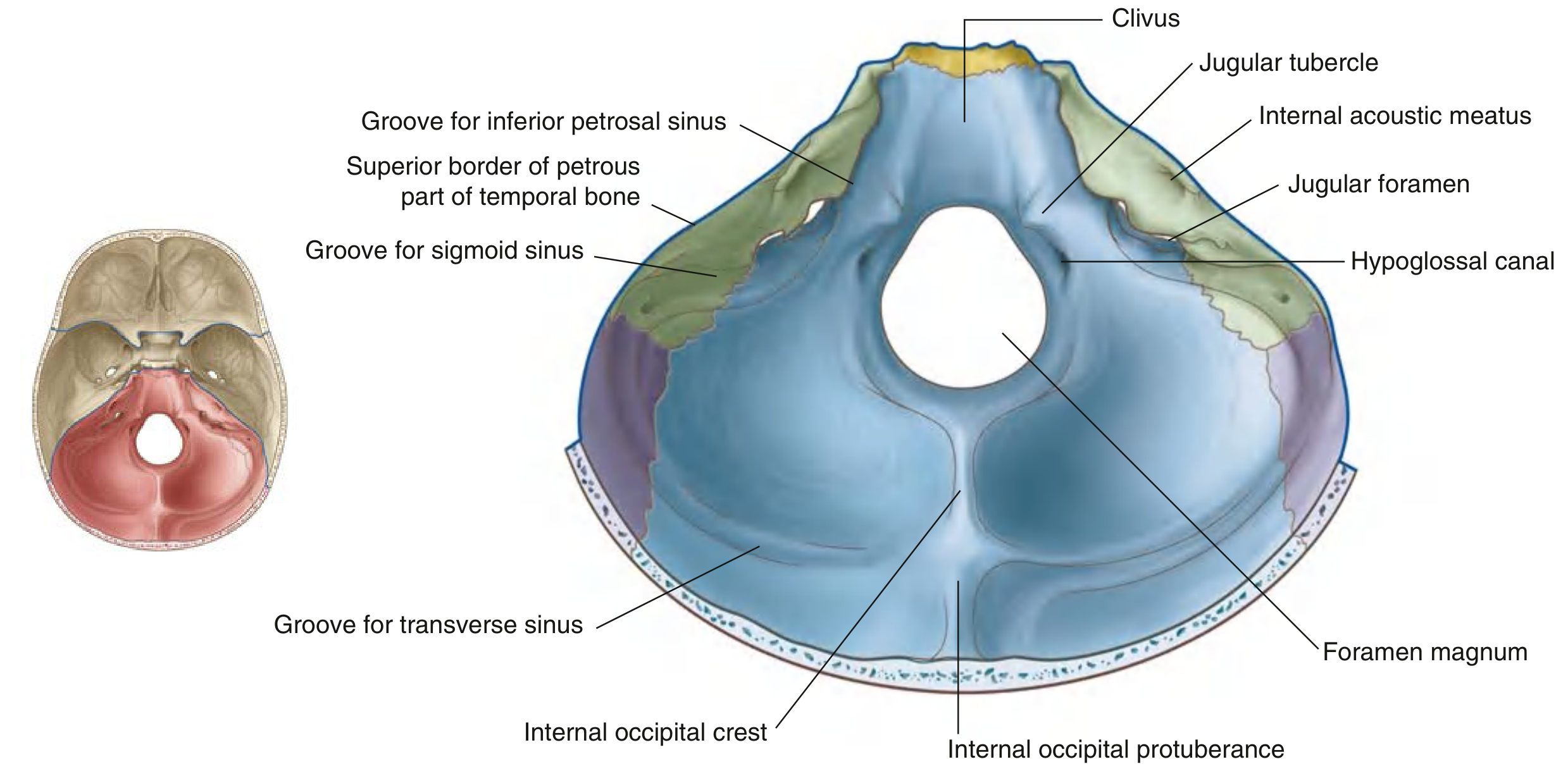
Definition
Boundaries
| Position | Structure |
|---|---|
| Anterior | Basilar part of the occipital bone (basiocciput) |
| Posterolateral (each side) | Lateral parts (condylar parts) of the occipital bone |
| Posterior | Squamous part of the occipital bone |
Shape and Size
- Oval/ovoid in shape (anteroposterior diameter > transverse diameter)
- Anteroposterior diameter: ~35 mm; Transverse diameter: ~30 mm
Structures Passing Through the Foramen Magnum
From Above Downward (Endocranial to Extracranial):
| Structure | Notes |
|---|---|
| Medulla oblongata / brainstem | Continuous with the spinal cord at the level of C1 |
| Meninges (all three layers) | Dura, arachnoid, pia mater |
| Vertebral arteries (paired) | Enter the skull, unite to form the basilar artery |
| Anterior and posterior spinal arteries | Arise from vertebral arteries just above/within the foramen |
| Spinal roots of accessory nerve [XI] | Ascend through the foramen to join the cranial root |
| Tectorial membrane | Continuation of posterior longitudinal ligament |
| Cruciate ligament / apical ligament of dens | Ligamentous structures |
Relations
- Anteriorly: Clivus slopes upward from the anterior rim of the foramen
- Laterally: Occipital condyles (articulate with atlas C1), hypoglossal canals, jugular foramina
- Posteriorly: Internal occipital crest (attachment for falx cerebelli)
Clinical Significance (Vishram Singh emphasis)
- Tonsillar herniation (coning): Raised intracranial pressure can push the cerebellar tonsils through the foramen magnum, compressing the brainstem - a life-threatening emergency
- Foramen magnum syndrome: Space-occupying lesions at or near the foramen (meningioma, neurofibroma, metastases) produce a characteristic pattern of lower cranial nerve palsies + upper cervical cord compression
- Arnold-Chiari malformation: Herniation of cerebellar tonsils and brainstem through the foramen
- McRae line: A diagnostic line drawn across the foramen magnum (anterior to posterior tip); the tip of the odontoid should be below this line - if above, it indicates basilar invagination
- Atlanto-occipital dislocation: Trauma can disrupt the osseoligamentous structures at this level
Quick Comparison Summary
| Feature | MacEwen's Triangle | Foramen Magnum |
|---|---|---|
| Location | Lateral temporal bone, mastoid region | Central posterior cranial fossa |
| Bone | Temporal (mastoid part) | Occipital bone |
| Key overlying structure | Mastoid antrum | Medulla/spinal cord junction |
| Main clinical use | Surgical landmark for mastoidectomy | Herniation, cord compression, basilar invagination |
| Named after | Sir William MacEwen (Scottish surgeon) | Descriptive Latin ("great foramen") |
Create a visual comparison poster: MacEwen's triangle vs foramen magnum anatomy
A detailed medical anatomy comparison poster titled "MacEwen's Triangle vs Foramen Magnum" designed in a clean academic style with two side-by-side panels. LEFT PANEL - MacEwen's Triangle: - Title: "MacEwen's Triangle (Suprameatal Triangle)" - A clear anatomical illustration of the lateral surface of the temporal bone showing: - The triangle outlined in bright yellow/gold with bold dashed lines - Superior boundary labeled: "Temporal Line (Linea Temporalis)" - Posterior boundary labeled: "Tangent to posterior EAC wall" - Anteroinferior boundary labeled: "Posterosuperior rim of EAC" - Interior of triangle labeled: "Fossa Mastoidea / Cribrose Area" - Spine of Henle marked as a small dot - Mastoid antrum shown as a shaded area deep to the triangle with arrow: "Mastoid antrum lies 12-15mm deep" - Small perforating vessel dots inside triangle - Key facts box below: Location: Lateral mastoid | Bone: Temporal | Overlies: Mastoid antrum | Use: Mastoidectomy landmark - Surgical note: "Cortical mastoidectomy entry point" RIGHT PANEL - Foramen Magnum: - Title: "Foramen Magnum (Largest Skull Foramen)" - A clear anatomical illustration of the skull base (inferior view) showing: - The foramen magnum as a large oval opening in the center, outlined in red - Four surrounding parts of occipital bone labeled with arrows: Basilar part (anterior), Lateral parts (sides), Squamous part (posterior) - Occipital condyles marked on each side - Hypoglossal canals labeled - Contents listed with arrows pointing into foramen: Medulla oblongata, Meninges, Vertebral arteries (paired), Spinal root of CN XI, Anterior & posterior spinal arteries - Key facts box: Location: Posterior cranial fossa | Bone: Occipital | Contains: Medulla + arteries | Size: 35 x 30mm - Clinical note: "McRae line - odontoid must be below" BOTTOM COMPARISON TABLE spanning full width: A neat comparison table with columns: Feature | MacEwen's Triangle | Foramen Magnum Rows: Location | Bone | Key Structure | Clinical Significance | Named After Overall design: White background, navy blue headers, color-coded panels (blue-tinted left, red-tinted right), clean medical illustration style with clear labels and arrows, suitable for medical students studying anatomy by Vishram Singh
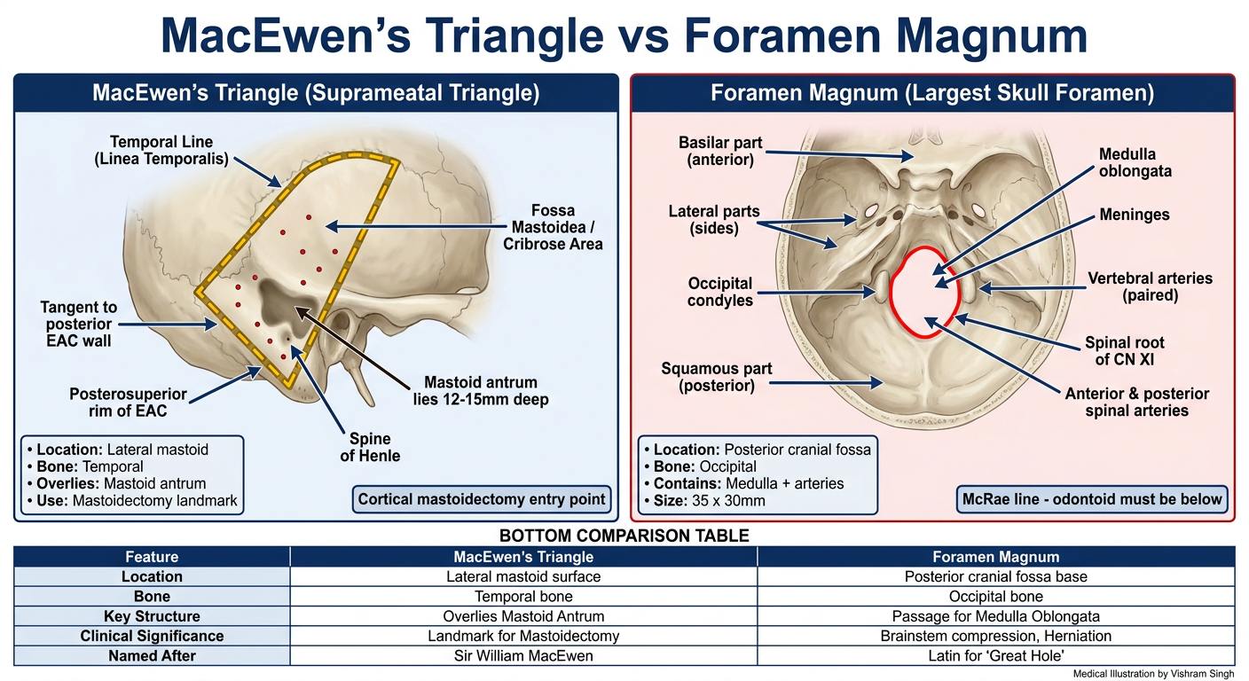
- Triangle boundaries marked on the lateral temporal bone
- Depth to mastoid antrum (12-15 mm)
- Cribrose/cribriform area noted
- Surgical use for cortical mastoidectomy
- Skull base (inferior view) with the oval foramen in the center
- Four parts of occipital bone labeled around it
- Contents: medulla, meninges, vertebral arteries, spinal root CN XI
- McRae line clinical note
Cervical rib syndrome by vishram singh
Cervical Rib Syndrome
Definition
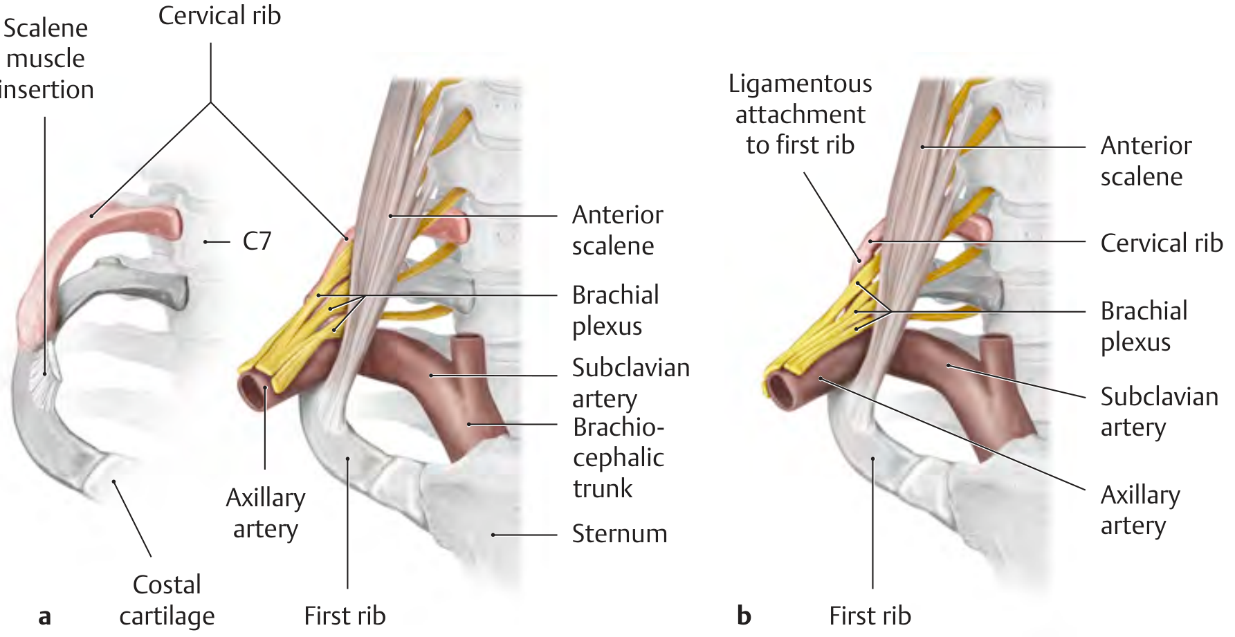
Anatomy of the Problem
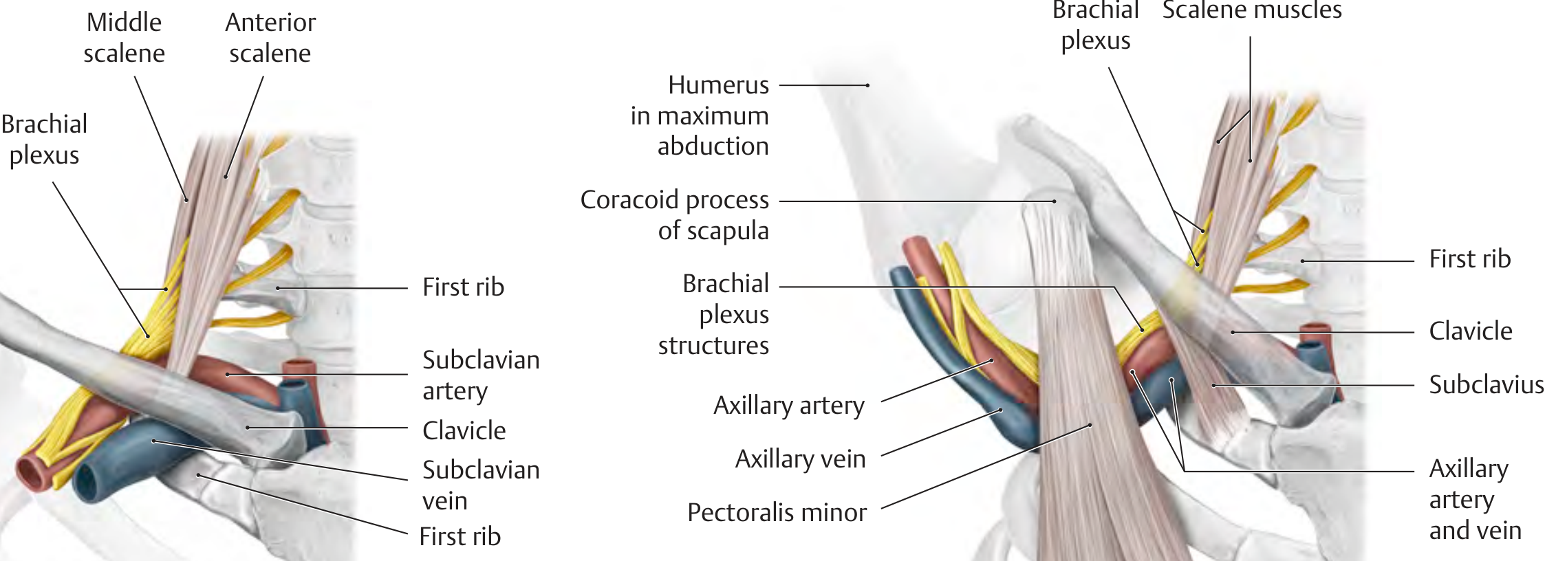
- Anteriorly: Scalenus anterior muscle
- Posteriorly: Scalenus medius muscle
- Inferiorly: First rib
Who Gets It? (Epidemiology - Vishram Singh emphasis)
- Young women are the usual victims
- Symptoms typically appear at puberty due to sagging (drooping) of the shoulder girdle as body habitus changes
- Sometimes appear later in life due to progressive weakness of shoulder girdle muscles
- Only 10% of those with a cervical rib become symptomatic - symptoms only appear when shoulder girdle muscles weaken enough to let the artery and nerve trunk compress on the rib
Three Disease Entities Produced
1. Neurogenic (Most Common - ~95%)
| Type | Symptoms |
|---|---|
| Sensory | Tingling, numbness, pain along medial side of forearm and hand (ulnar distribution) |
| Motor | Loss of power of the hand; wasting of thenar and hypothenar eminences leading ultimately to claw hand |
| Vasomotor | Excessive sweating of hand; circulatory impairment; in severe cases, gangrene of fingertips |
2. Arterial (Vascular) (~5%)
- Post-stenotic dilatation - eddy currents form
- Platelet aggregation and thrombus formation
- Distal embolization - emboli travel distally to occlude digital vessels
- Ischaemic changes in the hand and fingers (black areas of skin = embolic infarcts)
- Hand pale on elevation, cyanotic on dependent position
- Radial pulse feeble or absent on affected side
- Pain in forearm worsened by exercise
3. Venous
- Axillary vein thrombosis (effort thrombosis / Paget-Schroetter syndrome)
- Oedema, venous distension, pain, cyanosis of the upper limb
Local Signs
- Hard, fixed lump in the lower part of the posterior triangle of the neck (the rib itself, palpable above the clavicle)
- Local pain and tenderness in the supraclavicular region
- Occasionally a pulsatile mass (dilated subclavian artery) in the supraclavicular fossa
Clinical Tests
Adson's Test (most important)
- Patient keeps arm at the side, hyperextends and rotates the neck to the affected side, takes a deep breath and holds it
- Positive: Radial pulse diminishes or disappears
- Mechanism: Tenses the scalene muscles, further narrowing the interscalene space
- Also useful for Scalenus anticus syndrome
Other Tests
- Costoclavicular compressive manoeuvre: Bring shoulders back and down - reproduces symptoms
- Hyperabduction manoeuvre (Wright's test): Maximum abduction of arm - pulse diminishes (for pectoralis minor compression)
- Roos sign: Heaviness and fatigue in hands after holding arms above head for 3 minutes
Causes of Thoracic Outlet Syndrome (TOS) - Differential Approach
- Cervical rib (bony or fibrous band)
- Scalenus anticus syndrome - pull of scalenus anterior alone compresses the bundle
- Costoclavicular syndrome - narrowing between first rib and clavicle
- Pectoralis minor syndrome (hyperabduction syndrome) - compression under coracoid
- Wide first thoracic rib
- Fracture of first rib or clavicle (callus formation)
Investigations
| Investigation | Finding |
|---|---|
| X-ray neck (AP view) | Shows cervical rib if bony; fibrous band NOT visible |
| Doppler ultrasound | Stenosis of subclavian artery; thrombus; abnormal flow |
| CT angiography / MR angiography | Best for arterial involvement |
| Nerve conduction studies | Confirm lower trunk brachial plexus compression |
| Chest X-ray | Rules out Pancoast tumour (important DDx) |
Differential Diagnosis (Vishram Singh / S Das)
| Condition | Distinguishing feature |
|---|---|
| Scalenus anticus syndrome | Adson's test positive; no rib on X-ray |
| Pancoast (superior sulcus) tumour | Horner's syndrome; apical lung shadow on CXR |
| Carpal tunnel syndrome | Median nerve; wrist involvement; Tinel/Phalen positive |
| Cervical spondylosis | Multi-level; neck movements reproduce symptoms |
| Ulnar nerve entrapment at elbow | Ulnar distribution; cubital tunnel signs |
| Raynaud's phenomenon | Bilateral; triggered by cold; no neurological deficit |
Treatment
Conservative
- Physiotherapy to strengthen shoulder girdle muscles (often sufficient if symptoms are mild/neurogenic only)
- Postural correction
- Avoid carrying heavy loads on shoulders
Surgical (indicated for arterial/vascular TOS or failed conservative therapy)
- Excision of cervical rib (the definitive treatment)
- Scalenectomy (division of scalenus anterior) - for scalenus anticus syndrome
- First rib resection - via transaxillary or supraclavicular approach
- If thrombus present: embolectomy + anticoagulation + rib excision
Key Exam Points (Vishram Singh Style)
- Cervical rib articulates with C7 (not C6 or C8)
- Present in 1% of population; only 10% are symptomatic
- Young women most commonly affected; onset at puberty due to shoulder girdle sagging
- Compresses lower trunk of brachial plexus (C8, T1) - hence ulnar distribution of symptoms
- Fibrous band is equally important as bony rib - not visible on X-ray
- Adson's test - rotate head to the affected side (not away)
- Treatment = excision of rib + division of fibrous band
Difference between male and female bony pelvis Compare and contrast direct and indirect inguinal hernia By vishram singh
Part 1: Differences Between Male and Female Bony Pelvis
Comprehensive Comparison Table
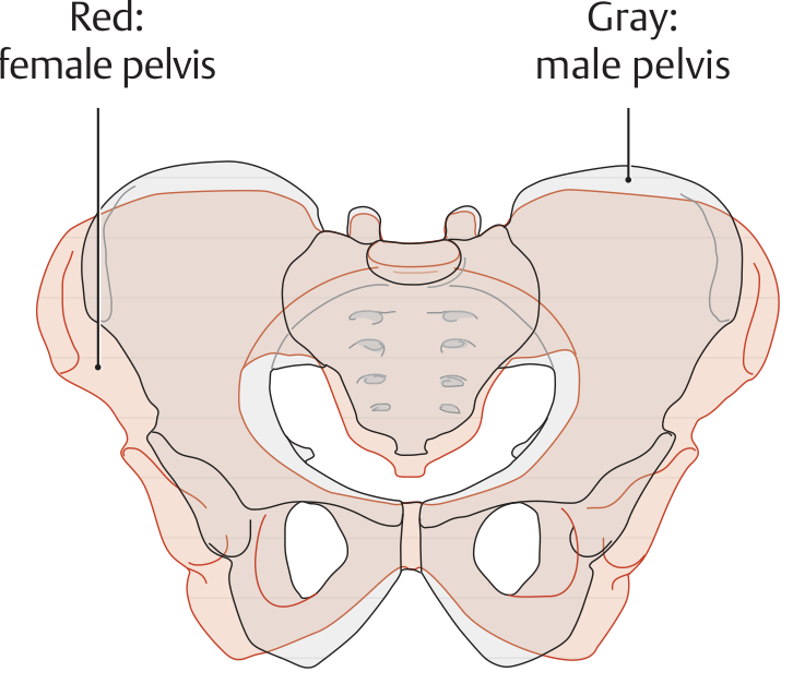
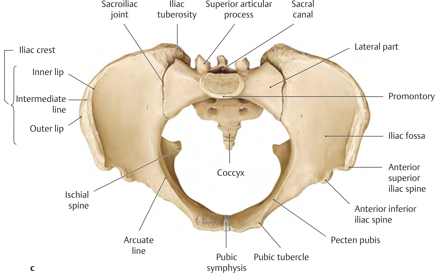
| Feature | Male Pelvis | Female Pelvis |
|---|---|---|
| General build | Deep, funnel-shaped; massive, rough | Shallow, bowl-shaped; less massive, smooth |
| True pelvis (lesser pelvis) | Narrow, deep, funnel-shaped | Wide, shallow, cylindrical |
| Pelvic brim (inlet) | Heart-shaped (sacral promontory projects more) | Circular / oval (wider transversely) |
| Pelvic outlet | Smaller | Larger |
| Subpubic angle | 70°-75° (acute); arch is V-shaped | 90°-100°; arch is U-shaped |
| Ilium | More sloped, curve more prominent | Less sloped, less prominent curve, smoother margins |
| Iliac crest | More prominent | Less prominent |
| ASIS | Not widely separated | Widely separated |
| Pre-auricular sulcus | Narrow, shallow, not prominent | Broad, deep, prominent (marker of parturition) |
| Acetabulum | Wide and deep; diameter ~52 mm | Small and narrow; diameter ~46 mm |
| Obturator foramen | Large, oval with base upward | Small, triangular with apex forward |
| Greater sciatic notch | Smaller, deeper, narrower; less than right angle | Wider, larger, shallower; approximately right angle |
| Sciatic notch index (Width/Depth) | 4-5 | 5-6 |
| Ischial tuberosity | More or less inverted | Everted (turned outward) |
| Pubic body | Narrow and triangular | Broad and square |
| Symphysis pubis | Higher, bigger in depth, narrow in width; pubic arch margins everted; no parturition pits | Lower, wider, rounded; arch margins not everted; parturition pits present on dorsal border |
| Pelvic index (AP/Transverse ×100) | More | Less |
| Kell index (acetabulum surface/ilium surface ×100) | More | Less |
| Ischiopubic index (ischial length/pubic length ×100) | 73-94 | 91-115 |
| Weight | Heavier | Lighter |
Differences in the Sacrum (between male and female)
| Feature | Male Sacrum | Female Sacrum |
|---|---|---|
| Shape | Long and narrow | Shorter and wider |
| Sacral promontory | Well-marked, projects forward more | Less prominent |
| Anterior curvature | Uniformly curved along its length | Straight in upper half, sharply curved in lower half |
| Sacroiliac articular surface | Large; extends up to 2.5-3 vertebral bodies | Small; extends up to 2-2.5 vertebral bodies |
| Coccyx | Less movable | More movable (for parturition) |
| Sacral index (Breadth of base/Anterior length ×100) | 112 | 116 |
| Corporo-basal index | 45 | 40.5 |
Key Points for Exams (Vishram Singh emphasis)
- Subpubic angle is the single most reliable sex indicator: V-shape (male) vs U-shape (female)
- Greater sciatic notch: right angle or more = female; less than right angle = male
- Pre-auricular sulcus: present and prominent in females (childbirth-related)
- Obturator foramen: oval in male, triangular in female
- Parturition pits: on the dorsal border of symphysis pubis in females who have given birth
- The female pelvis is WIDER but SHALLOWER; the male pelvis is NARROWER but DEEPER
- Sex can be determined with 95% accuracy from the pelvis alone
Part 2: Direct vs Indirect Inguinal Hernia
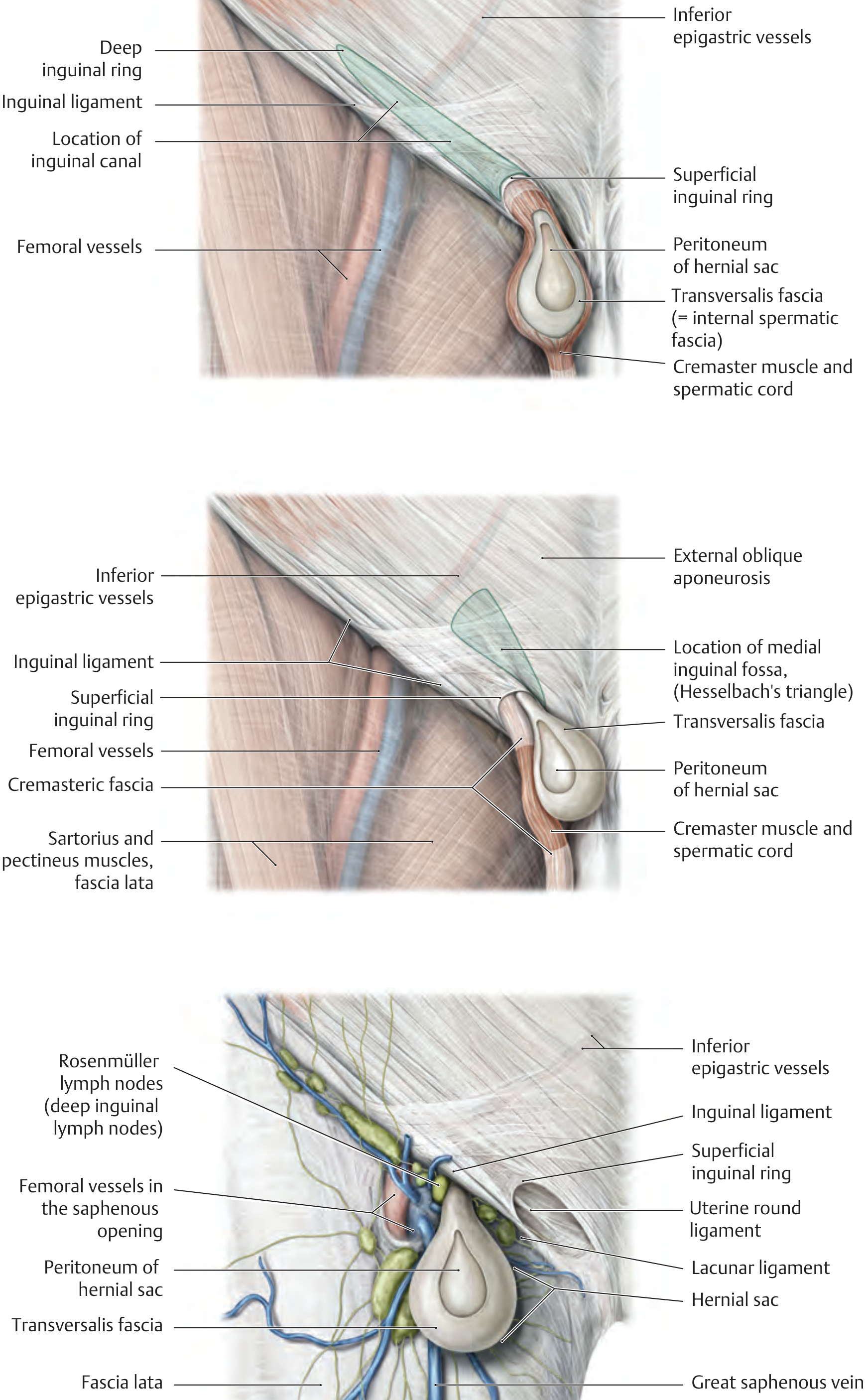
Hesselbach's (Inguinal) Triangle - The Key to Understanding
- Medially: Lateral border of rectus abdominis / rectus sheath
- Superolaterally: Inferior epigastric vessels
- Inferiorly: Inguinal ligament
Rule: Indirect hernia = LATERAL to inferior epigastric vessels; Direct hernia = MEDIAL to inferior epigastric vessels (within Hesselbach's triangle)
Comprehensive Comparison Table
| Feature | Indirect Inguinal Hernia | Direct Inguinal Hernia |
|---|---|---|
| Also called | Oblique inguinal hernia | Medial inguinal hernia |
| Nature | Congenital OR acquired | Always acquired |
| Pathology | Due to persistence of processus vaginalis | Due to weakness of the posterior wall of inguinal canal (transversalis fascia) |
| Site of origin | Lateral to inferior epigastric vessels | Medial to inferior epigastric vessels |
| Internal ring | Exits through the deep inguinal ring (expanded) - lateral to epigastric vessels | Does NOT pass through the deep inguinal ring; exits through Hesselbach's triangle (medial to epigastric vessels) |
| Path | Passes through the full length of the inguinal canal (indirect/oblique path) - hence "oblique" | Passes directly (perpendicularly) forward through the posterior wall of the inguinal canal - hence "direct" |
| External ring | Exits through the superficial inguinal ring | Also exits through the superficial inguinal ring |
| Scrotum | May descend into the scrotum (scrotal hernia) | Rarely enters the scrotum (stops at superficial ring usually) |
| Sac coverings | Covered by ALL three layers of spermatic cord coverings (internal spermatic fascia, cremasteric fascia, external spermatic fascia) | Covered by external spermatic fascia only (medial to cord coverings) |
| Neck of sac | Narrow neck (risk of strangulation is HIGH) | Wide neck (strangulation is RARE) |
| Age of presentation | Any age; common in children and young adults | Usually middle-aged and elderly men |
| Sex | Both males and females (but predominantly male) | Almost exclusively males |
| Laterality | Right > left (right processus vaginalis closes later) | Usually bilateral |
| Relation to spermatic cord | Sac is within the spermatic cord | Sac lies medial to the spermatic cord |
| Reducibility | May be irreducible; prone to strangulation | Usually easily reducible; strangulation is uncommon |
Clinical Tests to Differentiate
1. Ring Occlusion Test (Zieman's Test / most important)
- Reduce the hernia; press thumb firmly over the deep inguinal ring (½ inch above the midpoint of the inguinal ligament, i.e., mid-inguinal point)
- Ask patient to cough
- Indirect hernia: controlled - does NOT appear (it comes through the deep ring which is occluded)
- Direct hernia: NOT controlled - bulge appears medial to the occluding thumb (it does not pass through the deep ring)
2. Invagination Test (Finger in Canal)
- Invaginate the scrotal skin and push the finger into the superficial inguinal ring toward the deep ring
- Ask patient to cough
- Direct hernia: impulse felt on the pulp of finger (hernia comes directly forward)
- Indirect hernia: impulse felt on the tip of finger (hernia comes down the canal from above)
Anatomical Diagram
Summary Mnemonic
"MDs get Direct" - Middle-aged, Degenerative, Directly through the wall, Medial to epigastrics"Young lads get Indirect" - Young age, processus vaginalis, Lateral to epigastrics, down Into scrotum
Comparison at a Glance
| Indirect | Direct | |
|---|---|---|
| Epigastric vessels | Lateral to them | Medial to them |
| Type | Congenital/acquired | Acquired only |
| Neck | Narrow | Wide |
| Into scrotum | Yes | Rarely |
| Strangulation | Common | Rare |
| Age | Young | Elderly |
| Ring occlusion test | Controlled | NOT controlled |