
Bronchial Muscle name and diagram and bronchial tree
labeled anatomical diagram of bronchial tree right and left main bronchi lobar segmental bronchi
bronchial smooth muscle anatomy
bronchial tree anatomy bronchial smooth muscle official educational resource
bronchial tree anatomy diagram main bronchi lobar bronchi segmental bronchi labeled
Bronchial muscle
- It is involuntary smooth muscle, not skeletal muscle.
- In larger bronchi it forms a near-continuous circular layer; in smaller bronchi it has a more spiral arrangement.
- Its contraction narrows the airway, called bronchoconstriction. Relaxation widens the airway, called bronchodilation.
- Unlike bronchi, bronchioles have no cartilage plates, but they still have prominent smooth muscle that controls their diameter.
Junqueira's Basic Histology - Text and Atlas, 17e, pp. 870-871.
Bronchial tree diagram
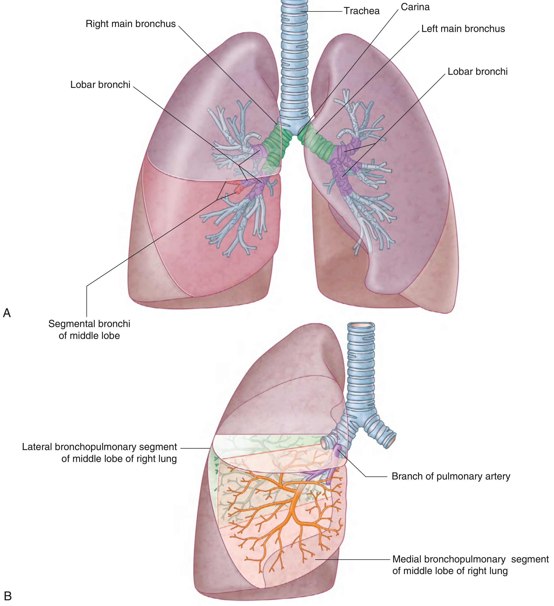
Bronchial tree: order of branches
Trachea
  │
  └─ Carina
      ├─ Right main (primary) bronchus
      │    ├─ Superior lobar bronchus
      │    ├─ Middle lobar bronchus
      │    └─ Inferior lobar bronchus
      │         └─ Segmental (tertiary) bronchi
      │
      └─ Left main (primary) bronchus
           ├─ Superior lobar bronchus
           └─ Inferior lobar bronchus
                └─ Segmental (tertiary) bronchi

Segmental bronchi
  → smaller bronchi
  → bronchioles
  → terminal bronchioles
  → respiratory bronchioles
  → alveolar ducts
  → alveolar sacs and alveoli
Important points
- The right main bronchus is wider, shorter, and more vertical than the left. Aspirated foreign bodies therefore commonly enter the right bronchial tree.
- Lobar bronchi supply lung lobes: 3 on the right and 2 on the left.
- Segmental bronchi supply bronchopulmonary segments.
- The trachea divides at the carina, usually around vertebral level T4-T5.
Show a labeled diagram of bronchial smooth muscle
labeled histology diagram bronchial wall smooth muscle muscularis cartilage epithelium
bronchial smooth muscle anatomy
labeled bronchial wall smooth muscle histology diagram
Labeled diagram: bronchial smooth muscle
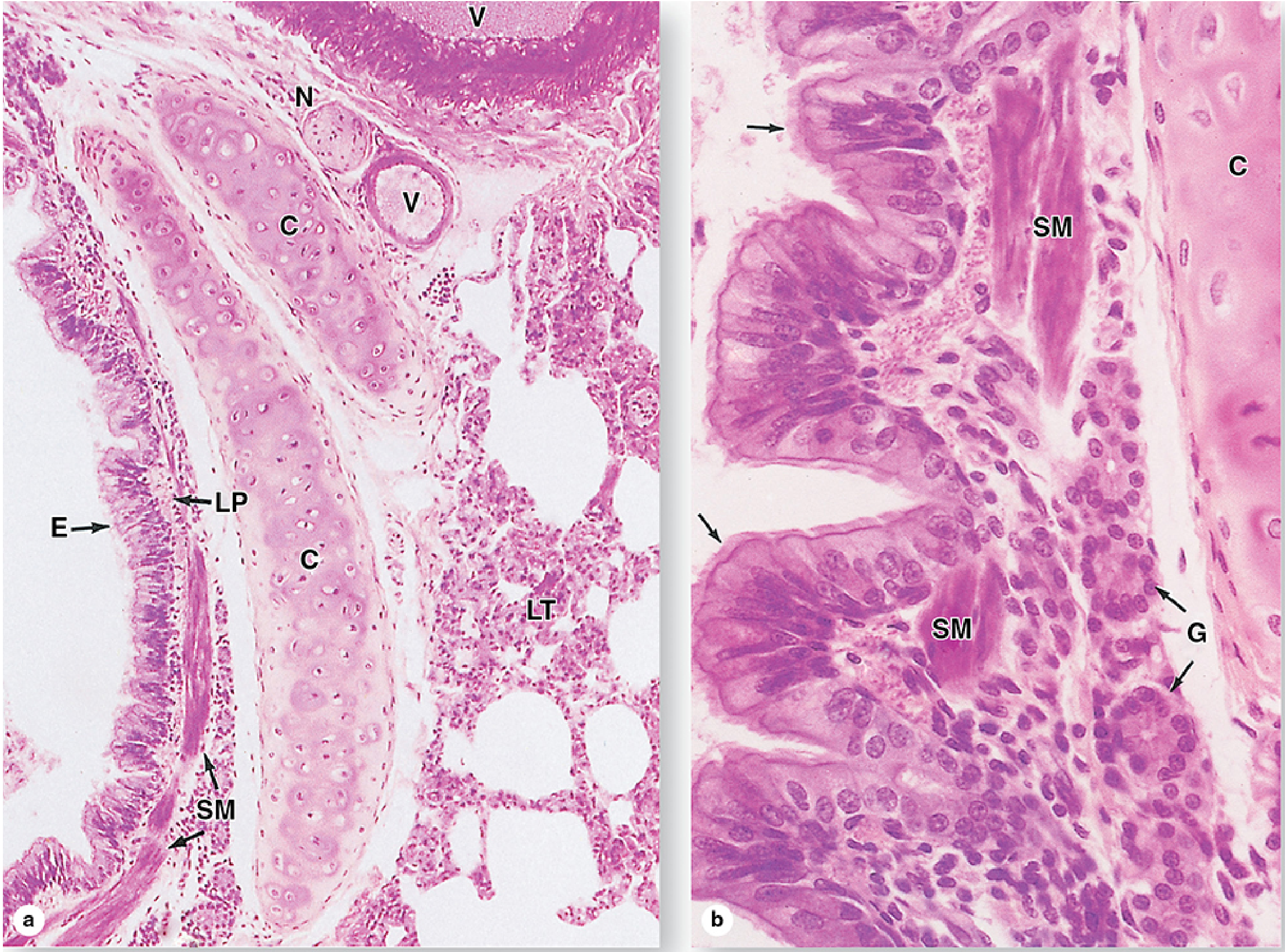
- SM: Bronchial smooth muscle or muscularis. It forms a circular to spiral layer around the airway and changes airway diameter.
- E: Respiratory epithelium, mainly pseudostratified ciliated columnar epithelium.
- C: Hyaline cartilage plates that support the bronchial wall.
- V: Blood vessels.
- G: Seromucous gland, where visible.
Muscle of bronchial wall
bronchial smooth muscle anatomy
bronchial smooth muscle anatomy
- It lies beneath the mucosa, forming a mainly circular or spiral layer around the bronchus.
- In larger bronchi it is more continuous; in smaller bronchi it appears as spirally arranged bundles.
- Contraction narrows the bronchial lumen: bronchoconstriction.
- Relaxation widens the lumen: bronchodilation.
- It is controlled involuntarily by the autonomic nervous system. Beta-2 receptor stimulation relaxes it.
Gray's Anatomy bronchial wall muscle page around 1160 all information and page photo of bronchial wall muscle.
bronchial smooth muscle anatomy
Gray's Anatomy bronchial wall smooth muscle page 1160
Bronchial wall muscle: key information
- Respiratory epithelium
- Lamina propria
- Smooth-muscle layer (muscularis)
- Submucosa with seromucous glands
- Hyaline cartilage plates
- Adventitia and surrounding lung tissue
- Smooth muscle forms a generally circular, often spirally arranged layer around the bronchial lumen.
- In larger bronchi it is a more continuous layer; it becomes thinner and less organized in smaller bronchi.
- Contraction decreases lumen diameter, causing bronchoconstriction and greater airway resistance.
- Relaxation increases lumen diameter, causing bronchodilation.
- Cartilage plates support the bronchi, while smooth muscle regulates their diameter. Cartilage is absent in bronchioles, where smooth muscle has a larger role in controlling airway caliber.
Labeled bronchial-wall histology image
- SM = smooth muscle
- E = respiratory epithelium
- C = hyaline cartilage
- V = blood vessel

In which book which page number I get this diagram and information
"Key Smooth Muscle Components and Functional Arrangement" "Bronchoalveolar"
"Inner Helical Smooth Muscle Layer" "Outer Helical Smooth Muscle Layer" bronchi
airway smooth muscle anatomy bronchial tree
- The heading has an error: “Functi tional Arrangement Wall.”
- Labels 5 and 7 are duplicated.
- It presents unusually specific named layers, such as “inner helical” and “outer helical” smooth-muscle layers, which are not standard labels used in the major undergraduate anatomy and histology texts.
Closest reliable textbook sources for the same information
| What you need | Book | Page |
|---|---|---|
| Bronchial tree, primary, lobar, and segmental bronchi | Gray's Anatomy for Students | pp. 208-209 |
| Bronchial wall, smooth muscle, cartilage, glands, and a labeled micrograph | Junqueira's Basic Histology: Text and Atlas, 17th ed. | pp. 870-871 |
| Bronchial smooth-muscle layer or muscularis and its function in airway diameter | Histology: A Text and Atlas with Correlated Cell and Molecular Biology | p. 1791 |
| Bronchioles and relatively greater smooth muscle in bronchiolar walls | Histology: A Text and Atlas with Correlated Cell and Molecular Biology | p. 1792 and Plate 19.4 |
Best source for a labeled bronchial-wall image
- Figure 17-7: tertiary or segmental bronchus
- Figure 17-8: bronchial wall
- Labels include:
- SM: smooth muscle
- E: epithelium
- C: cartilage
- G: seromucous glands
- V: blood vessels
Standard description to write in notes
The bronchial wall has a smooth-muscle layer, called the muscularis, arranged in interlacing circular and spiral bundles beneath the mucosa. It becomes relatively more prominent as bronchi become smaller. Contraction narrows the airway lumen and produces bronchoconstriction. Cartilage changes from rings in large bronchi to discontinuous plates in intrapulmonary bronchi, and is absent in bronchioles.