
Spaces of larynx
spaces of larynx anatomy paraglottic pre-epiglottic diagram
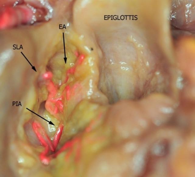
This clinical photograph displays a superior view of a fresh-frozen human cadaver larynx prepared for anatomical study. The dissection shows the epiglottis and the paraglottic space, with the overlying fat tissue partially removed to reveal the underlying vasculature. The arteries have been injected with a bright red bicomponent silicone to enhance visualization and contrast against the yellowish, translucent paraglottic fat and pale mucosal surfaces. Specific vascular structures are labeled: the Superior Laryngeal Artery (SLA), the Epiglottic Artery (EA), and the Postero-Inferior Artery (PIA). This image serves as an educational resource for understanding laryngeal surgical anatomy, particularly the arterial distribution and anastomotic networks within the paraglottic space, which is critical for minimizing hemorrhagic risk during laryngological procedures.
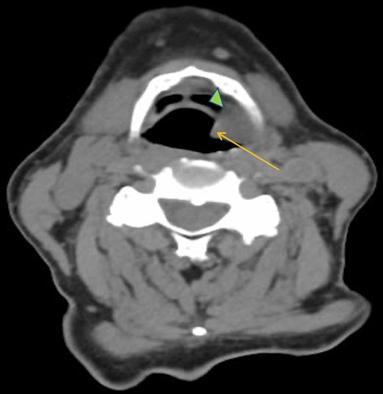
This diagnostic image is an axial contrast-enhanced computed tomography (CT) scan of the neck at the level of the supraglottic larynx. The image demonstrates a prominent, hyperdense soft tissue mass (indicated by a yellow arrow) originating from the left piriform sinus and vallecula (indicated by a green arrowhead). The mass exhibits significant local mass effect, causing partial occlusion and distortion of the laryngeal airway. Anatomical landmarks visible include the hyoid bone anteriorly, the cervical vertebra posteriorly, and the surrounding neck musculature and vasculature. The visual findings are consistent with an invasive neoplastic process, specifically squamous cell carcinoma (SCC) of the larynx. The image provides critical clinical visualization of tumor staging and airway compromise, highlighting the primary site of origin and its relationship to the paraglottic and pre-epiglottic spaces.
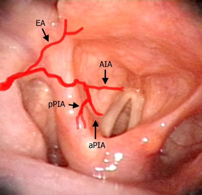
This clinical photograph displays a laryngeal videoendoscopy, simulating a transoral surgical perspective of the human larynx. The image features a healthy, pinkish-red mucosal surface with anatomical landmarks including the epiglottis, arytenoids, and vocal folds. Superimposed schematic red lines illustrate the branching patterns of the laryngeal arteries. The epiglottic artery (EA) originates superiorly, branching downward. The antero-inferior artery (AIA) follows a horizontal course across the paraglottic space. The postero-inferior artery bifurcates into the anterior branch (aPIA), which descends vertically toward the vocal process and paraglottic space, and the posterior branch (pPIA), which is situated lateral and posterior to the arytenoid. This educational graphic serves to demonstrate the surgical vascular anatomy relevant to transoral laryngeal procedures, highlighting the relationship between major arterial branches and endolaryngeal structures.
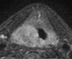
**Imaging Modality:** Magnetic Resonance Imaging (MRI), Axial plane. **Anatomical Region:** Larynx at the glottic level. **Observed Pathology:** An asymmetric, intermediate-signal soft tissue mass is localized to the right glottis. The lesion demonstrates significant mass effect, resulting in the medial displacement and narrowing of the laryngeal airway. **Characteristic Visual Features:** - **Mass Distribution:** The tumor involves the right vocal cord and extends posteriorly toward the arytenoid region. - **Invasion Patterns:** There is evidence of posterior extension toward the submucosa of the pyriform sinus and the posterior commissure. The mass involves the area of the posterior cricoarytenoid muscles, suggestive of a T3 stage glottic carcinoma with vocal cord fixation. - **Signal Intensity:** The lesion appears relatively isointense to slightly hyperintense compared to surrounding musculature on this sequence. **Diagnostic Differentiating Features:** The critical finding is the posterior and lateral transglottic spread, crossing the midline at the posterior commissure and involving the cricoarytenoid unit, which distinguishes it from localized T1 or T2 glottic lesions. This configuration is consistent with advanced primary laryngeal malignancy requiring detailed assessment of the paraglottic and pre-epiglottic spaces.
Spaces of the Larynx
1. Pre-Epiglottic Space (PES)
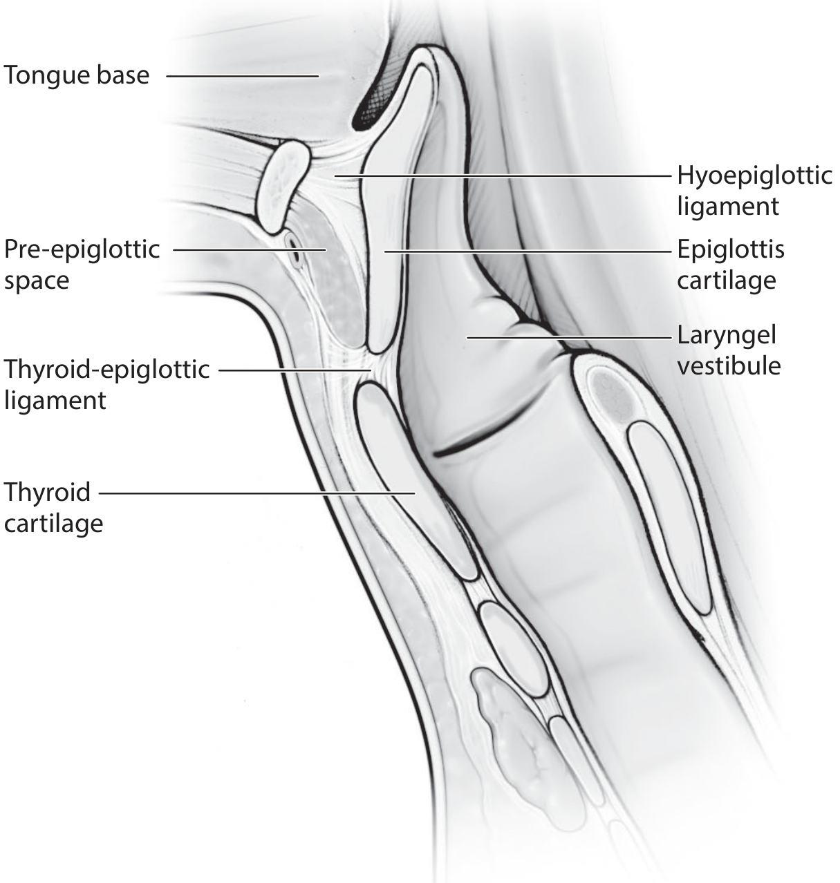
- Anterior: Thyrohyoid membrane and hyoid bone
- Posterior: Anterior surface of the epiglottis
- Superior: Hyoepiglottic ligament (connects epiglottis to hyoid bone)
- Inferior: Thyroepiglottic ligament / thyroid cartilage notch
- Lateral: Continuous with the paraglottic space (no distinct anatomical boundary separates the two)
- Tumour spreads into this space through small perforations (fenestrations) in the epiglottic cartilage or directly through the hyoepiglottic ligament.
- Its involvement upgrades a supraglottic tumour to at least T3 (in AJCC staging).
- It is the primary reason supraglottic carcinomas tend to carry a worse prognosis than glottic carcinomas.
2. Paraglottic Space (PGS)
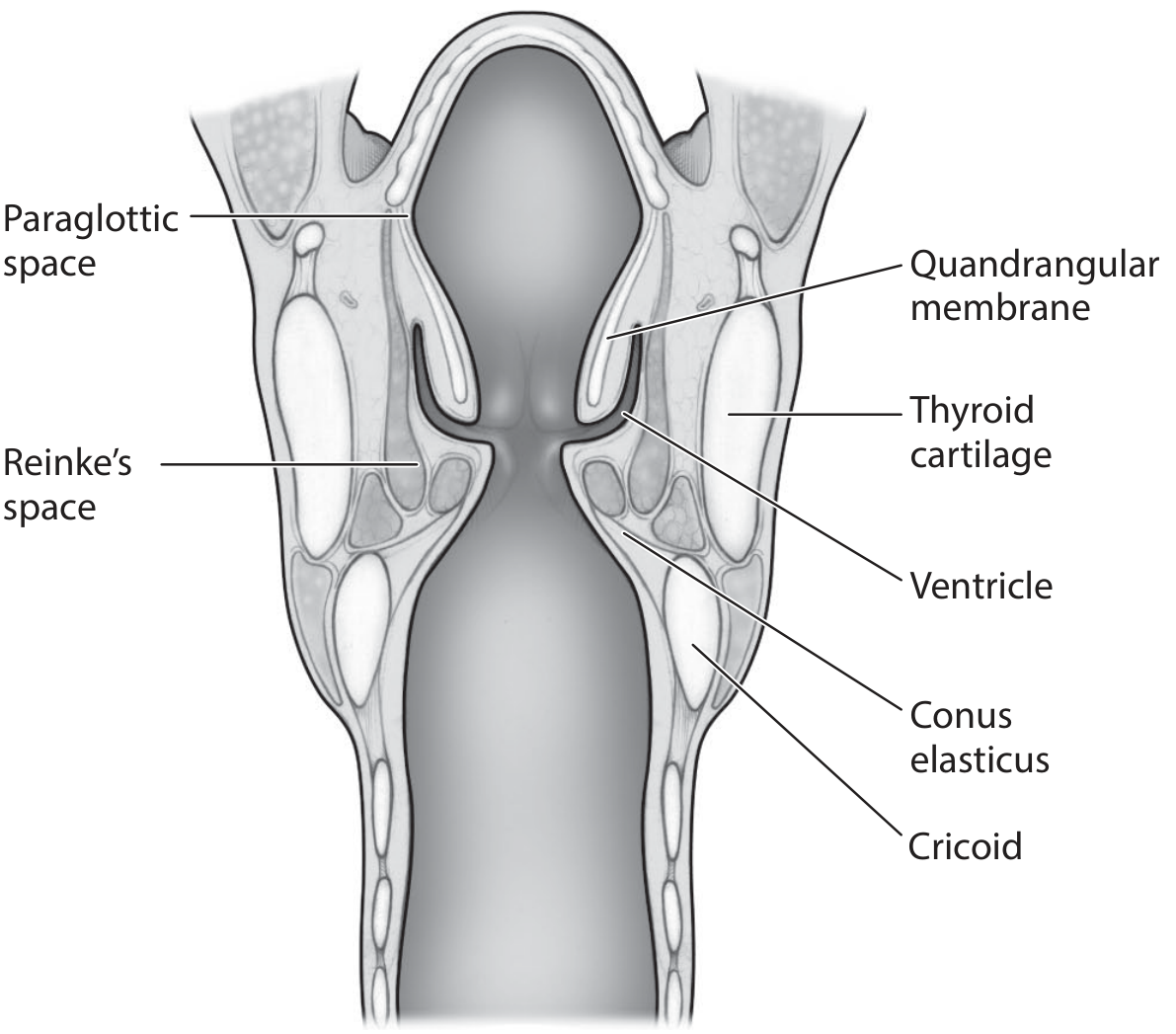
- Lateral: Inner surface of the thyroid cartilage and its perichondrium
- Medial: Conus elasticus (inferiorly) and quadrangular membrane (superiorly)
- Posterior: Mucosa of the piriform fossa
- It is the key pathway for transglottic spread of tumour - a cancer originating in the supraglottis can spread submucosally through the paraglottic space to the glottis and vice versa, often with minimal or no mucosal changes visible on endoscopy.
- The conus elasticus acts as a partial barrier, limiting lateral spread of glottic cancers.
- It is continuous with the pre-epiglottic space anterosuperiorly - there is no anatomical barrier between the two.
- Invasion of this space also upgrades a tumour to T3.
3. Reinke's Space
- Superior/Inferior: Arcuate lines at the superior and inferior edges of the membranous vocal fold
- Medial: Vocal fold epithelium
- Deep: Vocal ligament (middle + deep layers of lamina propria)
- Reinke's oedema (polypoid degeneration): Fluid accumulates in this space due to chronic smoking and voice abuse. The entire membranous vocal fold from anterior commissure to vocal process becomes diffusely, bilaterally swollen. Treatment is surgical entry into Reinke's space (Hirano technique), aspiration of fluid, and excision of excess mucosa.
- The space does not cross the anterior or posterior commissure, explaining why Reinke's oedema tends to be bilateral but confined to the membranous fold.
- On stroboscopy, pathology within Reinke's space (sulcus, oedema, polyp) manifests as abnormal mucosal wave.
Summary Table
| Space | Location | Contents | Key Clinical Significance |
|---|---|---|---|
| Pre-epiglottic | Anterior to epiglottis, posterior to thyrohyoid membrane | Fibrofatty tissue | Supraglottic tumour spread; T3 staging |
| Paraglottic | Between thyroid cartilage and conus elasticus/quadrangular membrane | Fibrofatty tissue, ventricle, saccule | Transglottic tumour spread; T3 staging |
| Reinke's | Superficial lamina propria of true vocal fold | Loose ECM, hyaluronic acid | Reinke's oedema; mucosal wave vibration |
Key Relationships
- The pre-epiglottic and paraglottic spaces are directly continuous with each other laterally - this allows supraglottic tumours to spread to the paraglottic space and then inferiorly across the glottis (transglottic spread).
- The conus elasticus acts as a natural barrier that limits downward and lateral spread of glottic cancers into the subglottis.
- Broyles' tendon (insertion of the vocalis muscle into the thyroid cartilage) lacks perichondrium and represents a potential route for anterior commissure tumours to invade the thyroid cartilage.