

Screenshot_20260714_092153.jpg
Ans these for exam
Reading File
The image shows General Physiology exam questions. Let me search the medical library for all topics simultaneously.
Finding Sources
Finding Sources
Finding Sources
Finding Sources
Finding Sources
Finding Sources
Reading File
Reading File
Finding Sources
Reading File
Finding Sources
Reading File
Reading File
Now I have more than enough material. Let me also fetch the Na+/K+ pump diagram image and get the cell membrane structure section.
Reading File
Loading Image
I now have comprehensive material from Guyton & Hall and Costanzo Physiology. Here are complete exam answers for all questions:
General Physiology - Exam Answers
Q1. Structure and Function of the Cell Membrane (5 marks)
Structure
The cell membrane (plasma membrane) is a lipid bilayer approximately 7-10 nm thick, composed of:
1. Lipid Bilayer
- Two layers of phospholipid molecules, each with a hydrophilic (water-loving) head and two hydrophobic (water-repelling) fatty acid tails
- The tails face inward (away from water), heads face outward
- This arrangement forms a fluid, selectively permeable barrier
- Lipid-soluble substances (O2, CO2, steroids, alcohol) can diffuse directly through it
2. Proteins (constitute ~55% of membrane mass)
- Integral (transmembrane) proteins - span the full width of the membrane; act as:
- Channel proteins - have watery pores that allow ions/water to pass through (e.g., Na+, K+, Ca2+ channels)
- Carrier proteins - bind specific molecules, undergo conformational change to transport them (e.g., glucose transporters)
- Pump proteins - ATPase enzymes (e.g., Na+/K+ ATPase)
- Receptor proteins - bind hormones/ligands to trigger intracellular signaling
- Peripheral proteins - attached to inner or outer surface; act as enzymes or structural anchors
3. Carbohydrates
- Attached to outer surface as glycoproteins and glycolipids
- Form the glycocalyx - involved in cell recognition, immunity, and cell-cell adhesion
4. Cholesterol
- Embedded in the lipid bilayer
- Regulates fluidity - prevents membrane from becoming too rigid (cold) or too fluid (heat)
Functions
- Selective barrier - controls what enters/exits the cell
- Transport - moves nutrients, ions, waste products
- Cell recognition - glycocalyx identifies "self" vs "non-self"
- Signal reception - receptor proteins receive hormonal and chemical signals
- Structural support - maintains cell shape
- Electrical excitability - generates and propagates action potentials via ion channels
(Guyton and Hall Textbook of Medical Physiology, Ch. 4)
Q2. Transport Mechanisms Across the Cell Membrane (5 marks)
Transport occurs via two fundamental processes: Diffusion and Active Transport.
A. PASSIVE TRANSPORT (No energy required - moves down concentration gradient)
1. Simple Diffusion
- Random molecular movement from high to low concentration
- Lipid-soluble molecules (O2, CO2, fatty acids, steroids) diffuse directly through the lipid bilayer
- Very small uncharged molecules (water, urea) also pass through
- Rate depends on: concentration gradient, membrane permeability, surface area, temperature
2. Diffusion Through Protein Channels (Channel-mediated diffusion)
- Ions (Na+, K+, Ca2+, Cl-) are water-soluble and cannot cross the lipid bilayer directly
- They pass through specific channel proteins that form water-filled pores
- Channels are selective (ion-specific) and may be:
- Voltage-gated (open on depolarization - nerve/muscle)
- Ligand-gated (open on binding of neurotransmitter)
- Mechanically-gated (open on physical deformation)
3. Facilitated Diffusion
- Water-soluble molecules (glucose, amino acids) move down their concentration gradient via carrier proteins
- Carrier binds substrate, undergoes conformational change, releases substrate on the other side
- No energy (ATP) required, but is saturable and specific
- Example: GLUT transporters for glucose in red blood cells
4. Osmosis
- Net movement of water through a semipermeable membrane from low solute to high solute concentration (from high water to low water)
- Driven by the osmotic pressure gradient
B. ACTIVE TRANSPORT (Requires energy - moves against concentration gradient)
1. Primary Active Transport
- Energy comes directly from ATP hydrolysis
- Example: Na+/K+ ATPase pump (see below)
- Other examples: Ca2+ pump (SERCA), H+/K+ ATPase (stomach)
2. Secondary Active Transport
- Uses the electrochemical gradient created by primary active transport (especially Na+ gradient) as the energy source
- Co-transport (symport) - Na+ and glucose/amino acid move in the same direction (e.g., Na+-glucose transporter in intestinal epithelium)
- Counter-transport (antiport) - Na+ moves inward while another ion/molecule moves outward (e.g., Na+-Ca2+ exchanger in heart muscle)
C. VESICULAR TRANSPORT
Endocytosis - cell membrane engulfs extracellular material
- Phagocytosis (large particles, bacteria)
- Pinocytosis (fluid droplets)
- Receptor-mediated endocytosis (LDL cholesterol)
Exocytosis - vesicles fuse with membrane to release contents (e.g., neurotransmitter release, hormone secretion)
(Guyton and Hall, Ch. 4)
Q3. Cell Membrane (Short note, 3 marks)
The cell membrane is a fluid mosaic of phospholipids and proteins. It is a 7-10 nm thick lipid bilayer in which protein molecules are embedded (Singer-Nicolson Fluid Mosaic Model, 1972).
Key components:
- Phospholipids - amphipathic; hydrophilic heads face ECF/ICF, hydrophobic tails form the core barrier
- Proteins - integral (channel, carrier, receptor, pumps) and peripheral
- Cholesterol - stabilizes fluidity
- Glycocalyx - sugar coat on outer surface for recognition
Functions: selective permeability, transport, signal transduction, cell adhesion, immune recognition, and generation of resting membrane potential.
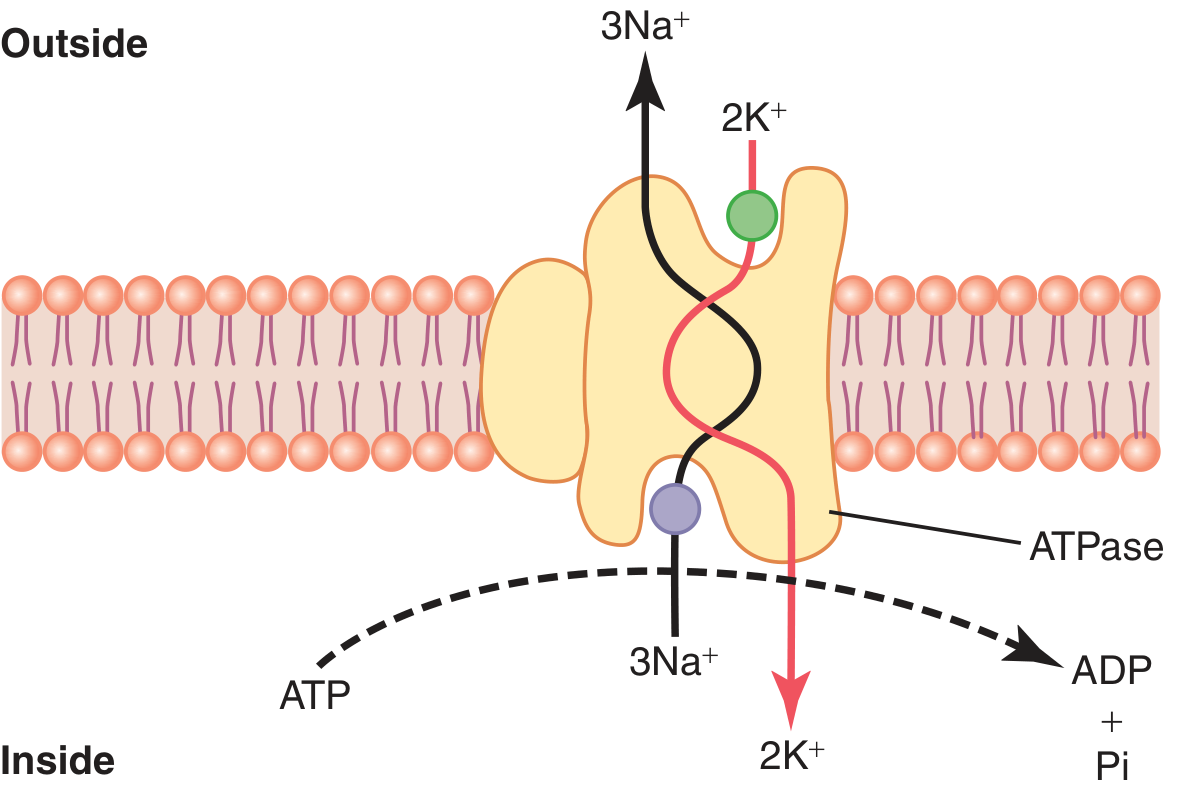
Q4. Na+/K+ Pump (Na+/K+ ATPase)
The Na+/K+ pump is the primary active transport mechanism in virtually all body cells.
Structure
It is a carrier protein complex with:
- α subunit (mol. wt ~100,000) - the functional unit with:
- 3 binding sites for Na+ (on the intracellular side)
- 2 binding sites for K+ (on the extracellular side)
- ATPase activity (near the Na+ binding sites)
- β subunit (mol. wt ~55,000) - anchors the complex in the membrane
Mechanism
- 3 Na+ ions bind to the intracellular face of the α subunit
- 2 K+ ions bind to the extracellular face
- ATP is cleaved to ADP + Pi, releasing energy
- This energy causes a conformational change in the protein
- 3 Na+ are pumped OUT and 2 K+ are pumped IN per cycle
- Net result: 1 positive charge moves out per cycle → electrogenic (contributes to negative resting membrane potential)
Importance
- Maintains high intracellular K+ and low intracellular Na+ (essential for cell excitability)
- Generates the resting membrane potential (-70 mV in neurons)
- Controls cell volume (prevents osmotic swelling)
- Creates the Na+ gradient that powers secondary active transport
- Is the basis for nerve impulse conduction
(Guyton and Hall, Ch. 4)
Q5. Passive Diffusion
Passive diffusion is the movement of molecules from a region of high concentration to low concentration (down a concentration gradient) without expenditure of cellular energy.
Types
1. Simple diffusion - molecules pass directly through the lipid bilayer
- Limited to lipid-soluble molecules (O2, CO2, N2, alcohol, steroid hormones) and very small uncharged molecules
- Rate governed by Fick's Law of Diffusion: Rate ∝ (Concentration gradient × Surface area × Permeability coefficient) / Membrane thickness
2. Diffusion through ion channels - ions move through protein-lined pores down their electrochemical gradient
- Selective permeability (each channel type is specific to one ion)
- Governed by both concentration gradient AND electrical gradient (electrochemical gradient)
3. Facilitated diffusion - carrier-protein mediated, still down the gradient
- Examples: glucose (GLUT transporters), amino acids, nucleosides
Characteristics
- No ATP required
- Movement is down the concentration/electrochemical gradient
- Reaches equilibrium (net diffusion stops when gradient is zero)
- Rate is proportional to the concentration difference
- Temperature-dependent (higher temp = faster diffusion)
Q6. Positive (+ve) Feedback Mechanism
In a positive feedback system, the output of a system amplifies or reinforces the original stimulus, producing a self-sustaining, escalating (explosive) response. It is the opposite of negative feedback.
Characteristics
- Self-augmenting (not self-limiting like negative feedback)
- Rare in normal physiology but important in specific situations
- Leads to an explosive or irreversible event
Physiological Examples
1. Action Potential (nerve impulse)
- Membrane depolarization → voltage-gated Na+ channels open → Na+ rushes in → more depolarization → more Na+ channels open → explosive upstroke of action potential
- This is the classic positive feedback loop producing the rapid, all-or-none spike
2. Ovulation - LH Surge (Hormonal)
- Rising estrogen (follicular phase) → stimulates anterior pituitary to release LH and FSH → LH/FSH cause more estrogen secretion and follicle maturation → rapid burst (surge) of LH → ovulation
- The explosive event = LH surge triggering ovulation
3. Parturition (Oxytocin feedback)
- Cervical dilation → posterior pituitary releases oxytocin → uterine contractions → more cervical dilation → more oxytocin → stronger contractions
- The explosive event = delivery of the fetus
4. Blood Clotting
- Activation of clotting factors amplifies itself through the cascade, leading to rapid clot formation
5. Irreversible hemorrhagic shock (pathological positive feedback)
- Severe bleeding → reduced cardiac output → reduced coronary blood flow → weaker heart → even less output → death (a "vicious cycle")
Key Point
Positive feedback is useful when the body needs a rapid, all-or-nothing response (action potential, ovulation, labor). When it becomes uncontrolled (as in shock or fever), it is pathological.
(Costanzo Physiology 7th Edition; Guyton and Hall Textbook of Medical Physiology)
Sources: Guyton and Hall Textbook of Medical Physiology (14th Ed.), Costanzo Physiology (7th Ed.)
Along with ans put diagram for better understanding
Let me fetch all the relevant diagrams from the textbooks simultaneously.
Loading Image
Loading Image
Loading Image
Now let me fetch the remaining diagrams - cell membrane structure and positive feedback.
Finding Sources
Finding Sources
Loading Image
Loading Image
Finding Sources
Finding Sources
Finding Sources
Loading Image
Loading Image
All diagrams are now collected. Here are the complete answers with diagrams from the textbooks:
General Physiology - Exam Answers with Diagrams
Q1. Structure and Function of the Cell Membrane (5 marks)
Structure - Fluid Mosaic Model (Singer-Nicolson, 1972)
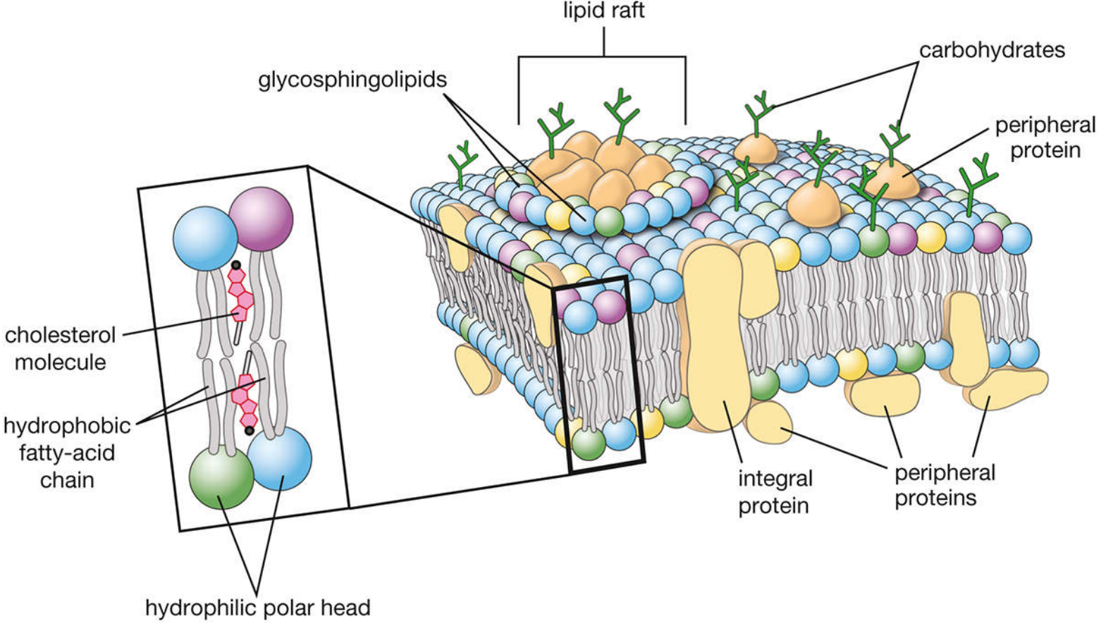
Fig: Fluid mosaic model of plasma membrane - showing lipid bilayer with hydrophilic polar heads (outer), hydrophobic fatty-acid chains (inner), cholesterol, integral proteins, peripheral proteins, glycosphingolipids and carbohydrate chains.
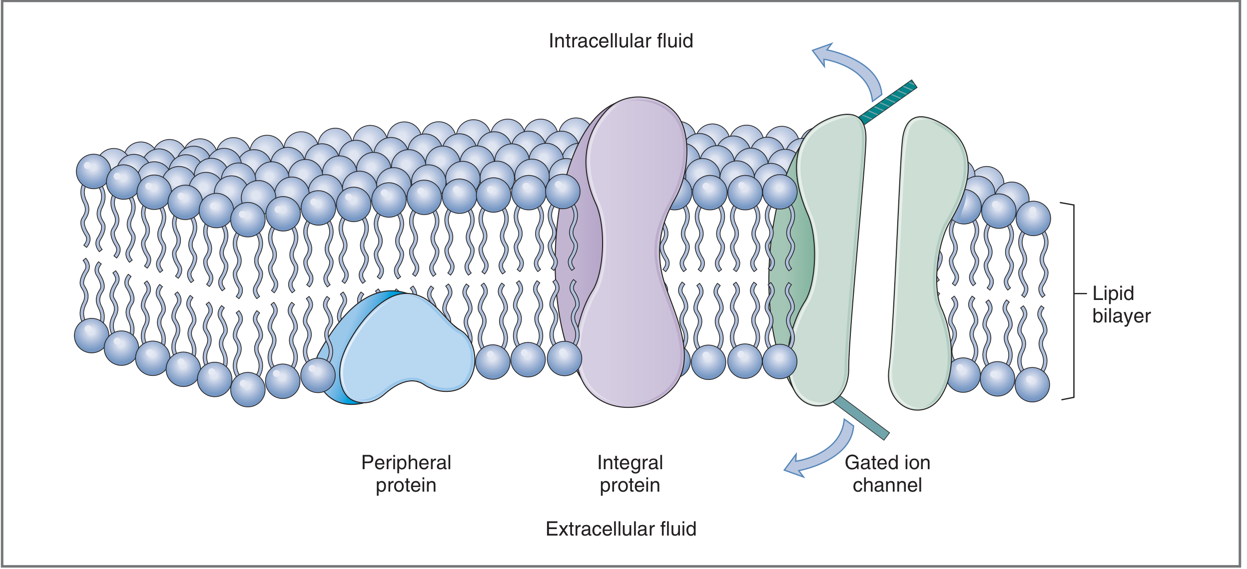
Fig: Simplified cross-section showing peripheral protein, integral transmembrane protein, and gated ion channel embedded in the lipid bilayer.
Components:
| Component | Location | Function |
|---|---|---|
| Phospholipid bilayer | Main scaffold | Selectively permeable barrier |
| Integral proteins | Span the membrane | Channels, carriers, pumps, receptors |
| Peripheral proteins | Inner/outer surface only | Enzymes, structural anchors |
| Cholesterol | Within bilayer | Regulates fluidity |
| Glycocalyx (glycoproteins/glycolipids) | Outer surface | Cell recognition, immunity |
Functions of cell membrane:
- Selectively permeable barrier between ECF and ICF
- Transport of ions, nutrients, waste
- Cell recognition and communication
- Reception of hormonal signals
- Generation and propagation of action potentials
- Regulation of cell volume
(Guyton & Hall, Ch. 4; Costanzo Physiology)
Q2. Transport Mechanisms Across Cell Membrane (5 marks)
Ion concentrations maintained by these transport mechanisms:
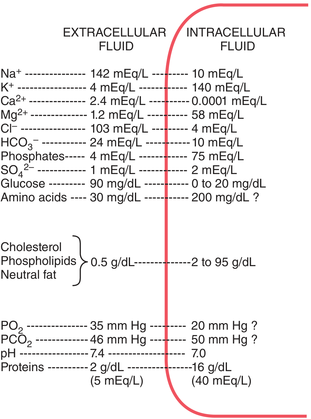
Fig: Chemical composition of ECF vs ICF - note high Na+ outside and high K+ inside, maintained by active transport.
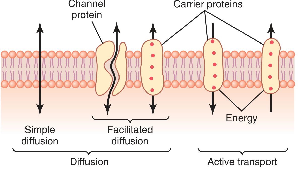
Fig: Three transport mechanisms - Simple diffusion (lipid soluble), Facilitated diffusion (carrier protein, no energy), Active transport (carrier protein + energy).
A. Passive Transport (No ATP, moves DOWN concentration gradient)
1. Simple Diffusion
- Lipid-soluble molecules (O2, CO2, steroids, alcohol) pass directly through lipid bilayer
- Governed by Fick's Law: Rate ∝ Concentration gradient × Area / Thickness
2. Diffusion Through Ion Channels
- Ions (Na+, K+, Ca2+, Cl-) pass through protein-lined pores
- Channels are selective and may be voltage-gated, ligand-gated, or mechanically-gated
3. Facilitated Diffusion
- Glucose, amino acids move via carrier proteins, still down gradient
- Saturable, specific, no ATP needed (e.g., GLUT transporters)
4. Osmosis
- Net movement of water through semipermeable membrane from low to high solute (high to low water concentration)
B. Active Transport (Requires ATP, moves AGAINST gradient)
1. Primary Active Transport
- Uses ATP directly (e.g., Na+/K+ ATPase, Ca2+ pump, H+/K+ pump)
2. Secondary Active Transport
- Uses Na+ gradient created by Na+/K+ pump
- Symport (co-transport): Na+ + glucose/amino acids move same direction (intestinal absorption)
- Antiport (counter-transport): Na+ in, Ca2+ out (Na+/Ca2+ exchanger)
C. Vesicular Transport
- Endocytosis: phagocytosis, pinocytosis, receptor-mediated (LDL)
- Exocytosis: hormone/neurotransmitter secretion
(Guyton & Hall, Ch. 4)
Q3. Cell Membrane (Short note, 3 marks)
The cell membrane (plasma membrane) is a 7-10 nm thick fluid mosaic of phospholipids and proteins:
- Phospholipid bilayer: amphipathic molecules - hydrophilic heads face ECF and ICF; hydrophobic tails form the impermeable core
- Proteins: integral (channels, carriers, pumps, receptors) and peripheral (enzymes, structural)
- Cholesterol: embedded in bilayer, stabilizes membrane fluidity
- Glycocalyx: outer sugar coat for cell-cell recognition and immunity
Functions: selective permeability, transport, signal transduction, cell adhesion, generation of resting membrane potential (-70 mV).
Q4. Na+/K+ Pump
Fig: Na+/K+ pump - 3 Na+ pumped OUT of the cell, 2 K+ pumped IN per cycle, using 1 ATP. The alpha subunit has ATPase activity. Net 1 positive charge leaves the cell per cycle (electrogenic).
Structure
- α subunit (100,000 Da): 3 intracellular Na+ binding sites + 2 extracellular K+ binding sites + ATPase activity
- β subunit (55,000 Da): anchors the complex in the membrane
Mechanism (Step-by-step)
- 3 Na+ bind to intracellular sites of α subunit
- 2 K+ bind to extracellular sites
- ATP is cleaved → ADP + Pi (energy released)
- Conformational change in carrier protein
- 3 Na+ extruded OUTSIDE; 2 K+ brought INSIDE
- Net: 1 positive charge leaves → electrogenic pump
Importance
- Maintains Na+ low (10 mEq/L) and K+ high (140 mEq/L) inside cells
- Creates resting membrane potential (-70 mV)
- Prevents cell swelling (controls osmotic balance)
- Na+ gradient drives secondary active transport (glucose, amino acids)
- Basis for nerve impulse conduction
(Guyton & Hall, Ch. 4)
Q5. Passive Diffusion
Passive diffusion is movement of molecules down their concentration gradient without expenditure of energy (no ATP required).
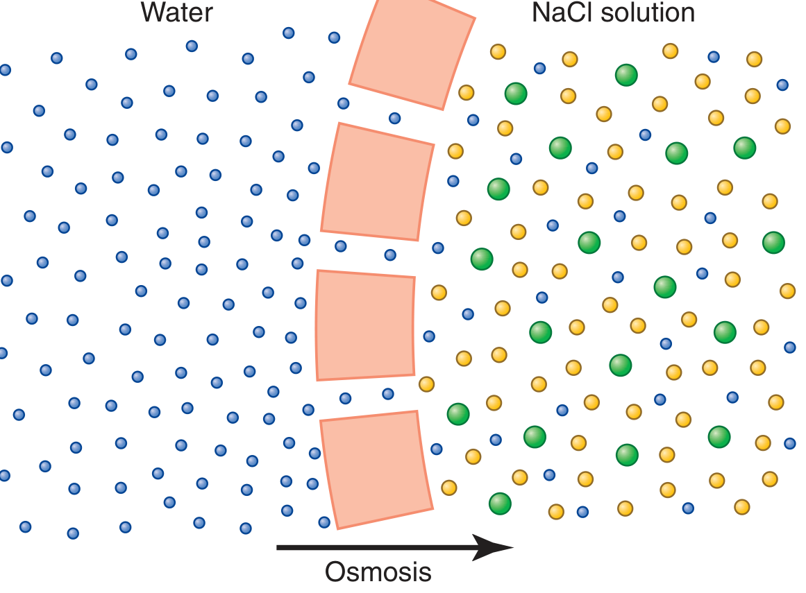
Fig: Osmosis - a special form of passive diffusion. Water moves from pure water (high water concentration) through the selectively permeable membrane toward the NaCl solution (lower water concentration).
Types
1. Simple diffusion - through lipid bilayer
- O2, CO2, N2, fatty acids, steroids, alcohol
- Rate described by Fick's Law: J = DA(C1 - C2)/L
- D = diffusion coefficient, A = area, C = concentration, L = thickness
2. Channel-mediated diffusion - through ion channel proteins
- Na+, K+, Ca2+, Cl- move down electrochemical gradients
- Driven by both concentration AND electrical gradients
3. Facilitated diffusion - via carrier proteins, still passive
- Glucose (GLUT1-4), amino acids, nucleosides
- Saturable (maximum transport rate = Tmax)
4. Osmosis - passive movement of water
- Water moves from hypotonic to hypertonic solution
- Osmotic pressure opposes further osmosis
Key Features of Passive Diffusion
| Feature | Detail |
|---|---|
| Energy | Not required |
| Direction | High → Low concentration |
| Equilibrium | Reached when gradient = 0 |
| Rate | Proportional to concentration gradient |
| Temperature | Higher temp = faster diffusion |
Q6. Positive (+ve) Feedback Mechanism
In positive feedback, the output amplifies the original stimulus - it is self-augmenting (opposite of negative feedback). Positive feedback is rare in normal physiology but produces explosive, all-or-nothing events.
Comparison
| Feature | Negative Feedback | Positive Feedback |
|---|---|---|
| Effect on stimulus | Opposes/reduces it | Amplifies/reinforces it |
| System behavior | Self-limiting, stable | Self-augmenting, unstable |
| Common or rare | Very common | Rare |
| Result | Homeostasis | Explosive event |
Examples with Diagrams
1. Action Potential (most important non-hormonal example)
Depolarization → voltage-gated Na+ channels open → Na+ enters cell → MORE depolarization → MORE Na+ channels open → EXPLOSIVE upstroke of action potential
Depolarization
     ↓
Na⁺ channels open
     ↓
Na⁺ enters → more depolarization
     ↓
More Na⁺ channels open (POSITIVE FEEDBACK)
     ↓
Explosive action potential spike
2. LH Surge and Ovulation (hormonal positive feedback)
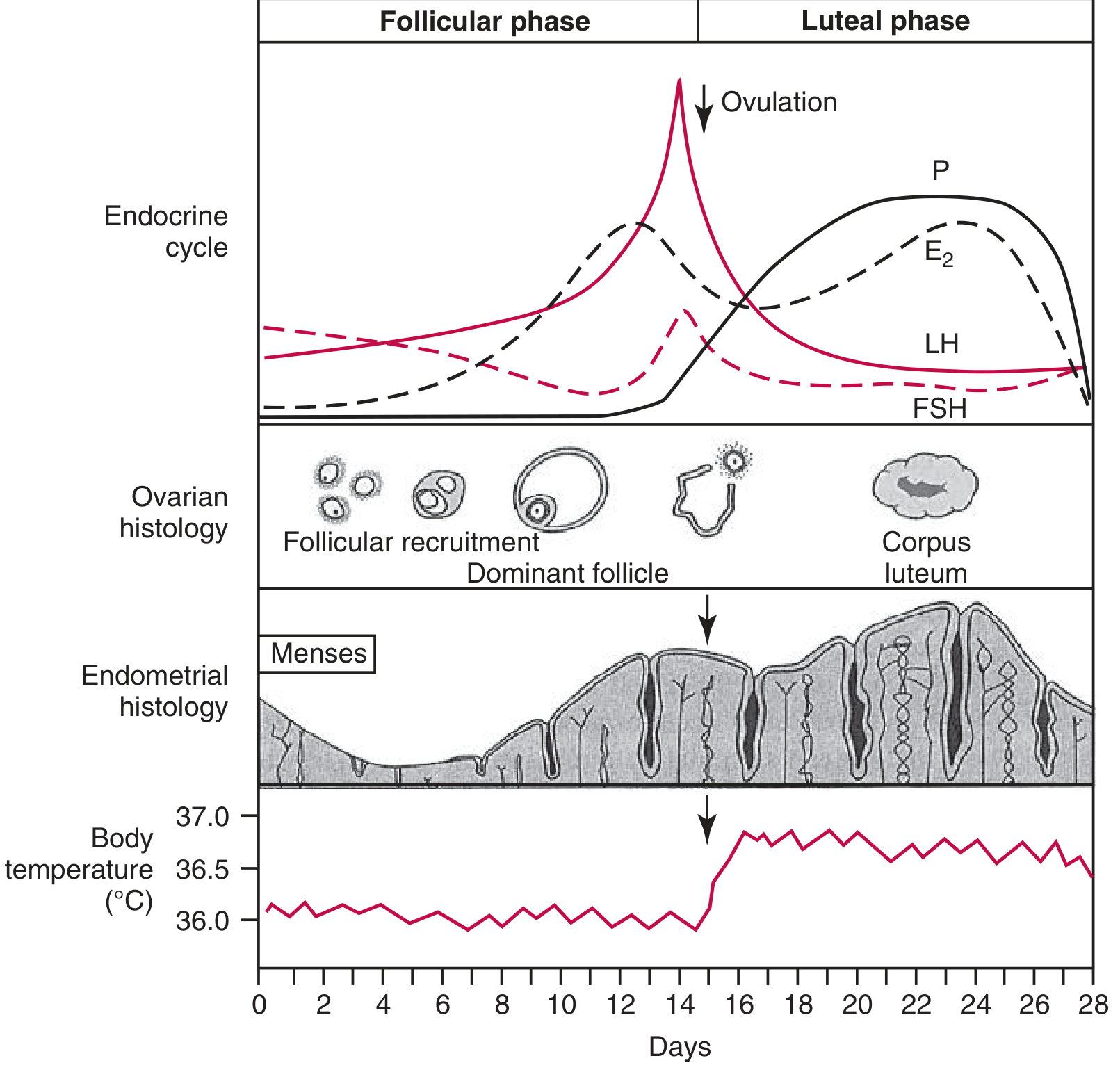
Fig: The explosive LH surge at day 14 (shown as the sharp peak in the red curve) is triggered by rising estrogen (E2) - a classic positive feedback. This surge causes ovulation.
- Rising estrogen → anterior pituitary releases LH/FSH → ovaries secrete more estrogen → more LH/FSH → explosive LH surge → ovulation
3. Oxytocin during Parturition (labor)
- Cervical dilation → oxytocin released → uterine contractions → more cervical dilation → more oxytocin → explosive delivery of fetus
4. Blood Clotting Cascade
- Factor activation triggers more factor activation → rapid clot formation
5. Pathological: Hemorrhagic Shock (vicious cycle)
- Severe bleeding → low cardiac output → poor coronary perfusion → weaker heart → even lower output → death
Key Point
Positive feedback is beneficial when a rapid, decisive event is needed (action potential, ovulation, childbirth). When uncontrolled (hemorrhagic shock, high fever), it becomes life-threatening.
(Costanzo Physiology 7th Ed.; Guyton and Hall Textbook of Medical Physiology 14th Ed.)
https://orris.care/chat/c0e64ea3-f9c6-4220-8dcc-4e8c369e12ed
That link points to a previous Orris chat session. Unfortunately, I can't access or retrieve content from other chat sessions or URLs - each conversation is independent and I don't have the ability to browse or log into the Orris platform.
What you can do:
- Copy and paste the questions or content from that chat directly into this conversation
- Take a screenshot of the questions and share it here (like you did with your physiology notes)
- Type out what you need answered
I'll then answer them fully with diagrams from the medical textbooks, just like the General Physiology questions above!
Transport mechanism across cell membrane not explained in simple language
Here's the full explanation in simple, easy language with all diagrams:
Transport Mechanisms Across Cell Membrane
(Simple Language - Exam Ready)
First, understand the basic idea:
Think of the cell membrane as a security gate around the cell. Not everything can enter or exit freely. The cell uses different "methods" to move substances in and out. These methods are divided into 2 big groups:
TRANSPORT ACROSS CELL MEMBRANE
         |
    _____|_____
    |         |
PASSIVE    ACTIVE
(No energy) (Needs energy/ATP)
GROUP 1 - PASSIVE TRANSPORT
"Lazy transport" - No ATP needed, substances move on their own from HIGH → LOW concentration
1. Simple Diffusion - "Walk through the wall"
Simple idea: Imagine a room full of perfume on one side of a door. The smell naturally spreads to the other side. That's diffusion.
- Substances move from more concentrated → less concentrated area
- They pass directly through the lipid bilayer (the fatty layer of membrane)
- Only fat-soluble (lipid-soluble) substances can do this
What crosses by simple diffusion?
| Substance | Why it can cross |
|---|---|
| O2 (oxygen) | Small, fat-soluble |
| CO2 (carbon dioxide) | Small, fat-soluble |
| Alcohol | Fat-soluble |
| Steroid hormones | Fat-soluble |
| Urea | Very small, uncharged |
Key rule: The bigger the concentration difference, the faster the diffusion (Fick's Law)
2. Osmosis - "Water's version of diffusion"
Simple idea: Water always moves toward the side with MORE dissolved stuff (solutes).
Fig: Water molecules (blue dots) move from the pure water side → toward the NaCl solution (which has Na+ green and Cl- yellow ions). Water moves from LOW solute to HIGH solute side.
- Only water moves this way
- It moves through aquaporins (special water channels in the membrane)
- Movement is from hypotonic → hypertonic solution
- The pressure needed to STOP osmosis = osmotic pressure
Quick memory trick:
Water is lazy - it moves toward where it's needed most (where solutes are concentrated)
3. Facilitated Diffusion - "Moving with a helper, still downhill"
Simple idea: Glucose is water-soluble - it cannot cross the fatty membrane on its own. So it uses a protein carrier as a helper. But it still moves from HIGH → LOW concentration (no energy needed).
- Uses carrier proteins (e.g., GLUT transporters for glucose)
- Still moves down the concentration gradient
- No ATP required
- But it is saturable - too much glucose = all carriers are busy = transport slows down
Examples:
- Glucose entering RBCs (GLUT1)
- Glucose entering muscle and fat cells (GLUT4)
- Amino acid transport
4. Ion Channel Diffusion - "Ions rushing through open gates"
Simple idea: Ions like Na+, K+, Ca2+ are charged - they can't cross the fatty membrane. They use protein channels (like tunnels) that can open and close.
- These channels are selective (Na+ channel only lets Na+ through)
- They are controlled (gated):
- Voltage-gated - open when membrane voltage changes (nerve impulse)
- Ligand-gated - open when a chemical (neurotransmitter) binds
- Mechanically-gated - open when physically stretched
Examples:
- Na+ rushing into nerve cells during action potential
- K+ moving out during repolarization
- Ca2+ entering muscle cells to trigger contraction
GROUP 2 - ACTIVE TRANSPORT
"Hard work transport" - Needs ATP, moves substances from LOW → HIGH concentration (uphill, against the gradient)
5. Primary Active Transport - "Direct use of ATP"
Simple idea: Like pumping water uphill using a motor. The "motor" here is ATP.
Fig: The Na+/K+ pump - the most important primary active transporter. Every cycle: 3 Na+ pumped OUT of cell, 2 K+ pumped IN, 1 ATP used. This keeps Na+ high outside and K+ high inside.
The Na+/K+ ATPase Pump (Most important example):
Step by step (very simple):
STEP 1: 3 Na+ (inside cell) bind to the pump protein
         ↓
STEP 2: 2 K+ (outside cell) bind to the pump protein
         ↓
STEP 3: ATP is broken down → ADP + energy released
         ↓
STEP 4: Pump changes shape
         ↓
STEP 5: 3 Na+ thrown OUTSIDE, 2 K+ pulled INSIDE
         ↓
STEP 6: Pump resets → cycle repeats
Result:
- Na+ = HIGH outside (142 mEq/L) | LOW inside (10 mEq/L)
- K+ = LOW outside (4 mEq/L) | HIGH inside (140 mEq/L)
- Creates resting membrane potential (-70 mV)
- Pumps out more + charges than it brings in → electrogenic
Other examples of primary active transport:
- Ca2+ pump (keeps calcium low inside cells)
- H+/K+ ATPase (stomach acid secretion)
6. Secondary Active Transport - "Stealing energy from Na+ gradient"
Simple idea: The Na+/K+ pump creates a "spring" (Na+ gradient - Na+ wants to rush back into the cell). The cell uses this "spring energy" to carry other substances against THEIR gradient. No direct ATP used - borrows from Na+ gradient.
Two types:
a) Co-transport / Symport - same direction
- Na+ and glucose BOTH move INTO the cell together
- Na+ goes down its gradient (pulling glucose along)
- Glucose goes AGAINST its gradient (piggybacks on Na+)
OUTSIDE → [Na+ + Glucose] → carrier protein → INSIDE
          Na+ going downhill drags glucose uphill
Example: Glucose absorption from intestine and kidney tubules (SGLT transporters)
b) Counter-transport / Antiport - opposite directions
- Na+ moves IN while another substance moves OUT
- Na+ going downhill pushes the other substance uphill
OUTSIDE ← [Ca2+] ← carrier protein ← [Na+] ← INSIDE
         Na+ going inward pushes Ca2+ outward
Example: Na+/Ca2+ exchanger in heart muscle
7. Vesicular Transport - "Bulk transport by packaging"
Simple idea: For large things (proteins, bacteria, hormones) that can't cross through channels or pumps, the cell wraps them in a membrane "bag" (vesicle) to move them.
a) Endocytosis - "Cell eating/drinking" (bringing things IN)
- Phagocytosis ("cell eating") - engulfs large particles like bacteria, dead cells
- Pinocytosis ("cell drinking") - engulfs droplets of fluid
- Receptor-mediated endocytosis - specific molecules (LDL cholesterol, insulin) bind receptors, then get engulfed
b) Exocytosis - "Cell secreting" (sending things OUT)
- Vesicles fuse with cell membrane and release contents
- Examples: Insulin secretion from pancreas, neurotransmitter release from nerve terminals, mucus secretion
Full Summary Diagram
Fig: From left to right - Simple diffusion (directly through lipid bilayer), Facilitated diffusion (via carrier protein, no energy), Active transport (via carrier protein + energy = ATP).
Quick Revision Table
| Type | Energy | Direction | Carrier? | Examples |
|---|---|---|---|---|
| Simple diffusion | ❌ No | High→Low | ❌ No | O2, CO2, steroids |
| Osmosis | ❌ No | High→Low water | ❌ No (aquaporins) | Water |
| Facilitated diffusion | ❌ No | High→Low | ✅ Yes | Glucose (GLUT) |
| Ion channel | ❌ No | High→Low | ✅ Channel | Na+, K+, Ca2+ |
| Primary active | ✅ ATP | Low→High | ✅ Yes | Na+/K+ pump |
| Secondary active | ✅ Na+ gradient | Low→High | ✅ Yes | Glucose (intestine) |
| Endocytosis | ✅ Yes | Into cell | Vesicle | Bacteria, LDL |
| Exocytosis | ✅ Yes | Out of cell | Vesicle | Insulin, neurotransmitters |
Memory Trick for Exam
"SOFT PAV E"
- Simple diffusion
- Osmosis
- Facilitated diffusion
- Tunnel (ion channels)
- Primary active transport
- Antiport/Symport (secondary active)
- Vesicular (endo/exocytosis)
- Exocytosis
(Source: Guyton & Hall Textbook of Medical Physiology, 14th Ed.)
Make quike rivision table small so i can take screenshot
Quick Revision - Transport Across Cell Membrane
| Type | ATP? | Direction | Example |
|---|---|---|---|
| Simple diffusion | ❌ | High→Low | O2, CO2 |
| Osmosis | ❌ | High→Low (water) | Water |
| Facilitated diffusion | ❌ | High→Low | Glucose (GLUT) |
| Ion channel | ❌ | High→Low | Na+, K+, Ca2+ |
| Primary active | ✅ | Low→High | Na+/K+ pump |
| Secondary active | ✅* | Low→High | Glucose (intestine) |
| Endocytosis | ✅ | Into cell | Bacteria, LDL |
| Exocytosis | ✅ | Out of cell | Insulin, neurotransmitters |
✅ = uses Na+ gradient (not direct ATP)
Cell membrane explain simple so i can remember
Cell Membrane - Super Simple Explanation 🧠
Think of it like a SANDWICH 🥪
🍞 Top bread    = Hydrophilic heads (love water, face ECF)
🧈 Butter layer = Hydrophobic tails (hate water, face each other)
🍞 Bottom bread = Hydrophilic heads (love water, face ICF)
That's your phospholipid bilayer - the main wall of the cell.
4 Main Components - "PPCC"
| Letter | Component | Simple Role |
|---|---|---|
| P | Phospholipids | Main wall/barrier |
| P | Proteins | Gates, channels, pumps |
| C | Cholesterol | Controls fluidity (not too stiff, not too loose) |
| C | Carbohydrates | ID card of the cell (recognition) |
Proteins - 2 Types Only
INTEGRAL protein  = Goes ALL THE WAY through the membrane
                    (like a pipe through a wall)
                    → Acts as channels, pumps, receptors

PERIPHERAL protein = Sits on the SURFACE only
                    (like a sticker on a wall)
                    → Acts as enzymes, structural support
5 Functions - "STERC"
| Letter | Function |
|---|---|
| S | Selective barrier (controls entry/exit) |
| T | Transport (moves substances) |
| E | Electrical potential (creates -70mV) |
| R | Reception (receives hormones/signals) |
| C | Cell recognition (glycocalyx - immune) |
One Line to Remember Everything:
"A fatty sandwich with protein doors, cholesterol springs, and a sugar coat outside"
Explain sodium potassium pump well simple so i remember and also how to write in exam
Na⁺/K⁺ Pump - Simple + Exam Ready
First, WHY does this pump exist?
The cell needs Na+ LOW inside and K+ HIGH inside. But both want to leak back to where they came from. So the cell has a pump that constantly fixes this - 24/7, non-stop.
The Simple Story 🔄
Think of it like a revolving door security guard who:
- Throws out 3 Na+ every time
- Lets in 2 K+ every time
- Charges 1 ATP as fee each cycle
INSIDE CELL                    OUTSIDE CELL
                    PUMP
   3 Na+  ────────────────→   3 Na+ (OUT)
   2 K+   ←────────────────   2 K+  (IN)
   ATP    ────→  ADP + Pi
         (energy used)
Structure (2 parts only)
| Subunit | Size | Job |
|---|---|---|
| α subunit | Large (100,000 Da) | Main worker - has Na+ sites, K+ sites, ATPase |
| β subunit | Small (55,000 Da) | Anchor - holds pump in membrane |
α subunit has 3 features:
- 3 binding sites for Na+ (inside face)
- 2 binding sites for K+ (outside face)
- ATPase activity (breaks ATP for energy)
How it Works - Step by Step
Step 1️⃣  →  3 Na+ (inside) bind to pump
Step 2️⃣  →  2 K+ (outside) bind to pump
Step 3️⃣  →  ATP breaks → ADP + Pi + ENERGY
Step 4️⃣  →  Pump changes shape (conformational change)
Step 5️⃣  →  3 Na+ pushed OUT of cell
Step 6️⃣  →  2 K+ pulled IN to cell
Step 7️⃣  →  Pump resets → repeat forever
Result of this pump
| Ion | Inside cell | Outside cell |
|---|---|---|
| Na+ | 10 mEq/L (LOW) | 142 mEq/L (HIGH) |
| K+ | 140 mEq/L (HIGH) | 4 mEq/L (LOW) |
Diagram
Why is it ELECTROGENIC?
3 positive charges go OUT, only 2 come IN Net = 1 extra positive charge leaves every cycle This makes the inside of cell more negative = -70mV resting membrane potential
Importance - "NERVE CAPS"
| Letter | Importance |
|---|---|
| N | Nerve impulse conduction |
| E | Electrogenic - creates -70mV RMP |
| R | Resting membrane potential maintenance |
| V | Volume control (prevents cell swelling) |
| E | Energy for secondary active transport |
| C | Concentration gradients of Na+ and K+ |
| A | Action potential generation |
| P | Pump runs in reverse if gradients reversed |
| S | Secondary active transport (glucose, amino acids) |
✍️ HOW TO WRITE IN EXAM
Q: Write a note on Na+/K+ pump (5 marks)
Introduction (1-2 lines):
The Na+/K+ ATPase pump is a primary active transport mechanism present in virtually all body cells. It maintains the electrochemical gradient essential for cellular excitability.
Structure (3-4 lines):
The pump is a carrier protein complex consisting of two subunits:
- α subunit (mol. wt. ~100,000): contains 3 Na+ binding sites (intracellular), 2 K+ binding sites (extracellular), and ATPase activity
- β subunit (mol. wt. ~55,000): anchors the complex in the lipid bilayer
Mechanism (write as numbered steps):
- Three Na+ ions bind to intracellular sites of α subunit
- Two K+ ions bind to extracellular sites
- ATP is hydrolyzed → ADP + Pi, releasing energy
- Conformational change occurs in the carrier protein
- Three Na+ are extruded outside; two K+ are transported inside
- Pump resets and cycle repeats
Draw the diagram (same as above)
Significance (bullet points):
- Maintains Na+ low inside (10 mEq/L) and K+ high inside (140 mEq/L)
- Generates resting membrane potential (-70 mV) - electrogenic pump
- Controls cell volume by regulating osmotic balance
- Creates Na+ gradient used for secondary active transport
- Essential for nerve impulse and muscle contraction
Concluding line:
The Na+/K+ pump is thus one of the most vital active transport mechanisms, consuming nearly 20-30% of total cellular ATP, reflecting its physiological importance.
3-Mark version (short note):
Write: Introduction + Structure (α and β) + Mechanism in 5 steps + Diagram + 3 points of significance
5-Mark version:
Write: Everything above in full
Passive diffusion same as last question explain
Passive Diffusion - Simple + Exam Ready
First, WHY is it called "PASSIVE"?
Because the substance does the work on its own. No energy (ATP) needed. It just flows downhill - from where there is MORE → to where there is LESS.
The Simple Story 🌬️
Imagine you spray perfume in one corner of a room. The smell automatically spreads to the whole room. Nobody pushed it. It moved on its own. That's passive diffusion.
3 Types - "SIF"
PASSIVE DIFFUSION
       |
  _____|_____________________
  |          |              |
Simple    Ion Channel   Facilitated
Diffusion  Diffusion    Diffusion
(through   (through     (through
lipid)     pores)       carrier)
Type 1 - Simple Diffusion
"Walk straight through the wall"
- Substance crosses directly through the lipid bilayer
- Only fat-soluble (lipid-soluble) substances can do this
- No protein needed
What crosses?
| Substance | Why? |
|---|---|
| O2, CO2 | Small + fat-soluble |
| N2 | Small + fat-soluble |
| Alcohol | Fat-soluble |
| Steroid hormones | Fat-soluble |
| Urea | Very small, uncharged |
Type 2 - Ion Channel Diffusion
"Rush through an open gate"
- Ions are charged → cannot cross fatty membrane
- They use protein channels (tunnels) = selective pores
- Still passive - ions rush through down their electrochemical gradient
Types of channels:
| Channel Type | Opens when... | Example |
|---|---|---|
| Voltage-gated | Membrane voltage changes | Na+ channel in nerve |
| Ligand-gated | Chemical (neurotransmitter) binds | Acetylcholine receptor |
| Mechanically-gated | Physical stretch/pressure | Touch receptors |
What crosses?
Na+, K+, Ca2+, Cl-
Type 3 - Facilitated Diffusion
"Moving downhill with a helper"
- Substance is water-soluble → can't cross lipid layer alone
- Uses a carrier protein as helper
- Still moves HIGH → LOW concentration
- No ATP needed
- BUT it is saturable (too much substance = carriers get full = slows down)
What crosses?
| Substance | Carrier |
|---|---|
| Glucose | GLUT 1-4 transporters |
| Amino acids | Amino acid carriers |
| Nucleosides | Nucleoside transporters |
Osmosis - Bonus type (water's diffusion)
- Water moves from LOW solute → HIGH solute side
- Through aquaporins (water channels)
- No ATP needed
Fick's Law (Rate of Diffusion) - Just remember the concept
Rate of diffusion depends on:
Rate ∝  Concentration difference × Surface area
        ─────────────────────────────────────────
                  Membrane thickness
More concentration difference = Faster diffusion
Thicker membrane = Slower diffusion
Quick Comparison Table
| Feature | Simple | Ion Channel | Facilitated |
|---|---|---|---|
| Pathway | Lipid bilayer | Protein pore | Carrier protein |
| ATP needed | ❌ | ❌ | ❌ |
| Direction | High→Low | High→Low | High→Low |
| Saturable | ❌ | ❌ | ✅ |
| Examples | O2, CO2 | Na+, K+ | Glucose |
✍️ HOW TO WRITE IN EXAM
Q: Write a note on Passive Diffusion (5 marks)
Introduction (2 lines):
Passive diffusion is the movement of molecules across the cell membrane from a region of higher concentration to lower concentration, down the concentration gradient, without the expenditure of metabolic energy (ATP).
Types - write as headings with explanation:
1. Simple Diffusion
Lipid-soluble substances (O2, CO2, steroids, alcohol) diffuse directly through the phospholipid bilayer. The rate is governed by Fick's Law of Diffusion: Rate ∝ (Concentration gradient × Surface area) / Membrane thickness
2. Diffusion Through Ion Channels
Water-soluble ions (Na+, K+, Ca2+, Cl-) pass through specific protein-lined pores called ion channels. Channels are selective and may be voltage-gated, ligand-gated, or mechanically-gated.
3. Facilitated Diffusion
Water-soluble molecules like glucose and amino acids move down their concentration gradient via specific carrier proteins (e.g., GLUT transporters). This process is saturable and specific but requires no ATP.
4. Osmosis
Water moves across a semipermeable membrane from a region of low solute concentration (high water concentration) to high solute concentration (low water concentration), through aquaporins.
Characteristics (bullet points):
- No energy (ATP) required
- Movement is always down the concentration gradient
- Net diffusion stops when equilibrium is reached
- Rate is proportional to the concentration difference
- Rate increases with temperature
Diagram (draw transport diagram - simple, facilitated, active side by side)
Concluding line:
Passive diffusion is the simplest and most energy-efficient method of membrane transport, playing a vital role in gas exchange, nutrient absorption, and maintenance of ionic gradients across cell membranes.
3-Mark version:
Write: Introduction + 3 types with one line each + 3 characteristics + diagram
5-Mark version:
Write: Everything above in full
One Line Memory Trick:
"Passive = No Payment (no ATP) - flows freely downhill" "SIF = Simple, Ion channel, Facilitated"
Again
Could you tell me what you mean by "Again"?
- 👉 Re-explain Passive Diffusion in an even simpler way?
- 👉 Re-explain Na+/K+ Pump again?
- 👉 Re-explain Cell Membrane again?
- 👉 Something else from your physiology notes?
Just let me know which topic and I'll redo it! 😊
Missing part
Your credits are exhausted. You can purchase a top-up or subscribe to a plan to continue using Orris. If you're already subscribed, your daily credits will reset tomorrow.
This is a shared conversation. Sign in to Orris to start your own chat.