The parts of brain in details I want to make exam oriented notes and also give me the blood supply of brain

Reading File
Finding Sources
Finding Sources
Finding Sources
Finding Sources
Finding Sources
Finding Sources
Finding Sources
Reading File
Reading File
Reading File
Searching Images

parts of brain cerebrum cerebellum brainstem diagram anatomy

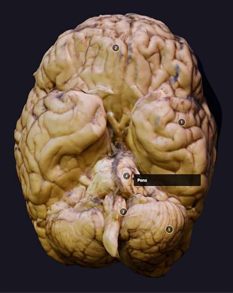
This clinical image shows a monoscopic, three-dimensional digital reconstruction of a postmortem human brain viewed from the inferior (ventral) aspect. The anatomical diagram highlights major neuroanatomical structures using numerical labels. The cerebrum (1) dominates the view, displaying characteristic gyri and sulci with a tan/pale-yellow coloration typical of preserved tissue; the frontal pole (2) is clearly oriented at the top. Located inferiorly and posteriorly, the cerebellum (5) is visible with its distinct, finely foliated surface. The brainstem (3) is positioned centrally, featuring the prominent, rounded protrusion of the pons (4) which serves as a major relay between the cerebrum and cerebellum. The model demonstrates the spatial relationships between the telencephalon, metencephalon, and myelencephalon. Some focal areas of darker discoloration are present on the cortical surface, likely representing postmortem vascular changes or processing artifacts. This visual is intended for neuroanatomical education, illustrating gross brain morphology and surface anatomy.

This clinical image shows a monoscopic, three-dimensional digital reconstruction of a postmortem human brain viewed from the inferior (ventral) aspect. The anatomical diagram highlights major neuroanatomical structures using numerical labels. The cerebrum (1) dominates the view, displaying characteristic gyri and sulci with a tan/pale-yellow coloration typical of preserved tissue; the frontal pole (2) is clearly oriented at the top. Located inferiorly and posteriorly, the cerebellum (5) is visible with its distinct, finely foliated surface. The brainstem (3) is positioned centrally, featuring the prominent, rounded protrusion of the pons (4) which serves as a major relay between the cerebrum and cerebellum. The model demonstrates the spatial relationships between the telencephalon, metencephalon, and myelencephalon. Some focal areas of darker discoloration are present on the cortical surface, likely representing postmortem vascular changes or processing artifacts. This visual is intended for neuroanatomical education, illustrating gross brain morphology and surface anatomy.

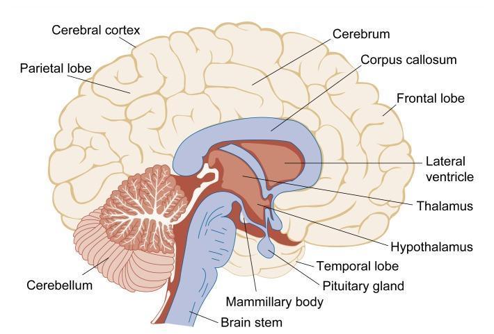
This medical anatomical diagram provides a schematic sagittal (lengthwise cross-section) view of the human brain, illustrating primary neuroanatomical structures. The cerebrum is depicted with prominent cortical folds, identifying the frontal, parietal, and temporal lobes and the cerebral cortex. Internally, the diagram highlights the components of the diencephalon and limbic system frequently affected by alcohol-related neurological disorders, including the thalamus, hypothalamus, and mammillary bodies. Key midline structures shown include the C-shaped corpus callosum, the lateral ventricle, and the pituitary gland. The posterior fossa contains the cerebellum, characterized by its distinct folia (fine surface ridges), positioned behind the brainstem. The brainstem is shown extending inferiorly toward the spinal cord. This illustration is designed for educational use in explaining neurological anatomy and the regional susceptibility of brain structures to toxic substances or metabolic changes, such as those occurring in chronic alcoholism.

This medical anatomical diagram provides a schematic sagittal (lengthwise cross-section) view of the human brain, illustrating primary neuroanatomical structures. The cerebrum is depicted with prominent cortical folds, identifying the frontal, parietal, and temporal lobes and the cerebral cortex. Internally, the diagram highlights the components of the diencephalon and limbic system frequently affected by alcohol-related neurological disorders, including the thalamus, hypothalamus, and mammillary bodies. Key midline structures shown include the C-shaped corpus callosum, the lateral ventricle, and the pituitary gland. The posterior fossa contains the cerebellum, characterized by its distinct folia (fine surface ridges), positioned behind the brainstem. The brainstem is shown extending inferiorly toward the spinal cord. This illustration is designed for educational use in explaining neurological anatomy and the regional susceptibility of brain structures to toxic substances or metabolic changes, such as those occurring in chronic alcoholism.

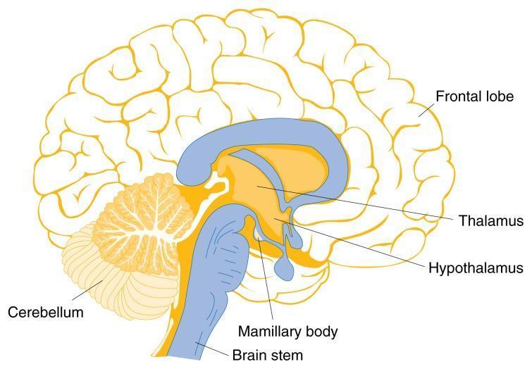
Anatomical Diagram: A mid-sagittal illustration of the human brain, highlighting regions vulnerable to thiamine deficiency and Wernicke-Korsakoff Syndrome. The diagram depicts the frontal lobe with its characteristic gyri and sulci occupying the anterior and superior portions of the cerebrum. Key labeled structures in the central and posterior regions include the thalamus and hypothalamus, located superior to the small, rounded mamillary body. The brain stem is shown extending inferiorly, connecting to the spinal cord. Posterior to the brain stem is the cerebellum, illustrating its distinctive arbor vitae branching pattern. The lateral ventricle is represented as a blue, C-shaped structure above the thalamus. This illustration serves an educational purpose in neurology and nutrition, demonstrating the spatial relationships and functional anatomy of subcortical and brainstem structures often involved in metabolic encephalopathies.

Anatomical Diagram: A mid-sagittal illustration of the human brain, highlighting regions vulnerable to thiamine deficiency and Wernicke-Korsakoff Syndrome. The diagram depicts the frontal lobe with its characteristic gyri and sulci occupying the anterior and superior portions of the cerebrum. Key labeled structures in the central and posterior regions include the thalamus and hypothalamus, located superior to the small, rounded mamillary body. The brain stem is shown extending inferiorly, connecting to the spinal cord. Posterior to the brain stem is the cerebellum, illustrating its distinctive arbor vitae branching pattern. The lateral ventricle is represented as a blue, C-shaped structure above the thalamus. This illustration serves an educational purpose in neurology and nutrition, demonstrating the spatial relationships and functional anatomy of subcortical and brainstem structures often involved in metabolic encephalopathies.

Reading File
Searching Images

circle of Willis cerebral arteries blood supply diagram

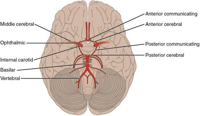
An anatomical diagram illustrating the arterial supply of the brain from an inferior (basal) perspective, specifically highlighting the Circle of Willis and its contributing vessels. The diagram shows the bilateral internal carotid arteries branching into the middle cerebral (MCA) and ophthalmic arteries. Centrally, the anterior cerebral arteries (ACA) are seen originating and coursing medially into the longitudinal fissure, connected by the anterior communicating artery (ACoA). The posterior circulation is depicted with the vertebral arteries merging into the basilar artery, which subsequently divides into the posterior cerebral arteries (PCA). Posterior communicating arteries (PCoA) are shown forming the critical anastomotic link between the internal carotid and posterior cerebral systems. The diagram is superimposed on the ventral surface of the brain, demonstrating the spatial relationship of these vessels to the frontal, temporal, and occipital lobes, as well as the cerebellum and brainstem. This illustration is an educational resource for understanding neurovascular anatomy and the territories susceptible to ischemic stroke.

An anatomical diagram illustrating the arterial supply of the brain from an inferior (basal) perspective, specifically highlighting the Circle of Willis and its contributing vessels. The diagram shows the bilateral internal carotid arteries branching into the middle cerebral (MCA) and ophthalmic arteries. Centrally, the anterior cerebral arteries (ACA) are seen originating and coursing medially into the longitudinal fissure, connected by the anterior communicating artery (ACoA). The posterior circulation is depicted with the vertebral arteries merging into the basilar artery, which subsequently divides into the posterior cerebral arteries (PCA). Posterior communicating arteries (PCoA) are shown forming the critical anastomotic link between the internal carotid and posterior cerebral systems. The diagram is superimposed on the ventral surface of the brain, demonstrating the spatial relationship of these vessels to the frontal, temporal, and occipital lobes, as well as the cerebellum and brainstem. This illustration is an educational resource for understanding neurovascular anatomy and the territories susceptible to ischemic stroke.

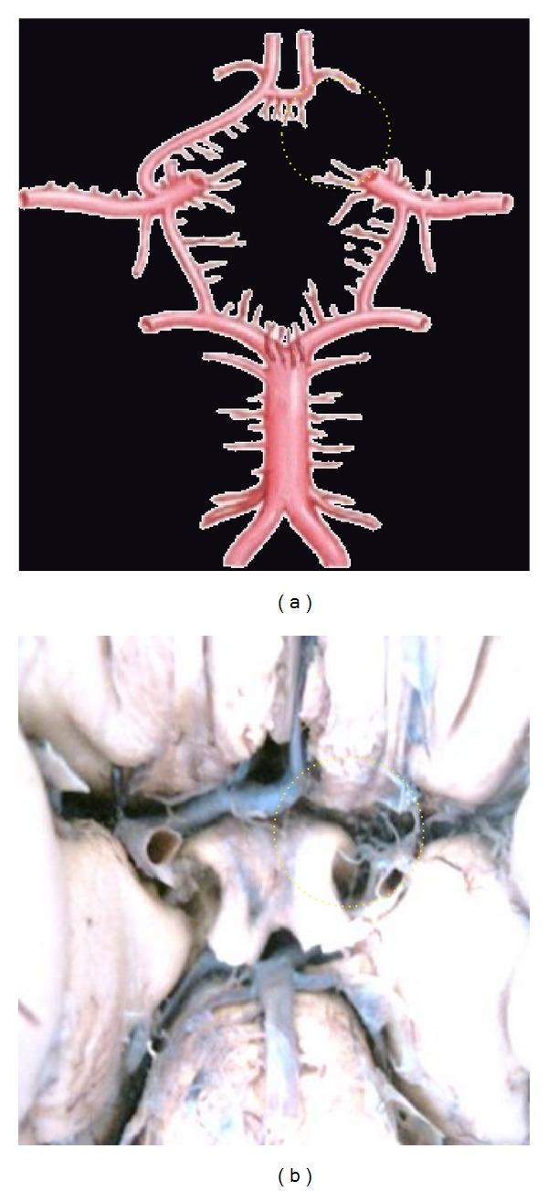
This medical visual content consists of an anatomical diagram (a) and a corresponding clinical photograph of a cadaveric dissection (b) illustrating neurovascular anatomy. The focus is on the Circle of Willis, specifically demonstrating a congenital vascular variation known as aplasia of the A1 segment of the anterior cerebral artery (ACA). In both panels, a yellow dotted circle highlights the area where the right A1 segment should connect the internal carotid artery to the anterior communicating artery (ACoA). The absence of this segment results in an incomplete Circle of Willis, which has clinical significance for collateral cerebral blood flow. The diagram (a) clearly depicts the major components of the cerebral arterial circle, including the basilar artery, posterior cerebral arteries, internal carotid arteries, and the intact left A1 and A2 segments. The clinical photograph (b) provides a real-world view of the same anatomical deficiency at the base of the brain. This material is designed for advanced medical education in neuroanatomy and radiology, emphasizing common vascular anomalies.

This medical visual content consists of an anatomical diagram (a) and a corresponding clinical photograph of a cadaveric dissection (b) illustrating neurovascular anatomy. The focus is on the Circle of Willis, specifically demonstrating a congenital vascular variation known as aplasia of the A1 segment of the anterior cerebral artery (ACA). In both panels, a yellow dotted circle highlights the area where the right A1 segment should connect the internal carotid artery to the anterior communicating artery (ACoA). The absence of this segment results in an incomplete Circle of Willis, which has clinical significance for collateral cerebral blood flow. The diagram (a) clearly depicts the major components of the cerebral arterial circle, including the basilar artery, posterior cerebral arteries, internal carotid arteries, and the intact left A1 and A2 segments. The clinical photograph (b) provides a real-world view of the same anatomical deficiency at the base of the brain. This material is designed for advanced medical education in neuroanatomy and radiology, emphasizing common vascular anomalies.

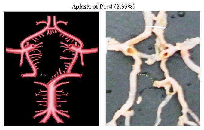
This educational image consists of a side-by-side comparison illustrating a vascular anatomical variation within the Circle of Willis. On the left is a medical illustration (Pathophysiology Diagram) of the cerebral arterial system, and on the right is a corresponding clinical photograph of a cadaveric specimen dissection. The focus is on aplasia of the P1 segment of the posterior cerebral artery (PCA). In both images, the basilar artery and the bifurcation into the superior cerebellar arteries are visible. However, there is a distinct discontinuity where the P1 segment should connect the basilar artery to the P2 segment of the PCA. A yellow dotted line is used as an annotation to indicate the expected anatomical trajectory of the missing P1 segment. This variation signifies a 'fetal-type' posterior circulation, where the distal PCA (P2 segment and beyond) receives its primary blood supply from the internal carotid artery via the posterior communicating artery rather than from the basilar artery. Such morphological variations are clinically significant in neurosurgery and the management of ischemic stroke or aneurysms.

This educational image consists of a side-by-side comparison illustrating a vascular anatomical variation within the Circle of Willis. On the left is a medical illustration (Pathophysiology Diagram) of the cerebral arterial system, and on the right is a corresponding clinical photograph of a cadaveric specimen dissection. The focus is on aplasia of the P1 segment of the posterior cerebral artery (PCA). In both images, the basilar artery and the bifurcation into the superior cerebellar arteries are visible. However, there is a distinct discontinuity where the P1 segment should connect the basilar artery to the P2 segment of the PCA. A yellow dotted line is used as an annotation to indicate the expected anatomical trajectory of the missing P1 segment. This variation signifies a 'fetal-type' posterior circulation, where the distal PCA (P2 segment and beyond) receives its primary blood supply from the internal carotid artery via the posterior communicating artery rather than from the basilar artery. Such morphological variations are clinically significant in neurosurgery and the management of ischemic stroke or aneurysms.

Excellent! I now have rich content from multiple sources. Let me compile the comprehensive exam-oriented notes.

🧠 Parts of the Brain - Exam-Oriented Notes

OVERVIEW: Major Divisions of the Brain

The brain is divided into 3 primary parts:
PartLatin MeaningKey Function
CerebrumLargest, rostral-mostThought, sensation, movement, consciousness
Cerebellum"Little brain"Movement coordination, balance
BrainstemStalk of the brainVital functions - breathing, consciousness, temperature

1. CEREBRUM

Basic Structure

  • Largest part of the brain (rostral-most)
  • Divided by the deep sagittal fissure into:
    • Right cerebral hemisphere - controls LEFT side of body (sensations + movements)
    • Left cerebral hemisphere - controls RIGHT side of body
  • Surface: Gyri (ridges), Sulci (grooves), Fissures (deep grooves)

Lobes of the Cerebrum (4 main lobes)

LobeLocationKey Functions
FrontalAnterior, behind foreheadMotor cortex, voluntary movement, speech (Broca's area), executive functions, personality
ParietalBehind central sulcusSomatosensory cortex, body awareness, spatial orientation
TemporalBelow lateral sulcusAuditory cortex, memory (hippocampus), language comprehension (Wernicke's area)
OccipitalPosterior/backPrimary visual cortex - vision processing
Exam tip: Broca's area (motor speech) = Frontal lobe. Wernicke's area (language comprehension) = Temporal lobe. Both typically LEFT hemisphere.

Key Landmarks

  • Central sulcus - separates frontal (motor) from parietal (sensory)
  • Lateral sulcus (Sylvian fissure) - separates temporal from frontal/parietal
  • Primary motor cortex - precentral gyrus (frontal lobe)
  • Primary somatosensory cortex - postcentral gyrus (parietal lobe)

Deep Structures of the Cerebrum

  • Corpus callosum - connects right and left hemispheres (commissural fibers)
  • Internal capsule - major white matter pathway; carries corticospinal tracts
  • Basal ganglia - caudate, putamen, globus pallidus; involved in motor control
  • Limbic system - hippocampus (memory), amygdala (emotion), cingulate gyrus

Cerebral Cortex

  • Gray matter on the outside (neuronal cell bodies)
  • Functional areas:
    • Primary areas - direct sensory/motor processing
    • Association areas - integration and higher-order processing

2. DIENCEPHALON (part of the "deep brain" / forebrain)

Often considered with the cerebrum:
StructureFunction
ThalamusRelay station - all sensory info (except smell) passes through here to cortex
HypothalamusHomeostasis - temperature, hunger, thirst, circadian rhythm, hormone regulation via pituitary
EpithalamusContains pineal gland (melatonin - sleep-wake cycle)
SubthalamusMotor function (subthalamic nucleus - implicated in Parkinson's)
Exam tip: "All roads lead to the thalamus" - it is the gateway to conscious perception.

3. CEREBELLUM

  • Located posterior and inferior to the cerebrum, above the brainstem
  • Contains as many neurons as both cerebral hemispheres combined (despite being much smaller)
  • IPSILATERAL control - unlike the cerebrum, LEFT cerebellum = LEFT body side

Functions

  • Coordination and fine-tuning of voluntary movement
  • Balance and equilibrium (via vestibular connections)
  • Motor learning and muscle tone
  • Timing and sequencing of movements

Structure

  • Two hemispheres + vermis (midline)
  • 3 lobes: Anterior, Posterior, Flocculonodular
  • Cerebellar cortex: 3 layers - molecular, Purkinje cell, granular

Cerebellar Connections (3 Peduncles)

PeduncleDirectionConnects to
Superior (SCP)Efferent (mainly output)Midbrain (red nucleus, thalamus)
Middle (MCP)Afferent (input)Pons (largest peduncle)
Inferior (ICP)BothMedulla + spinal cord
Exam tip: Cerebellar lesion signs = DANISH - Dysdiadochokinesia, Ataxia, Nystagmus, Intention tremor, Slurred speech (dysarthria), Hypotonia

4. BRAINSTEM

  • The most primitive but most vital part of the brain
  • Damage is usually fatal
  • Serves as relay between cerebrum, cerebellum, and spinal cord

3 Parts of the Brainstem (top to bottom):

PartLocationKey Structures/Functions
Midbrain (Mesencephalon)TopCN III, IV; superior/inferior colliculi (visual/auditory reflexes); red nucleus; substantia nigra (dopamine - Parkinson's)
PonsMiddleCN V, VI, VII, VIII; respiratory center; connects to cerebellum via MCP
Medulla OblongataBottomCN IX, X, XI, XII; cardiac, respiratory, vasomotor centers; pyramidal decussation
Exam tip: Cranial nerve nuclei 3-4 = Midbrain | CN 5-8 = Pons | CN 9-12 = Medulla

Key Brainstem Functions

  • Reticular Activating System (RAS) - consciousness and arousal
  • Vital centers in medulla: cardiac center, respiratory center, vomiting center
  • Decussation of pyramids in medulla (motor fibers cross sides here)

5. VENTRICULAR SYSTEM (Frequently Tested)

VentricleLocationCSF Flow
Lateral ventricles (1 & 2)Cerebral hemispheres→ via foramen of Monro
3rd ventricleDiencephalon→ via cerebral aqueduct (Aqueduct of Sylvius)
4th ventricleBetween brainstem and cerebellum→ Foramina of Luschka (lateral) and Magendie (medial) into subarachnoid space
Exam tip: Blockage at cerebral aqueduct = obstructive hydrocephalus (commonest site)


🩸 BLOOD SUPPLY OF THE BRAIN

Two Circulations

The brain receives blood from two main sources:
SystemArteriesSupplies
Anterior (Carotid)Internal carotid arteries (ICA)Anterior 2/3 of brain (frontal, parietal, temporal)
Posterior (Vertebrobasilar)Vertebral arteries → Basilar arteryPosterior 1/3 (occipital, cerebellum, brainstem)

Circle of Willis (Circulus Arteriosus)

The anastomotic ring at the base of the brain formed by:
Circle of Willis diagram showing internal carotid arteries, anterior and middle cerebral arteries, posterior cerebral arteries, and communicating arteries

Components (go around the circle):

Anterior Communicating Artery (ACoA)
         |
ACA (left) --- ACoA --- ACA (right)
         |                   |
ICA (left)              ICA (right)
         |                   |
PCoA (left)             PCoA (right)
         |                   |
PCA (left) --- Basilar --- PCA (right)
                  |
          Vertebral arteries
ArteryAbbreviationOriginTerritory
Anterior Cerebral ArteryACAICAMedial surface of frontal & parietal lobes; leg area of motor/sensory cortex
Middle Cerebral ArteryMCAICA (largest branch)Lateral surface of frontal, parietal, temporal; arm & face motor/sensory; Broca's & Wernicke's areas
Posterior Cerebral ArteryPCABasilar arteryOccipital lobe (visual cortex), medial temporal, thalamus
Anterior Communicating ArteryACoAConnects two ACAsAnastomosis (most common site of Berry aneurysm)
Posterior Communicating ArteryPCoAICA to PCAConnects anterior & posterior circulations
Basilar ArteryBAVertebrals unitePons, midbrain, cerebellum, PCA

Arterial Territories - Stroke Mnemonics

ACA Stroke (leg symptoms dominant):

  • Contralateral leg weakness/sensory loss (ACA supplies "leg homunculus" = medial cortex)
  • Urinary incontinence, personality changes (frontal lobe)

MCA Stroke (most common - arm + face):

  • Contralateral arm and face weakness/sensory loss
  • Aphasia if left hemisphere (Broca's = motor aphasia, Wernicke's = sensory aphasia)
  • Contralateral homonymous hemianopia
  • Gaze deviation toward the side of lesion

PCA Stroke (visual symptoms):

  • Contralateral homonymous hemianopia with macular sparing
  • Visual hallucinations, alexia without agraphia

Vertebrobasilar System - Cerebellar Arteries

ArteryAbbreviationOriginSuppliesStroke Syndrome
Posterior Inferior Cerebellar A.PICAVertebralPosterior inferior cerebellum, lateral medullaWallenberg syndrome (lateral medullary syndrome)
Anterior Inferior Cerebellar A.AICABasilarAnterior inferior cerebellum, lateral ponsAICA syndrome (facial palsy + ipsilateral hearing loss)
Superior Cerebellar A.SCABasilarSuperior cerebellum, ponsCerebellar ataxia + CN IV palsy
Exam tip: PICA = PICA = Posterior Inferior Cerebellar Artery = Wallenberg syndrome - ipsilateral facial loss of pain/temp + contralateral body loss of pain/temp + dysphagia + hoarseness + Horner's syndrome

Venous Drainage of the Brain

  • Cerebral veins drain into dural venous sinuses
  • Main drainage: Internal jugular veins (almost entirely)
SinusDrains To
Superior sagittal sinusConfluence of sinuses
Inferior sagittal sinusStraight sinus
Transverse sinusSigmoid sinus → Internal jugular vein
Cavernous sinusImportant - drains orbit, receives CN III, IV, V1, V2, VI
Exam tip: Cavernous sinus thrombosis = proptosis, periorbital edema, ophthalmoplegia (CN III, IV, VI), fever

Internal Capsule Blood Supply (High-Yield!)

  • Anterior limb: Lenticulostriate arteries (MCA branches)
  • Genu: Lenticulostriate arteries
  • Posterior limb: Lenticulostriate (MCA) + anterior choroidal artery (ICA)
Exam tip: Lacunar infarcts in internal capsule (due to hypertension damaging small lenticulostriate arteries) = pure motor hemiplegia (most common lacunar stroke)

Autoregulation of Cerebral Blood Flow

  • Brain autoregulates blood flow between MAP 50-150 mmHg
  • Normal CBF: ~50 mL/100g/min
  • Below 50 mmHg MAP: autoregulation fails → ischemia
  • CO2 is the most potent vasodilator of cerebral vessels (hypercapnia → vasodilation → increased ICP)
  • Blood-brain barrier maintained by tight junctions of endothelial cells

Quick Revision Table - High-Yield Points

TopicKey Fact
Largest part of brainCerebrum
Most vital partBrainstem (medulla)
Relay station of sensory infoThalamus
Regulates homeostasisHypothalamus
IPSILATERAL controlCerebellum
CONTRALATERAL controlCerebral cortex
Decussation of pyramidsMedulla
Most common stroke territoryMCA
Most common Berry aneurysm siteACoA junction
Wallenberg syndrome arteryPICA
CSF obstruction common siteCerebral aqueduct

Sources: Neuroscience: Exploring the Brain (5th Ed.) | Neuroanatomy through Clinical Cases (3rd Ed.) | Medical Physiology (Boron & Boulpaep)
This is a shared conversation. Sign in to Orris to start your own chat.