
What is the Role of C-Peptide with glucose
C-peptide (connecting peptide) is a byproduct of insulin synthesis. When proinsulin is cleaved in the pancreatic beta cells, it produces one molecule of insulin and one molecule of C-peptide in equal molar amounts. This makes C-peptide a useful marker of endogenous insulin production.
Searching sources...
Reading File
Searching the Web
C-peptide role glucose metabolism insulin secretion clinical significance
Now let me search the medical library for detailed textbook content on C-peptide.
Finding Sources
Finding Sources
Reading File
Reading File
Reading File
Reading File
Reading File
Loading Image
Now let me get the detailed C-peptide clinical utility section and the monitoring therapy section:
Reading File
Reading File
I now have comprehensive information from multiple authoritative textbooks. Here is a full, well-structured answer:
Role of C-Peptide in Relation to Glucose
1. What is C-Peptide?
C-peptide ("connecting peptide") is a 31-amino acid polypeptide (MW ~3,600 Da) that links the alpha and beta chains of proinsulin. In the Golgi apparatus of pancreatic beta cells, proinsulin is proteolytically cleaved by proprotein convertases (PC2 and PC1/PC3) and carboxypeptidase H to yield one molecule each of insulin and C-peptide in equimolar amounts. Both are packaged into secretory granules and co-secreted into the portal circulation upon stimulation.
- Robbins Pathologic Basis of Disease, p. 1941
- Tietz Textbook of Laboratory Medicine, 7th Ed., p. 1720
- Henry's Clinical Diagnosis and Management, p. 265
2. How Glucose Triggers C-Peptide (and Insulin) Secretion
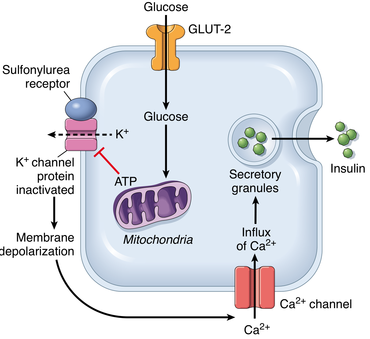
The key stimulus linking glucose to C-peptide release is illustrated below:
The sequence:
- Blood glucose rises → enters beta cells via GLUT-2 (insulin-independent)
- Glycolysis generates ATP → closes K+/ATP channels
- Membrane depolarizes → voltage-gated Ca2+ channels open
- Ca2+ influx triggers exocytosis of secretory granules → both insulin and C-peptide released simultaneously
Because they are released in equimolar amounts, C-peptide serves as a direct index of endogenous insulin secretion.
3. Why C-Peptide Levels Are Higher Than Insulin in Blood
Although secreted in equal molar amounts, fasting C-peptide concentrations are 5- to 15-fold higher than insulin:
- The liver extracts ~50% of insulin in first-pass but does not extract C-peptide
- C-peptide half-life is ~30-35 minutes vs. 4-9 minutes for insulin
- C-peptide is removed by the kidneys and a fraction is excreted unchanged in urine
This pharmacokinetic difference makes C-peptide a more stable and reliable marker of beta-cell secretory function than insulin itself.
4. Biological Activity of C-Peptide
C-peptide was long thought to be biologically inert, but emerging evidence shows it does have physiologic effects:
- May bind to cell-surface receptors (possibly GPCRs) and activate intracellular signaling
- Evidence for roles in microvascular circulation, renal tubular function, and nerve conduction - particularly relevant in diabetic complications
- Associated with glucose control: residual C-peptide levels correlate with better glycemic stability (especially in Type 1 DM)
- May modulate glucagon secretion - C-peptide is independently associated with glucagon levels
- Its therapeutic potential remains under active investigation (Frontiers in Endocrinology, 2023)
5. Clinical Significance in Glucose Disorders
A. Distinguishing Type 1 from Type 2 Diabetes
| Finding | Interpretation |
|---|---|
| Low/undetectable C-peptide + high glucose | Type 1 DM - near-total beta-cell destruction |
| Normal-high C-peptide + high glucose | Type 2 DM - insulin resistance with retained secretion |
| Peak stimulated C-peptide >1.8 ng/mL | Behaves clinically like Type 2 DM |
| Peak stimulated C-peptide <0.5 ng/mL | Behaves clinically like Type 1 DM |
B. Evaluating Fasting Hypoglycemia (Whipple's Triad)
C-peptide measurement is key in distinguishing causes of hypoglycemia:
- High insulin + HIGH C-peptide + low glucose → Insulinoma (endogenous hypersecretion)
- High insulin + LOW C-peptide + low glucose → Factitious hypoglycemia (exogenous insulin injection - commercial insulin contains no C-peptide and suppresses endogenous secretion)
C. Glucose Stimulation Tests
C-peptide is measured as part of:
- Oral Glucose Tolerance Test (OGTT) - 75 g glucose, samples at 0, 30, 60, 90, 120 min; correlates with insulin secretion in Type 2 DM
- Glucagon Stimulation Test - 1 mg IV glucagon; high sensitivity for residual beta-cell function
- Mixed Meal Tolerance Test (MMTT) - sensitive and reproducible for residual function in T1D
D. Monitoring Therapy and Transplant
- Should be undetectable after radical pancreatectomy
- Should rise after successful pancreas or islet cell transplant
- Used as an endpoint in immunomodulatory trials for Type 1 DM prevention
- 24-hour urine C-peptide correlates with fasting serum levels and post-glucose-load secretion
6. Reference Ranges
| Specimen | Normal Range |
|---|---|
| Fasting serum | 0.78 - 1.89 ng/mL (0.25 - 0.60 nmol/L) |
| Post-stimulation (glucose/glucagon) | 2.73 - 5.64 ng/mL (0.9 - 1.87 nmol/L) |
| Urine (24-hour) | 74 ± 26 μg/L (25 ± 8.8 μmol/L) |
Important: C-peptide must always be interpreted alongside a concurrent blood glucose level, as per Harrison's Principles of Internal Medicine, 22nd Ed.. A low C-peptide with elevated glucose confirms insulin deficiency; a high C-peptide with normal glucose indicates insulin resistance.
7. Summary Table
| Aspect | Role of C-Peptide |
|---|---|
| Biosynthesis | Released equimolarly with insulin from proinsulin cleavage |
| Glucose stimulus | Glucose → GLUT-2 → ATP → Ca2+ → co-secretion with insulin |
| Beta-cell surrogate | Best indirect measure of endogenous insulin production |
| Hypoglycemia workup | Distinguishes insulinoma vs. factitious hypoglycemia |
| Diabetes classification | Helps distinguish T1D (low) vs. T2D (normal/high) |
| Renal clearance | Cleared by kidneys - elevated in renal failure |
| Liver clearance | Negligible hepatic extraction (advantage over insulin measurement) |
| Biological activity | Emerging roles in microvascular and renal function |
Sources: Tietz Textbook of Laboratory Medicine 7th Ed. | Robbins Pathologic Basis of Disease | Henry's Clinical Diagnosis and Management | Harrison's Principles of Internal Medicine 22E | StatPearls: Biochemistry, C Peptide
This is a shared conversation. Sign in to Orris to start your own chat.