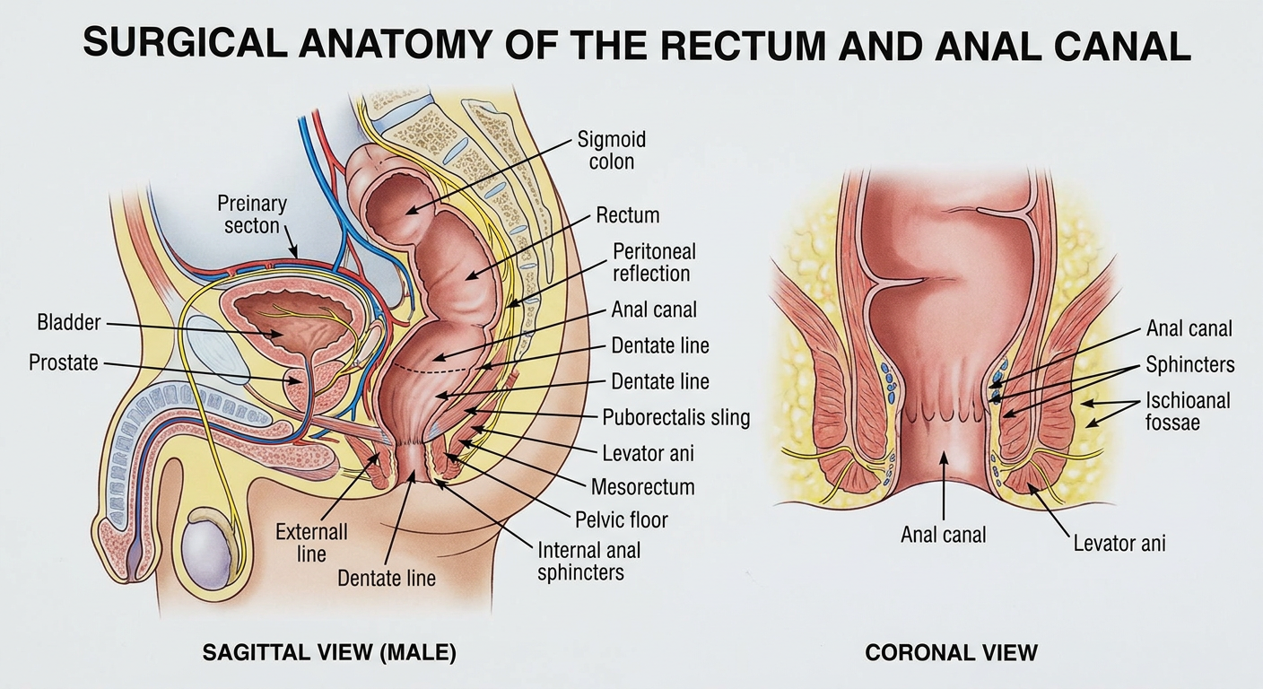
Generate a image of surgical anatomy of rectum and anal canal
A precise surgical anatomy illustration of the rectum and anal canal for medical education, showing a sagittal pelvic section and an adjacent coronal cross-sectional view. Clearly depict the rectum, sigmoid colon, anal canal, dentate line, internal and external anal sphincters, puborectalis sling, levator ani, mesorectum, pelvic floor, ischioanal fossae, peritoneal reflection, prostate and bladder in a male reference view, and relevant vessels and nerves. Use accurate labeled anatomy, clean textbook-style rendering, suitable for surgeons and trainees.
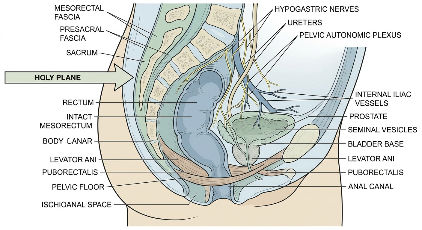
Create an annotated pelvic dissection diagram
Annotated surgical pelvic dissection diagram for colorectal surgery education, posterior and lateral pelvic dissection view in a male pelvis. Render as a clean, realistic textbook anatomical illustration, not a photograph. Show the rectum and intact mesorectum, presacral fascia over sacrum, hypogastric nerves, pelvic autonomic plexus, ureters, internal iliac vessels and branches, levator ani, puborectalis, pelvic floor, ischioanal space, prostate, seminal vesicles, bladder base, and anal canal. Include fine leader lines and clear readable labels for each structure. Emphasize correct surgical planes: holy plane between mesorectal fascia and presacral fascia, and lateral ligament region. Educational, clinically accurate, non-graphic.