
Turbid urine Post urination burning/pain ?
"urinary tract infection" AND "dysuria" AND diagnosis
turbid urine pyuria urinary tract infection microscopy
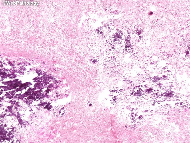
This is a histopathology slide of urinary bladder mucosa examined under light microscopy following hematoxylin and eosin staining. The primary subject is encrusted cystitis with deposition of calcium salts in the lamina propria of the bladder wall, consistent with infection by urease-splitting bacteria (e.g., Proteus mirabilis) that alkalinize urine and promote mineral precipitation on injured urothelium. The lamina propria contains scattered calcifications appearing as dark, basophilic deposits dispersed throughout the connective tissue matrix, often accompanied by a mixed inflammatory infiltrate and focal erosions of the urothelial surface. The urothelium may show reactive changes with papillary or irregular architecture, while the submucosa demonstrates edema and mild fibrosis in areas of chronicity. Calcific encrustations are multifocal and not limited to a discrete lesion, giving a 'salt-and-pepper' appearance on higher magnification. Clinically, encrusted cystitis signals a chronic bacterial infection with alkaline urine, requiring targeted antibiotics and correction of the underlying infectious milieu. The observed histology supports infection-driven mineralization rather than primary neoplastic processes. This image is relevant for educational scenarios addressing urinary tract infections, urease-producing bacteria, pathology of bladder mucosa, differential diagnosis with calcified cystitis and malakoplakia, and radiologic-pathologic correlation. Correlate with urine pH measurements and urease-positive bacterial cultures for targeted therapeutic planning.
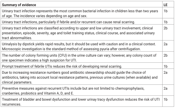
<table><thead><tr><th>Summary of evidence</th><th>LE</th></tr></thead><tbody><tr><td>Urinary tract infection represents the most common bacterial infection in children less than two years of age. The incidence varies depending on age and sex.</td><td>1b</td></tr><tr><td>Urinary tract infections, particularly if febrile and/or recurrent can cause renal scarring.</td><td>1b</td></tr><tr><td>Urinary tract infections are classified according to upper and low urinary tract involvement, clinical presentation, episode, severity, age and toilet training status, clinical course, and associated urinary tract abnormalities.</td><td>2b</td></tr><tr><td>Urinalysis by dipstick yields rapid results, but it should be used with caution and in a clinical context. Microscopic investigation is the standard method of assessing pyuria after centrifugation.</td><td>2a</td></tr><tr><td>The number of colony forming units (CFU) in the urine culture can vary; however, any colony count of one specimen indicates a high suspicion for UTI.</td><td>2b</td></tr><tr><td>Prompt treatment of febrile UTIs reduces the risk of developing renal scarring.</td><td>1b</td></tr><tr><td>Due to increasing resistance numbers good antibiotic stewardship should guide the choice of antibiotics, taking into account local resistance patterns, previous urine cultures (when available) and clinical parameters.</td><td>2a</td></tr><tr><td>Preventive measures against recurrent UTIs include but are not limited to chemoprophylaxis, cranberries, probiotics and Vitamin A, D, and E.</td><td>2a</td></tr><tr><td>Treatment of bladder and bowel dysfunction and lower urinay tracy dysfunction reduces the risk of UTI recurrences.</td><td>1b</td></tr></tbody></table>
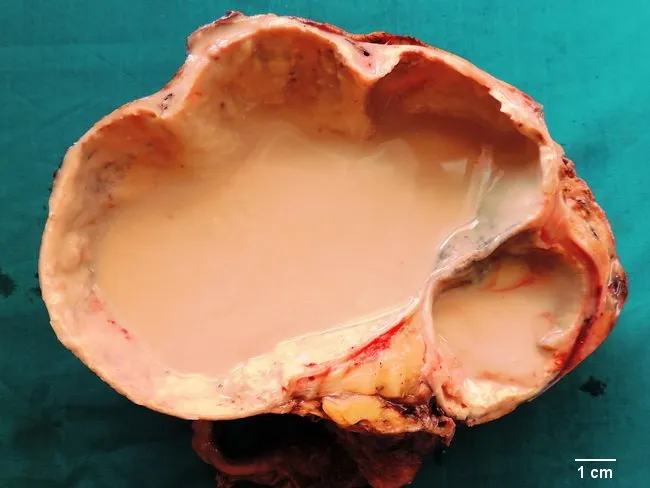
This gross pathology photograph depicts a right nephrectomy specimen with advanced hydronephrosis and pyonephrosis. The kidney is markedly enlarged and cut open to reveal an extensively dilated pelvicalyceal system filled with thick purulent material, while the renal cortex is exceedingly thinned, producing a paper-thin rim of parenchyma. The interior contains approximately three liters of creamy yellow pus and necrotic debris, reflecting suppurative destruction of the parenchyma and end-stage renal disease secondary to chronic obstructive uropathy. Histologic correlate shows xanthogranulomatous inflammation with foamy macrophages amid necrosis and granulomatous reaction, consistent with long-standing infection and obstruction. Urinary tract calculi are present in the ureter, and hydroureter accompanies the severely dilated collecting system. The gross features exemplify the classic evolution from obstructive uropathy to infected hydronephrosis, ultimately necessitating nephrectomy when function is lost. Clinically, the patient presented with high fever, flank pain, dysuria, and turbid urine with pyuria; urine cultures grew Escherichia coli. This image is valuable for teaching radiology-pathology correlation, surgical pathology training, and urologic management, illustrating the diagnostic significance of hydronephrosis complicated by infection and the rationale for drainage, antimicrobial therapy, and nephrectomy in non-functioning kidneys. It supports differential considerations including xanthogranulomatous pyelonephritis and chronic obstructive nephropathy.
Turbid Urine + Post-Urination Burning/Pain
What These Symptoms Mean
| Symptom | Pathophysiology |
|---|---|
| Turbid (cloudy) urine | Reflects pyuria (pus/WBCs in urine), bacteriuria, and sometimes hematuria. The cloudiness is caused by suspended leukocytes, bacteria, and cellular debris from an inflamed bladder wall. |
| Dysuria / burning on urination | Caused by bacterial toxins and inflammatory mediators irritating the urethral and bladder mucosa, especially during urine flow. The burning after voiding (post-micturition dysuria) is typical of cystitis. |
Classic Clinical Triad of Cystitis
- Dysuria (burning/pain during or after urination)
- Urinary frequency (need to urinate often, small volumes)
- Urgency (sudden, strong urge to void)
"Dysuria, frequency, and urgency are the classic clinical triad. Women presenting with at least one UTI symptom have a 50% chance of having a UTI. The combination of dysuria and frequency without vaginal symptoms increases the probability to 90%."
- Textbook of Family Medicine, 9e
Common Causes
- Staphylococcus saprophyticus (especially in young sexually active women)
- Klebsiella pneumoniae
- Proteus mirabilis
- Enterococcus faecalis
- Female sex (short urethra - bacteria travel easily to bladder)
- Sexual activity ("honeymoon cystitis")
- Use of spermicides or diaphragm
- Recent urinary instrumentation/catheterization
- Pregnancy, diabetes, immunosuppression
- Urinary tract abnormalities (complicated UTI)
Diagnosis
Urinalysis (first-line test)
| Finding | Significance |
|---|---|
| Pyuria (>5 WBC/hpf) | Infection/inflammation - nearly always present in cystitis |
| Bacteriuria | Confirms bacterial presence |
| Nitrites positive | Indicates gram-negative bacteria (e.g., E. coli) converting nitrates |
| Leukocyte esterase positive | Confirms WBCs present |
| Hematuria | Common in hemorrhagic cystitis |
Microscopic analysis should assess for pyuria, hematuria, and bacteriuria. The gold standard for diagnosis is urine culture. - Textbook of Family Medicine, 9e
- Urine culture with sensitivity - ordered to guide antibiotic selection, especially in recurrent or complicated cases
Red Flags Suggesting Upper Tract Involvement (Pyelonephritis)
- Fever / chills / rigors
- Flank pain or costovertebral angle (CVA) tenderness
- Nausea / vomiting
- Systemic illness
Treatment
Uncomplicated Cystitis (adult women, no structural abnormality)
| Antibiotic | Dose | Duration |
|---|---|---|
| Nitrofurantoin (Macrobid) | 100 mg twice daily | 5-7 days |
| Trimethoprim-Sulfamethoxazole (TMP-SMX) DS | 160/800 mg twice daily | 3 days |
| Fosfomycin | 3 g single dose | Single dose |
| Ciprofloxacin | 250 mg twice daily | 3 days |
Nitrofurantoin and fosfomycin are first-line choices because resistance rates are very low (~2-6%). - Tintinalli's Emergency Medicine
Supportive Measures
- Increase fluid intake (flushes bacteria from bladder)
- Phenazopyridine (urinary analgesic) - provides symptomatic relief of burning/pain but does NOT treat infection; turns urine orange
- Avoid caffeine, alcohol, and spicy foods (bladder irritants)
Differential Diagnoses to Consider
- Urethritis (Chlamydia, Gonorrhea) - especially if urethral discharge present or STI risk factors
- Vaginitis - dysuria often external, with vaginal discharge/itching (vaginal symptoms distinguish it)
- Urolithiasis (kidney/bladder stones) - colicky flank pain, hematuria without pyuria
- Interstitial cystitis - chronic pelvic pain, frequency, no bacterial growth on culture
- Bladder tumor - painless hematuria, older patients, no infection signs
When to Refer / Escalate
- Symptoms not improving after 48-72 hours of antibiotics
- Recurrent UTIs (3+ per year) - consider low-dose prophylaxis or post-coital antibiotics
- Men with UTI - always warrants further workup (rule out prostatitis, structural anomaly)
- Children with first UTI - imaging to rule out vesicoureteral reflux
- Pregnancy - even asymptomatic bacteriuria must be treated (risk of pyelonephritis and preterm birth)
- Fever + flank pain - consider IV antibiotics and hospitalization