
what is first pass metabolism in pharmacology
First pass metabolism (also called first pass effect) is the phenomenon where a drug absorbed from the gut passes through the portal vein to the liver before reaching systemic circulation, where it undergoes significant metabolic breakdown - reducing the amount of active drug that actually reaches the target tissues. This is why some drugs have very low oral bioavailability and must be given via other routes (sublingual, IV, transdermal, etc.).
Searching sources...
Reading File
Finding Sources
Searching the Web
first pass metabolism pharmacology mechanism bioavailability
Finding Sources
Reading File
Reading File
Reading File
Loading Image
Loading Image
Here is a thorough breakdown of first pass metabolism, drawn from Lippincott Pharmacology, Katzung, and Goodman & Gilman:
First Pass Metabolism (First Pass Effect)
Definition
First pass metabolism (also called the first pass effect or presystemic metabolism) is the biotransformation of a drug before it reaches systemic circulation. When a drug is swallowed and absorbed from the GI tract, it enters the portal vein and travels directly to the liver. The liver - and sometimes the gut wall itself - can metabolize a significant fraction of the drug during this initial transit, so that less active drug actually reaches the bloodstream and the target tissues.
"If the drug is rapidly metabolized in the liver or gut wall during this initial passage, the amount of unchanged drug entering the systemic circulation is decreased." - Lippincott Illustrated Reviews: Pharmacology
How It Happens - Step by Step
- Drug is swallowed and absorbed through the intestinal wall into mesenteric capillaries
- These drain into the portal vein, which goes directly to the liver
- Hepatic enzymes (primarily CYP450 enzymes, especially CYP3A4) metabolize a portion of the drug
- The gut wall also contributes (e.g., CYP3A4 and P-glycoprotein efflux)
- Only the fraction that survives this first passage enters the hepatic vein → inferior vena cava → systemic circulation
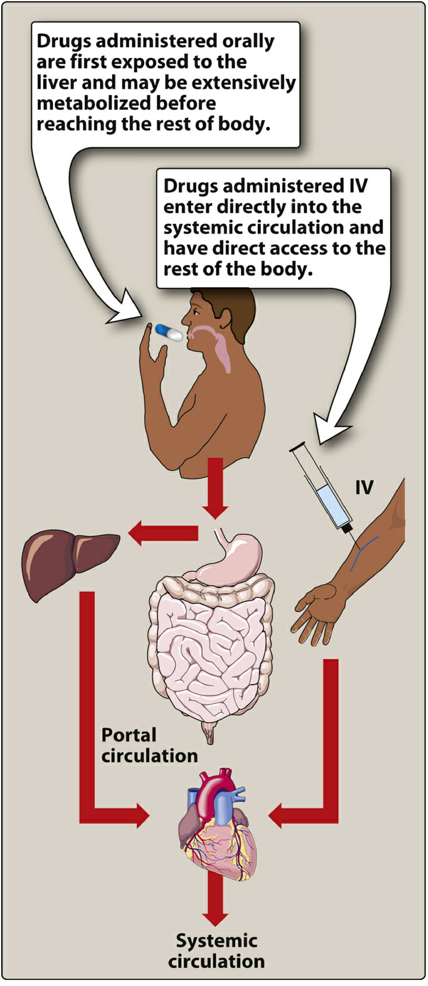
Figure: Oral drugs are exposed to the liver via portal circulation before reaching the body. IV drugs bypass this entirely. (Lippincott Pharmacology)
Effect on Bioavailability
Bioavailability (F) is the fraction of an administered dose that reaches systemic circulation unchanged. First pass metabolism is one of the key reasons oral bioavailability can be much lower than IV bioavailability.
F = AUC(oral) / AUC(IV) × 100%
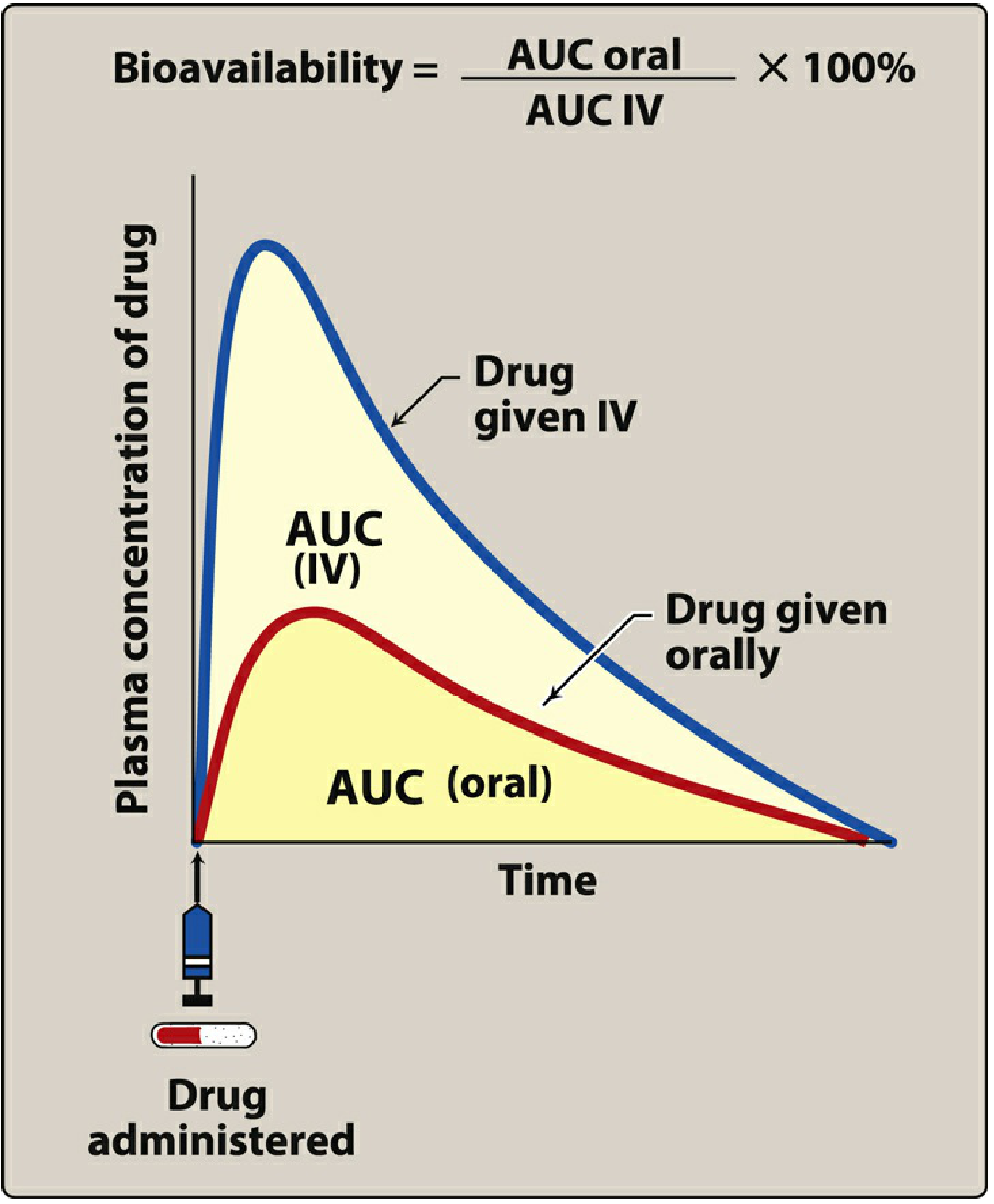
Figure: Oral drug (red curve) achieves a lower AUC than IV (blue curve) due to first pass metabolism. (Lippincott Pharmacology)
The extraction ratio is the proportion of drug removed by the liver per pass. Drugs with a high extraction ratio have very low oral bioavailability.
Classic Examples
| Drug | First Pass Effect | Clinical Implication |
|---|---|---|
| Nitroglycerin | >90% cleared | Must be given sublingually, transdermally, or IV - NOT orally |
| Lidocaine | Extensive (>60%) | Given IV only; oral metabolites cause CNS toxicity |
| Morphine | Significant | Oral dose must be much higher than IV dose |
| Propranolol | High | Large oral doses needed; wide interpatient variability |
| Naloxone | Extensive | Ineffective orally; given IV or IM |
| Isoniazid, Tricyclic antidepressants | High | Dose adjustments required |
| Codeine, Oxycodone | Low | Effective orally precisely because of reduced first pass |
Sites of First Pass Metabolism
The liver is the primary site, but first pass can occur in multiple locations:
- Gut wall - CYP3A4 and P-glycoprotein (e.g., benzylpenicillin, insulin degraded here)
- Liver - Main site; CYP450 enzymes (propranolol, lidocaine, nitroglycerin, morphine)
- Lung - For drugs given by non-GI parenteral routes; can serve as a site of first pass loss via excretion or metabolism
Routes That Bypass First Pass Metabolism
| Route | Bypass Mechanism |
|---|---|
| Intravenous (IV) | Enters systemic circulation directly |
| Sublingual | Absorbed into systemic veins (not portal), e.g., nitroglycerin |
| Transdermal | Bypasses portal circulation entirely |
| Inhalation | Bypasses hepatic first pass (though lungs may metabolize some drugs) |
| Rectal (suppository) | Lower rectum drains into inferior vena cava (~50% bypass); upper rectum drains into portal circulation |
Clinical Relevance
- Dose adjustment: Drugs with extensive first pass need much higher oral doses than IV doses (e.g., morphine oral:IV ratio is approximately 3:1)
- Inter-patient variability: Differences in CYP450 activity, hepatic blood flow, and genetic polymorphisms cause wide variation between individuals
- Liver disease: Cirrhosis with portosystemic shunting can dramatically increase bioavailability of high first-pass drugs (e.g., morphine, meperidine, midazolam, nifedipine oral bioavailability nearly doubles in cirrhosis - per Harrison's Principles of Internal Medicine)
- Drug interactions: One drug can inhibit or induce the enzymes involved, affecting the first pass metabolism of another drug (e.g., grapefruit juice inhibits CYP3A4)
- Prodrugs: Some drugs are designed to exploit first pass - they are inactive until metabolized by the liver (e.g., codeine → morphine via CYP2D6; enalapril → enalaprilat)
Summary Formula
Oral bioavailability (F) = Fraction absorbed × (1 - Extraction Ratio)
A drug with 100% GI absorption but a 0.9 extraction ratio ends up with only 10% bioavailability.
Sources: Lippincott Illustrated Reviews: Pharmacology | Katzung's Basic and Clinical Pharmacology, 16th Ed | Goodman & Gilman's The Pharmacological Basis of Therapeutics | Goldman-Cecil Medicine | Harrison's Principles of Internal Medicine 22E
Add a NOTE on DRUG ABSORPTION & FACTORS influencing it?
Drug absorption is the process by which a drug moves from its site of administration into the systemic circulation. Key factors include the drug's physicochemical properties (lipophilicity, molecular size, ionization), the route of administration, GI motility, blood flow to the absorption site, and formulation characteristics.
Searching sources...
Finding Sources
Finding Sources
Reading File
Reading File
Reading File
Finding Sources
Reading File
Loading Image
Now I have everything needed for a thorough, well-illustrated note. Here it is:
NOTE: Drug Absorption & Factors Influencing It
What is Drug Absorption?
Absorption is the transfer of a drug from its site of administration into the systemic circulation. It is the first step in the pharmacokinetic sequence - ADME (Absorption, Distribution, Metabolism, Excretion).
"Absorption from the site of administration permits entry of the drug (either directly or indirectly) into plasma." - Lippincott Illustrated Reviews: Pharmacology
Most drugs cross biological membranes by passive diffusion - moving down a concentration gradient. The rate depends on the concentration of free (unbound, uncharged) drug at the absorbing surface.
Mechanisms of Drug Absorption Across Membranes
| Mechanism | Energy | Saturable? | Notes |
|---|---|---|---|
| Passive diffusion | No | No | Most common; uncharged, lipophilic drugs prefer this |
| Facilitated diffusion | No | Yes | Uses carrier proteins; large molecules |
| Active transport | Yes (ATP) | Yes | Against concentration gradient; competitive inhibition possible |
| Endocytosis | Yes | No | Very large molecules (e.g., Vitamin B12 across gut wall) |
Factors Influencing Drug Absorption
1. pH and Drug Ionization (pKa)
This is one of the most important factors. Most drugs are weak acids or weak bases and exist in both ionized (charged) and un-ionized (uncharged) forms depending on the pH of the surrounding environment.
- Un-ionized form = lipid soluble → crosses membranes freely
- Ionized form = water soluble, charged → does NOT cross lipid membranes well
Henderson-Hasselbalch principle:
- Weak acids (e.g., aspirin, warfarin): better absorbed in acidic environments (stomach) where they are un-ionized
- Weak bases (e.g., morphine, codeine, atropine): better absorbed in alkaline environments (small intestine) where they are un-ionized
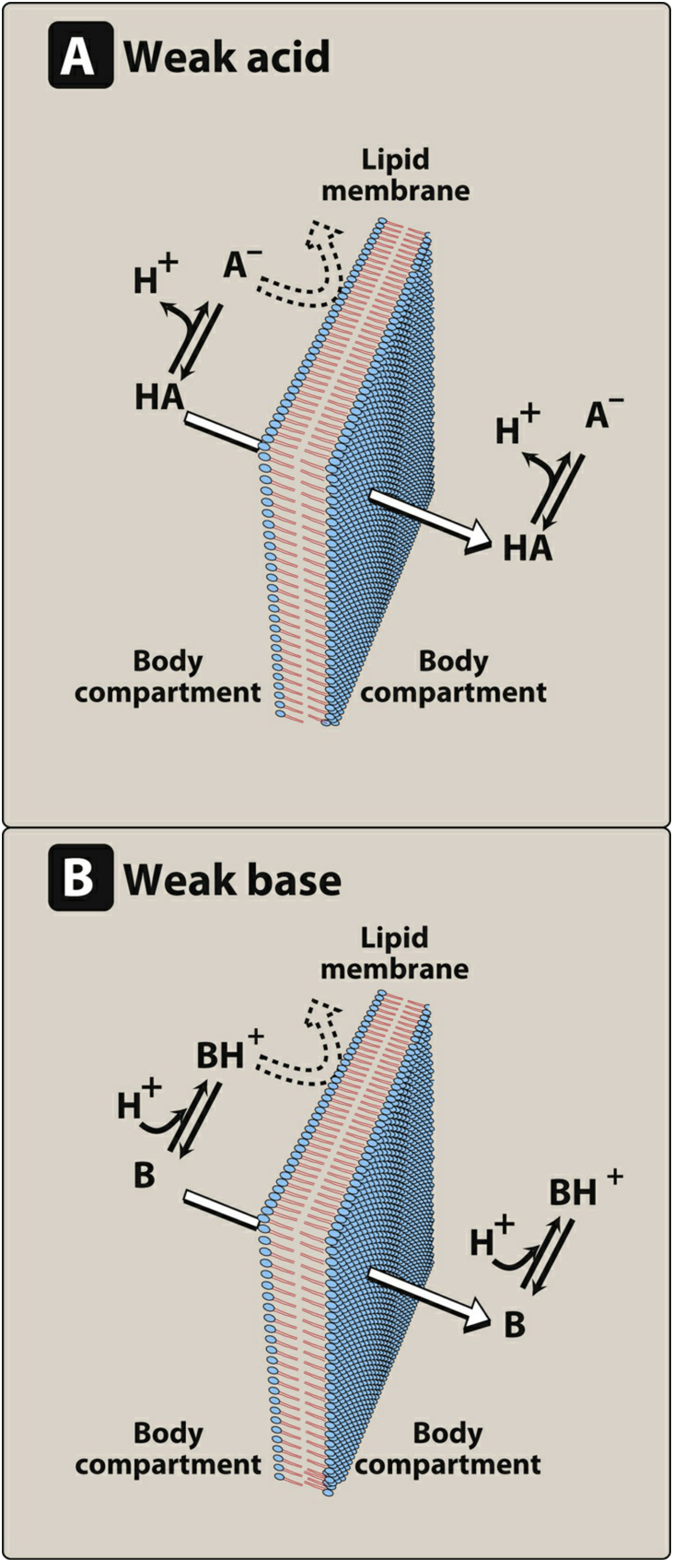
Figure: Only the uncharged (non-ionized) form of a drug permeates the lipid membrane. For weak acids (HA), the neutral form crosses; for weak bases (B), the free base form crosses. (Lippincott Pharmacology)
Ion trapping: When a drug passes into a compartment where it becomes ionized, it gets "trapped" there - it cannot diffuse back out. This is exploited clinically, e.g., alkalinizing urine to trap acidic drugs like aspirin for faster excretion in overdose.
2. Lipid Solubility
- Drugs must be lipophilic enough to dissolve in the lipid bilayer and cross membranes
- But must also have some water solubility to dissolve in GI fluids and reach the membrane surface
- Extremely hydrophilic OR extremely lipophilic drugs are both poorly absorbed
- This is why many drugs are weak acids or weak bases - they have partial lipid solubility
3. Route of Administration
The route determines which absorption barriers a drug faces and whether it undergoes first pass metabolism.
| Route | Onset | First Pass? | Notes |
|---|---|---|---|
| IV | Immediate | No | 100% bioavailability; gold standard |
| Sublingual/Buccal | Fast (minutes) | No | Directly into systemic veins |
| Oral | Slow-moderate | Yes | Most complex; affected by GI factors |
| Transdermal | Slow | No | Bypasses portal circulation |
| Inhalation | Fast | Minimal | Large surface area, rich blood supply |
| Rectal | Variable | ~50% bypass | Unpredictable absorption |
| IM / SC | Moderate | No | Depot effect possible |
4. GI Motility and Gastric Emptying
- Increased GI motility (e.g., diarrhea, metoclopramide) → faster gastric emptying → drug reaches small intestine sooner → may increase absorption rate but reduce total time for absorption
- Decreased motility (e.g., opioids, anticholinergics) → delayed gastric emptying → slower absorption onset
- Most oral absorption occurs in the small intestine (large surface area, rich vascular supply), so faster gastric emptying generally increases drug absorption
5. Blood Flow to the Absorption Site
- Greater blood flow maintains a high concentration gradient across the membrane, driving faster absorption
- IM injections absorb faster than SC because muscle has richer blood supply
- In shock states, peripheral absorption (IM, SC) is unreliable due to vasoconstriction - IV is preferred
- Sustained-release and depot preparations slow drug release to prolong absorption regardless of blood flow
6. Solubility and Drug Formulation
"Drug absorption may be altered by factors unrelated to the chemistry of the drug. For example, particle size, salt form, crystal polymorphism, enteric coatings, and the presence of excipients (such as binders and dispersing agents) can influence the ease of dissolution and absorption." - Lippincott Pharmacology
Key formulation factors:
- Particle size: smaller particles dissolve faster (more surface area)
- Salt form: e.g., sodium salts dissolve faster than free acid forms
- Crystal polymorphism: different crystal forms of the same drug dissolve at different rates
- Enteric coatings: protect acid-labile drugs (e.g., omeprazole) or irritating drugs (e.g., aspirin EC) - release occurs in the intestine
- Extended-release (ER/SR/XL) formulations: rate of dissolution controls rate of absorption; allows twice or once-daily dosing
7. Food and Drug Interactions
- Food can delay gastric emptying, dilute drug concentration, or chelate certain drugs (e.g., tetracyclines bind calcium in dairy)
- P-glycoprotein (P-gp): an efflux transporter in the gut wall that pumps some drugs back into the intestinal lumen, reducing absorption (e.g., digoxin). Inhibitors like verapamil increase digoxin bioavailability
- Grapefruit juice: inhibits intestinal CYP3A4 → increases absorption of many drugs (e.g., statins, calcium channel blockers)
- Antacids: raise gastric pH, altering ionization of drugs (e.g., reduces absorption of ketoconazole which needs acidic environment)
8. Drug Chemical Instability
- Some drugs are degraded before absorption:
- Penicillin G: unstable in acidic gastric environment
- Insulin: proteolytic enzymes in the GI tract destroy it completely - must be given by injection
- Nitroglycerin: destroyed by hepatic first pass (>90%) if swallowed
9. Patient-Specific Factors
| Factor | Effect on Absorption |
|---|---|
| Liver disease (cirrhosis) | Reduced first pass → higher bioavailability of high-extraction drugs |
| Age (elderly/neonates) | Altered gastric pH, GI motility, blood flow |
| CKD | Reduced first pass metabolism of some beta-blockers |
| Bowel disease (Crohn's, celiac) | Mucosal damage reduces absorption surface area |
| Obesity (post-bariatric surgery) | Malabsorptive procedures (e.g., RYGB) markedly alter drug absorption |
| Gender | Differences in GI motility and CYP enzyme activity affect bioavailability |
Summary Diagram of Key Concepts
Drug administered orally
↓
Dissolved in GI fluids (solubility)
↓
Un-ionized form (depends on pKa + pH) crosses gut wall
↓
P-gp efflux? CYP3A4 in gut wall? → reduced absorption
↓
Portal vein → LIVER → first pass metabolism
↓
Remaining drug → systemic circulation = BIOAVAILABILITY (F)
Sources: Lippincott Illustrated Reviews: Pharmacology | Goldman-Cecil Medicine | Katzung's Basic and Clinical Pharmacology, 16th Ed | Harrison's Principles of Internal Medicine 22E
Henderson-Hasselbalch principle:
The Henderson-Hasselbalch equation describes the relationship between pH, pKa, and the ratio of ionized to un-ionized forms of a drug. For acids: pH = pKa + log([A⁻]/[HA]), and for bases: pH = pKa + log([B]/[BH⁺]). This determines how much of a drug is in its absorbable (un-ionized) form at any given pH.
Searching sources...
Finding Sources
Finding Sources
Reading File
Reading File
Reading File
Reading File
Loading Image
Loading Image
Loading Image
Here is a complete deep-dive into the Henderson-Hasselbalch principle as it applies to pharmacology:
The Henderson-Hasselbalch Principle in Pharmacology
Background: Why It Matters
Most drugs are either weak acids or weak bases. They exist in two forms in body fluids:
- Un-ionized (uncharged) form - lipid soluble, crosses membranes freely
- Ionized (charged) form - water soluble, CANNOT cross lipid membranes
The Henderson-Hasselbalch equation tells you exactly what fraction of a drug is in each form at any given pH. This directly predicts absorption, distribution, and elimination.
The Equation - Derivation
Starting from the dissociation of a weak acid (HA):
HA ⇌ H⁺ + A⁻
The acid dissociation constant Ka is:
$$K_a = \frac{[H^+][A^-]}{[HA]}$$
Rearranging and taking the negative log of both sides gives the Henderson-Hasselbalch equation:
$$\boxed{pH = pK_a + \log\frac{[A^-]}{[HA]}}$$
Which can be rewritten as (Katzung's unified form applicable to both acids and bases):
$$\boxed{\log\frac{[\text{Protonated}]}{[\text{Unprotonated}]} = pK_a - pH}$$
"The lower the pH relative to the pKa, the greater will be the fraction of drug in the protonated form." - Katzung's Basic & Clinical Pharmacology
Applying the Equation to Drugs
For Weak ACIDS (e.g., aspirin, warfarin, furosemide):
| Form | Charge | Crosses membrane? |
|---|---|---|
| HA (protonated) | Neutral | ✅ YES |
| A⁻ (deprotonated) | Negative | ❌ NO |
- Acidic environment (low pH, stomach pH ~1-3): More drug stays as HA (un-ionized) → better absorbed
- Alkaline environment (high pH, intestine pH ~7.5-8): More drug becomes A⁻ (ionized) → less absorbed from that site
For Weak BASES (e.g., morphine, codeine, atropine, amphetamine):
| Form | Charge | Crosses membrane? |
|---|---|---|
| BH⁺ (protonated) | Positive | ❌ NO |
| B (deprotonated, free base) | Neutral | ✅ YES |
- Alkaline environment (intestine, blood pH 7.4): More drug stays as B (un-ionized) → better absorbed
- Acidic environment (stomach): More drug becomes BH⁺ (ionized) → less absorbed
The Key Rule (Memory Aid)
"Acids are absorbed in Acid; Bases are absorbed in Base"Or more precisely: un-ionized form = absorbed form
- Weak acid: un-ionized in acidic environment
- Weak base: un-ionized in alkaline environment
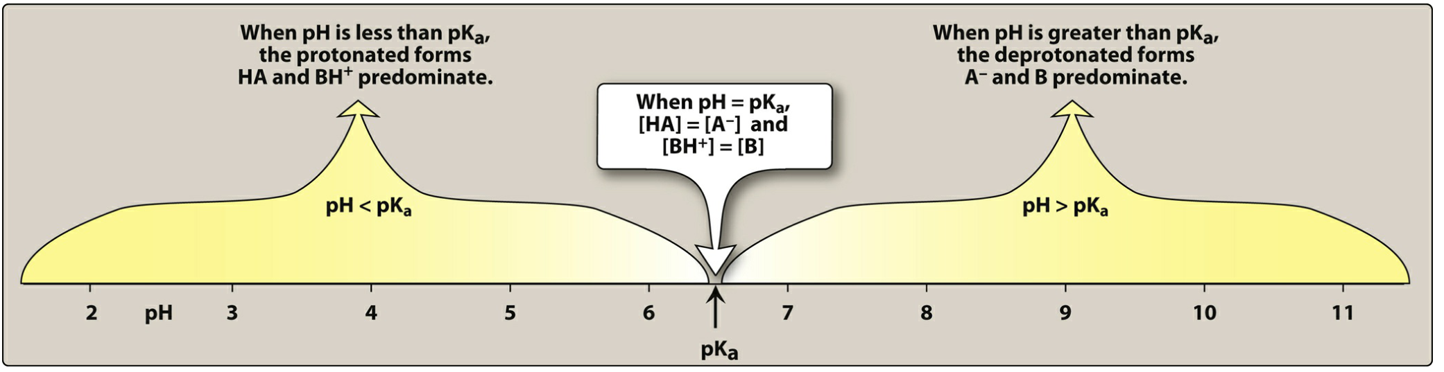
Figure: When pH = pKa, exactly 50% of the drug is ionized and 50% is un-ionized. When pH < pKa, protonated (HA, BH⁺) forms predominate. When pH > pKa, deprotonated (A⁻, B) forms predominate. (Lippincott Pharmacology)
The Golden Rule: When pH = pKa
At half-neutralization, [A⁻] = [HA], so:
$$pH = pK_a + \log(1) = pK_a + 0 = pK_a$$
When pH = pKa → exactly 50% ionized, 50% un-ionized
This is the midpoint of the titration curve and the point of maximum buffering capacity.
Worked Example
Drug: Aspirin (weak acid, pKa = 3.5)
Q: What fraction is un-ionized in the stomach (pH 1.0)?
Using: log([A⁻]/[HA]) = pH - pKa = 1.0 - 3.5 = -2.5
→ [A⁻]/[HA] = 10⁻²·⁵ = 1/316
→ For every 1 ionized molecule, there are 316 un-ionized → >99.7% un-ionized in stomach → excellent absorption from stomach
Q: Same drug in blood (pH 7.4)?
log([A⁻]/[HA]) = 7.4 - 3.5 = +3.9
→ [A⁻]/[HA] = 10³·⁹ ≈ 7943
→ ~99.99% ionized in blood → drug is "trapped" in blood as charged form → cannot easily re-enter cells
pKa Values of Common Drugs (Katzung)
Weak Acids:
| Drug | pKa |
|---|---|
| Aspirin | 3.5 |
| Furosemide | 3.9 |
| Warfarin | 5.0 |
| Phenobarbital | 7.4 |
| Phenytoin | 8.3 |
| Theophylline | 8.8 |
Weak Bases:
| Drug | pKa |
|---|---|
| Morphine | 7.9 |
| Codeine | 8.2 |
| Lidocaine | 7.9 |
| Amphetamine | 9.9 |
| Atropine | 9.7 |
| Chloroquine | 10.8 |
Clinical Application 1: Ion Trapping
When a drug enters a compartment with a different pH, it can become ionized and get trapped there - unable to diffuse back out.
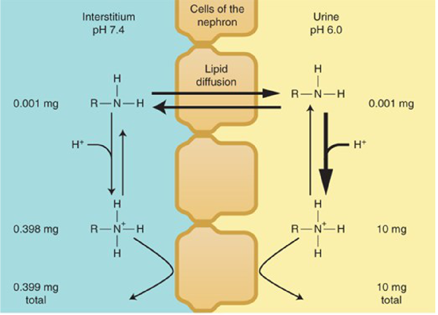
Figure: Ion trapping of a weak base (methamphetamine) in acidic urine. The uncharged form (0.001 mg) freely equilibrates across the membrane, but the ionized form (BH⁺) accumulates in urine to 10 mg total - 25x more than in blood. (Katzung)
Clinical Application 2: Urine Alkalinization / Acidification in Overdose
This is one of the most important clinical uses of Henderson-Hasselbalch:
| Drug Overdose | Type | Urine Treatment | Mechanism |
|---|---|---|---|
| Aspirin (salicylate) | Weak acid | Alkalinize urine (NaHCO₃) | Ionizes aspirin in urine → trapped → excreted faster |
| Phenobarbital | Weak acid | Alkalinize urine | Same as above |
| Amphetamine | Weak base | Acidify urine (ammonium chloride) | Ionizes amphetamine in urine → trapped → excreted faster |
| Methotrexate | Weak acid | Alkalinize urine | Prevents precipitation and aids excretion |
Rule: Weak acids are excreted faster in alkaline urine; weak bases are excreted faster in acidic urine
Clinical Application 3: pH Compartments in the Body
Different body compartments have different pH values, which determine drug distribution:
| Compartment | pH | Effect on weak bases |
|---|---|---|
| Stomach | 1.0-3.0 | Heavily ionized (trapped) |
| Small intestine | 7.5-8.0 | Un-ionized → absorbed well |
| Blood | 7.35-7.45 | Partial ionization |
| Urine (normal) | 4.5-8.0 | Variable; manipulable |
| Breast milk | 6.4-7.6 | Basic drugs concentrate here |
| Intracellular fluid | ~7.0 | Slightly more acidic than blood |
Note: Basic drugs (e.g., some antibiotics, opioids) tend to concentrate in breast milk (pH ~6.4-7.6) because the slightly acidic-to-neutral pH traps them in ionized form - important for drug safety in breastfeeding mothers.
Summary
Henderson-Hasselbalch: pH = pKa + log([ionized]/[un-ionized])
Un-ionized form = crosses membranes = absorbed
Ionized form = water soluble = trapped/excreted
Weak ACID: acid environment (low pH) → un-ionized → absorbed
Weak BASE: basic environment (high pH) → un-ionized → absorbed
When pH = pKa → 50:50 ionized:un-ionized
Each unit pH above pKa (for acids) → 10x more ionized
Each unit pH below pKa (for bases) → 10x more ionized
Sources: Katzung's Basic & Clinical Pharmacology 16th Ed | Lippincott Illustrated Reviews: Pharmacology | Harper's Illustrated Biochemistry 32nd Ed | Goldman-Cecil Medicine
katzung
Let me clarify what you need from Katzung before diving in.
Asking for Preferences
What would you like from Katzung's Basic & Clinical Pharmacology?
whats it and detailed info
Katzung's Basic and Clinical Pharmacology is one of the most widely used pharmacology textbooks, now in its 16th edition. It covers basic pharmacological principles, drug classes, and clinical applications in a comprehensive way used by medical and pharmacy students worldwide.
Searching sources...
Finding Sources
Searching the Web
Katzung Basic and Clinical Pharmacology 16th edition overview contents
Finding Sources
Finding Sources
Reading File
Finding Sources
Reading File
Finding Sources
Reading File
Finding Sources
Reading File
Finding Sources
Loading Image
Finding Sources
Finding Sources
Finding Sources
Here is a detailed overview of Katzung's Basic and Clinical Pharmacology, 16th Edition - what it is, who wrote it, what it covers, and how it's organized:
Katzung's Basic and Clinical Pharmacology, 16th Edition
What Is It?
Katzung's Basic and Clinical Pharmacology is one of the most authoritative and widely used pharmacology textbooks in the world. It is used by medical students, pharmacy students, nursing students, and practicing clinicians globally. Now in its 16th edition, it is published by McGraw-Hill and is available on platforms like AccessMedicine and AccessPharmacy.
Authors: Todd W. Vanderah & Bertram G. Katzung
- Bertram G. Katzung - MD, PhD; received his MD from State University of New York (1957) and PhD in Pharmacology from the University of California. He built the book from the ground up over decades.
- Todd W. Vanderah - co-author on the current edition, professor of pharmacology
What Is Pharmacology? (As Defined by Katzung)
"Pharmacology can be defined as the study of substances that interact with living systems through chemical processes. These interactions usually occur by binding of the substance to regulatory molecules and activating or inhibiting normal body processes."
It has two main branches:
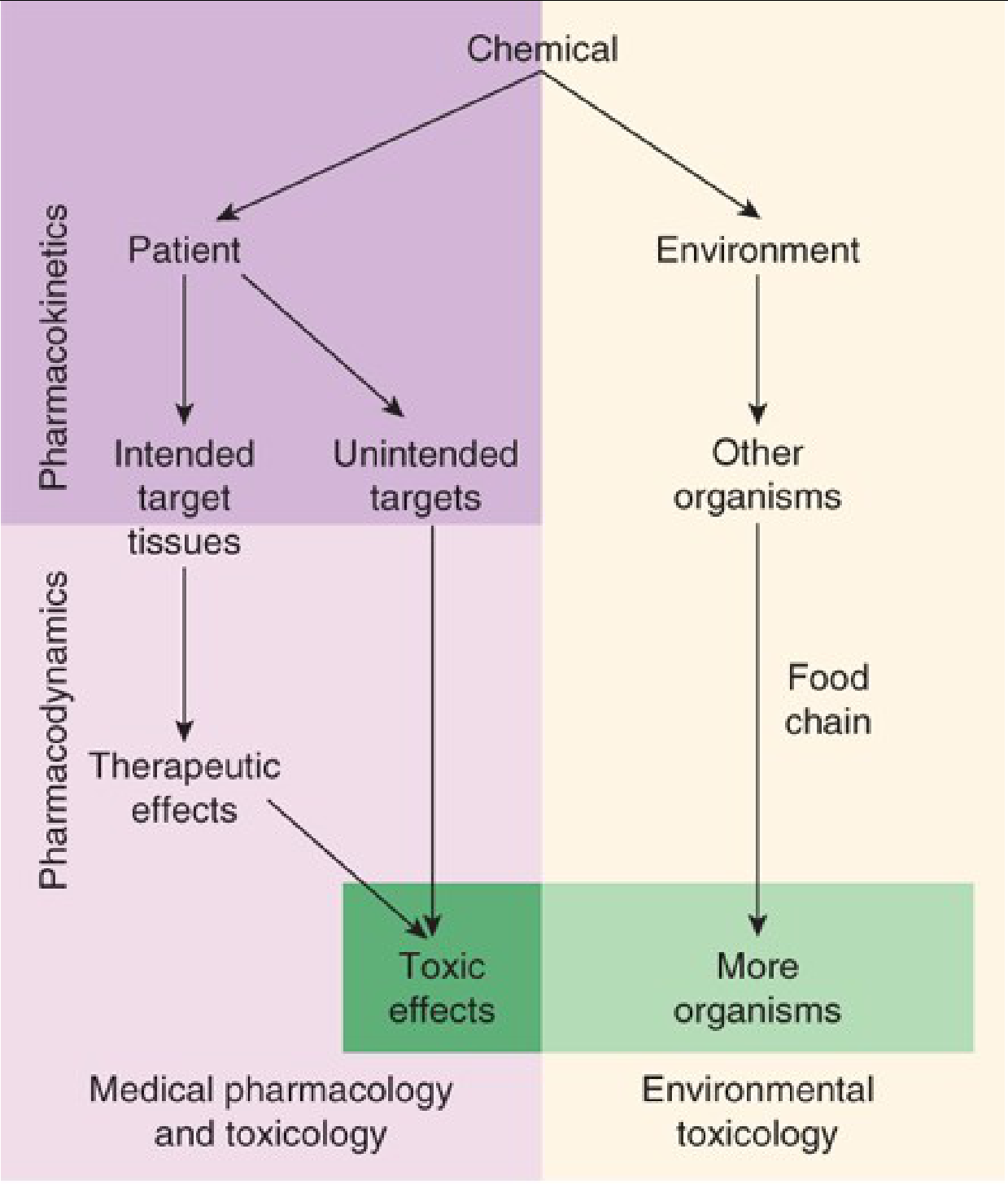
Figure 1-1: Major areas of study in pharmacology - Katzung 16th Ed
| Branch | Focus |
|---|---|
| Medical Pharmacology | Drug actions on individual organisms; therapeutic and toxic effects |
| Toxicology | Undesirable effects of chemicals on living systems |
| Pharmacokinetics | What the body does to the drug (absorption, distribution, metabolism, elimination) |
| Pharmacodynamics | What the drug does to the body (receptor interactions, dose-response) |
| Environmental Toxicology | Chemical effects on all organisms and ecosystems |
Structure & Organization (13 Blocks / Chapters)
The book is organized into major sections. Here is the full chapter map based on the textbook's table of contents:
SECTION I - Basic Principles
| Chapter | Title | Key Topics |
|---|---|---|
| Ch 1 | Introduction: The Nature of Drugs & Drug Development & Regulation | Drug size, shape, reactivity, receptor bonds, pharmacodynamics, pharmacokinetics, drug development pipeline, FDA, IND/NDA, clinical trials |
| Ch 2 | Drug Receptors & Pharmacodynamics | Receptor types, agonists, antagonists, partial agonists, inverse agonists, G proteins, second messengers (cAMP, IP3/Ca²⁺, cGMP), dose-response curves |
| Ch 3 | Pharmacokinetics & Pharmacodynamics | ADME, volume of distribution, clearance, half-life, bioavailability, extraction ratio, first pass effect, steady state |
| Ch 4 | Drug Biotransformation | Phase I (CYP450) and Phase II reactions, clinical relevance, drug-drug interactions, genetic factors in metabolism, gut microbiota effects |
| Ch 5 | Pharmacogenomics | CYP polymorphisms, phase II enzyme polymorphisms, transporter variations (OATP1B1, BCRP, OCT1), epigenomics |
| Ch 6 | Introduction to Autonomic Pharmacology | ANS anatomy, neurotransmission, cholinergic/adrenergic systems |
SECTION II - Autonomic Drugs
Covers cholinergic agonists/antagonists, adrenoceptor agonists/antagonists, antihypertensives, vasodilators
SECTION III - Cardiovascular & Renal Drugs
Cardiac glycosides, antiarrhythmics, diuretics, anticoagulants, lipid-lowering drugs
SECTION IV - CNS Drugs
Sedative-hypnotics, antiepileptics, antidepressants, antipsychotics, opioid analgesics, drug abuse
SECTION V - Drugs with Important Actions on Smooth Muscle
Histamine, serotonin, ergot alkaloids, NSAIDs, prostaglandins
SECTION VI - Endocrine Drugs
Thyroid, corticosteroids, reproductive hormones, antidiabetic drugs
SECTION VII - Chemotherapeutic Drugs
Antibacterials, antifungals, antivirals, antiparasitics, antineoplastics
SECTION VIII - Special Topics
Toxicology, poisoning management, special populations, herbal drugs, drug interactions
Key Features of the Book
1. Drug Groups & Prototypes Approach
"Almost all the several thousand drugs currently available can be arranged into about 70 groups... For most groups, one or two prototype drugs can be identified that typify the most important characteristics."
This is Katzung's signature teaching method - learn the prototype drug deeply, then understand other drugs in the group as variants.
2. Case Studies
Every chapter opens with a clinical case study that frames the pharmacology in real patient scenarios. For example, Chapter 1 opens with an aspirin overdose case that illustrates ion trapping and urine alkalinization using the Henderson-Hasselbalch principle.
3. Pharmacokinetic Principles (Chapter 3 Highlights)
Key concepts covered in depth:
| Concept | Formula | Meaning |
|---|---|---|
| Volume of Distribution (Vd) | Vd = Amount of drug / Plasma concentration | How widely a drug distributes in body tissues |
| Clearance (CL) | CL = Rate of elimination / Plasma concentration | Body's capacity to remove drug |
| Half-life (t½) | t½ = 0.693 × Vd / CL | Time to reduce drug concentration by 50% |
| Bioavailability (F) | F = AUC(oral) / AUC(IV) | Fraction of oral dose reaching systemic circulation |
| Extraction Ratio | ER = (CA - CV) / CA | Fraction of drug removed per pass through liver |
| Steady State | Achieved after ~4-5 half-lives | When drug input = drug elimination rate |
4. Biotransformation (Chapter 4)
Detailed coverage of:
- Phase I reactions: Oxidation, reduction, hydrolysis via CYP450 enzymes (CYP1A2, CYP2C9, CYP2C19, CYP2D6, CYP3A4)
- Phase II reactions: Glucuronidation, sulfation, acetylation, glutathione conjugation
- Enzyme induction vs inhibition and clinical drug-drug interactions
- Genetic polymorphisms: Poor vs. extensive metabolizers (e.g., CYP2D6 for codeine)
- Gut microbiota as a site of drug metabolism
5. Pharmacogenomics (Chapter 5)
A dedicated chapter on how genetic variation affects drug response - covering:
- CYP2C9 and warfarin dosing
- VKORC1 variants
- TPMT and thiopurine toxicity
- Transporter gene variations affecting drug absorption and excretion
Why It's Uniquely Valuable
| Feature | Details |
|---|---|
| Scope | Basic science + clinical application in one volume |
| USMLE relevance | High-yield content, USMLE-style questions in new edition |
| Drug tables | Extensive tables listing pKa values, CYP substrates/inhibitors/inducers, dosing |
| New in 16th Ed | New content on cannabinoids, updated biologics, mRNA vaccines, expanded pharmacogenomics |
| Drug development | Covers IND, NDA, Phase I-IV clinical trials, FDA process in detail |
| Illustrations | Rich diagrams of signaling pathways, receptor models, ion trapping, metabolism |
The Book's Approach to Drug-Body Interactions
Katzung structures all drug-body interactions into two master categories:
PHARMACOKINETICS = What the BODY does to the DRUG
Absorption → Distribution → Metabolism → Elimination
PHARMACODYNAMICS = What the DRUG does to the BODY
Receptor binding → Signal transduction → Physiologic effect → Dose-Response
Source: Katzung's Basic and Clinical Pharmacology, 16th Edition - Todd W. Vanderah & Bertram G. Katzung (McGraw-Hill) | Available in the medical library (ISBN: 9781260463309)
Short Note on SUPERANTIGENS.
Superantigens are microbial proteins that cause massive, non-specific T-cell activation by simultaneously binding MHC class II molecules on APCs and the Vβ region of T-cell receptors - bypassing normal antigen processing. This triggers a cytokine storm, leading to conditions like toxic shock syndrome.
Searching sources...
Finding Sources
Finding Sources
Reading File
Reading File
Reading File
Reading File
Loading Image
Loading Image
Reading File
Reading File
Loading Image
Here is a complete short note on Superantigens:
Short Note: SUPERANTIGENS
Definition
Superantigens (SAgs) are a family of microbial proteins - primarily exotoxins produced by bacteria such as Staphylococcus aureus and Group A Streptococcus (GAS) - that cause massive, non-specific polyclonal T-cell activation by bypassing normal antigen processing and presentation.
"Superantigens are proteins that are capable of stimulating large populations of T cells in a manner unrestricted by the class II major histocompatibility complex (MHC)." - Goldman-Cecil Medicine
Normal Antigen Presentation vs. Superantigen Action
| Feature | Normal Antigen | Superantigen |
|---|---|---|
| Processing required? | YES - proteolytic degradation | NO - binds intact |
| MHC binding site | Inside antigen-binding groove | Outside the groove (conserved residues) |
| TCR binding site | Vα + Vβ + CDR3 (specific) | Only the Vβ region (non-specific) |
| T cells activated | 0.001-0.01% of T cells | 5-20% of all T cells |
| Cytokine release | Controlled, targeted | Massive, systemic (cytokine storm) |
Mechanism of Action
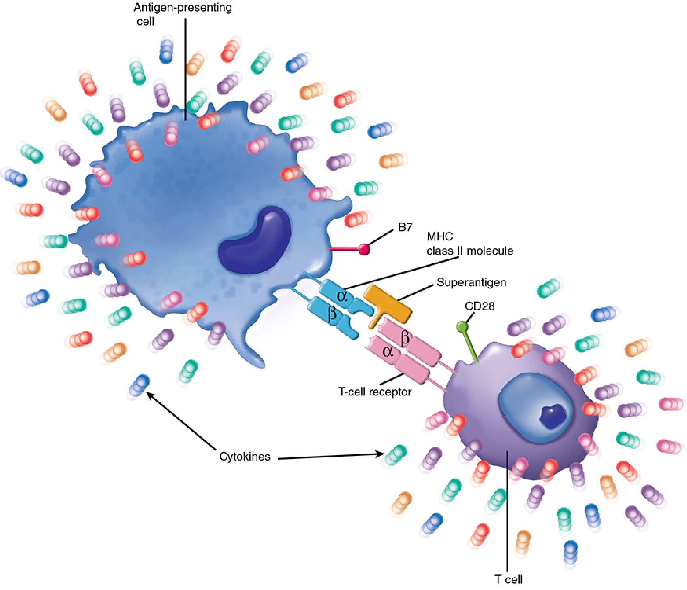
Figure: Superantigen binds simultaneously to MHC class II (outside the peptide groove) and the TCR Vβ region, cross-linking APC and T cell to trigger massive cytokine secretion. (Sherris & Ryan's Medical Microbiology)
Step-by-step mechanism:
- Superantigen is produced at an infected site and absorbed into the circulation
- It binds to MHC class II molecules on APCs (macrophages, dendritic cells) - but OUTSIDE the normal peptide-binding groove
- Simultaneously, it binds to the Vβ region of the T-cell receptor, independent of antigen specificity
- This cross-links APC and T cell, activating them without any need for antigen processing
- Up to 20% of the entire circulating T-cell pool is activated simultaneously
- Massive release of cytokines: IL-1, IL-2, TNF-α, TNF-β, IFN-γ
- This cytokine storm causes systemic effects - fever, hypotension, shock, organ failure
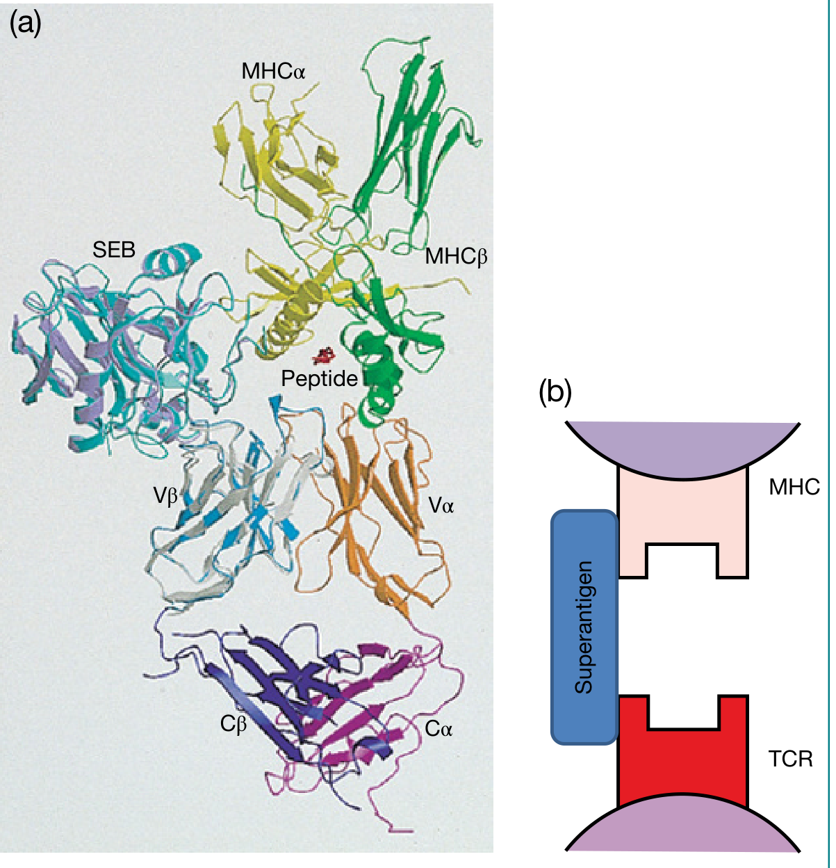
Figure: (a) Molecular model of SEB wedging between MHC and TCR Vβ, preventing normal TCR-peptide-MHC interaction. (b) Schematic of superantigen cross-linking MHC and TCR. (Roitt's Essential Immunology)
Cytokine Cascade
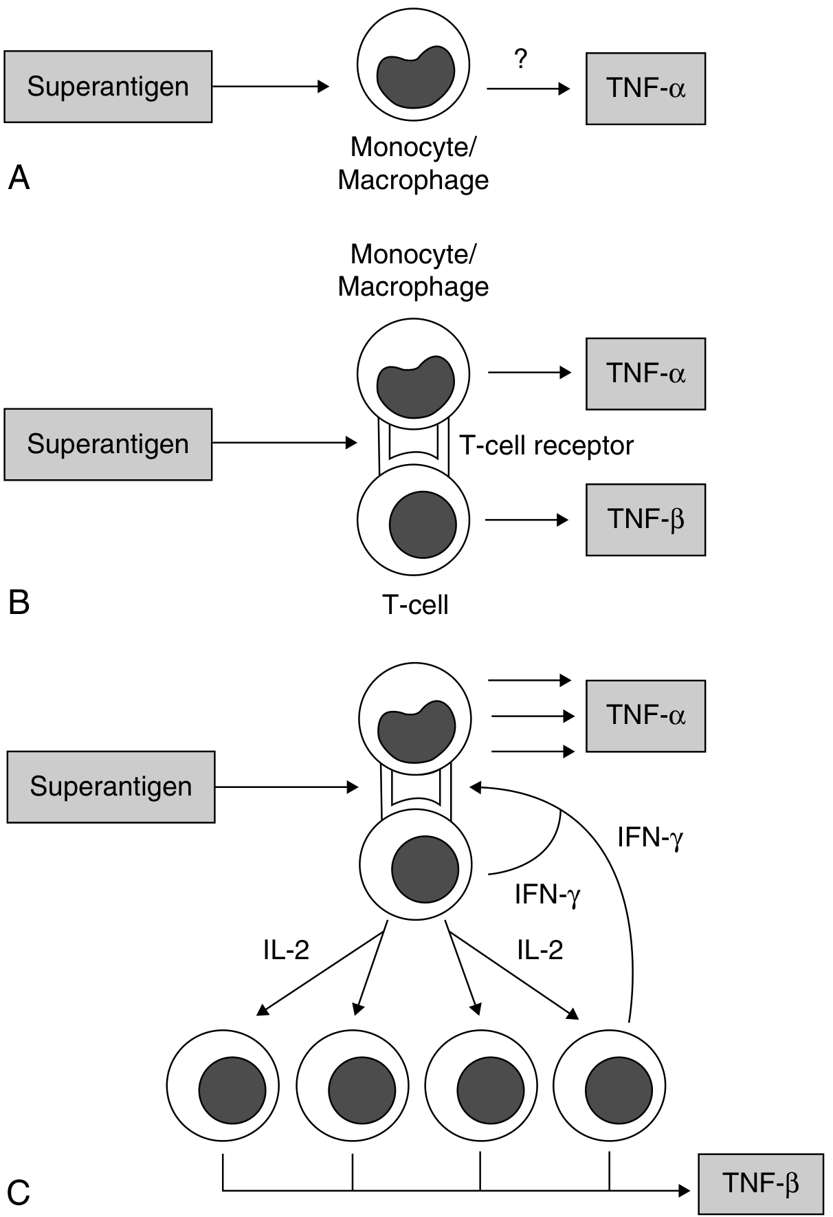
Figure: Cascade of superantigen-induced cytokine production. A - direct monocyte stimulation (TNF-α). B - bridging of T cell and APC releases TNF-α and TNF-β. C - IL-2 drives T cell proliferation; IFN-γ amplifies monocyte TNF-α, IL-1, and IL-6. (Goldman-Cecil Medicine)
Important Superantigens and Associated Diseases
Staphylococcal Superantigens
| Toxin | Disease |
|---|---|
| TSST-1 (Toxic Shock Syndrome Toxin-1) | Toxic Shock Syndrome (TSS) - especially menstrual TSS |
| SEA, SEB, SEC (Staphylococcal Enterotoxins A, B, C) | TSS (non-menstrual), food poisoning (vomiting via brainstem stimulation) |
| Exfoliative Toxins (ETs) | Staphylococcal Scalded Skin Syndrome (SSSS) |
Key fact: Staphylococcal enterotoxins are stable to boiling and gastric enzymes - so ingesting preformed toxin in food causes vomiting even without live bacteria.
Only <20% of S. aureus strains produce any SAg.
Streptococcal Superantigens
| Toxin | Disease |
|---|---|
| SpeA, SpeB, SpeC (Streptococcal Pyrogenic Exotoxins) | Streptococcal TSS, Scarlet Fever (rash), Necrotizing fasciitis |
Streptococcal SAgs also cause fever, rash (scarlet fever), T-cell proliferation, B-lymphocyte suppression, and heightened sensitivity to endotoxin. SpeB additionally has direct enzymatic activity - digesting tissue and extracellular matrix proteins.
Other Superantigens
| Source | SAg |
|---|---|
| Mycoplasma arthritidis | MAM (Mycoplasma arthritis mitogen) |
| Yersinia pseudotuberculosis | YPM |
| Rabies virus | Nucleocapsid protein (viral SAg) |
| B-cell superantigens | Staphylococcal Protein A (binds VH3 IgG), HIV gp120 |
Clinical Consequences: The Cytokine Storm
The massive cytokine release from superantigen activation produces:
Superantigen
↓
Massive T-cell activation (up to 20% of T-cell pool)
↓
IL-1, IL-2, TNF-α, TNF-β, IFN-γ flood released
↓
↑ Vascular permeability → fluid leak → hypotension
↑ Fever (pyrogenic cytokines)
↓ Blood pressure → SHOCK
Organ hypoperfusion → multiorgan failure
Toxic Shock Syndrome features:
- Sudden high fever (>38.9°C)
- Diffuse macular erythroderma (sunburn-like rash)
- Desquamation (1-2 weeks later, especially palms/soles)
- Hypotension (systolic BP <90 mmHg)
- Multisystem involvement (≥3 organ systems)
- Negative blood cultures (toxin circulates, not bacteria)
Why Superantigens Are Unique - Key Points
- No antigen processing needed - bind directly to intact MHC II without proteolysis
- Non-specific Vβ binding - one SAg activates ALL T cells sharing that Vβ segment, regardless of their normal antigen specificity
- Bivalent bridge - simultaneously binds APC and T cell
- Scale of activation - normal immune response activates 0.001-0.01% of T cells; SAgs activate up to 20%
- Toxin-mediated disease - only the toxin needs to circulate, not the bacteria (explains negative blood cultures in TSS)
- Post-activation anergy/deletion - after the massive activation, T cells bearing the stimulated Vβ segments undergo activation-induced cell death, causing transient immunosuppression
Association with Other Diseases
- Kawasaki disease: substantial attention has focused on TSST-1 as a potential trigger via the superantigen mechanism
- Granulomatosis with polyangiitis: superantigens postulated to play a role in disease flares
- Nasal polyposis: the staphylococcal superantigen hypothesis proposes that S. aureus exotoxins foster type 2 inflammation and polyposis in Caucasian patients (Cummings Otolaryngology)
Sources: Sherris & Ryan's Medical Microbiology 8th Ed | Goldman-Cecil Medicine | Roitt's Essential Immunology | Fitzpatrick's Dermatology | Cummings Otolaryngology
Define and classify Immunoglobulins. Describe Ig E,A,G, M under the following a) neat labeled diagram b) structure c) functions 6. Add a note on Monoclonal antibodies and their applications.
Immunoglobulins (antibodies) are glycoproteins produced by plasma cells that recognize and bind specific antigens. They share a basic Y-shaped structure of 4 polypeptide chains (2 heavy + 2 light) linked by disulfide bonds. There are 5 classes: IgG, IgA, IgM, IgD, and IgE, each with distinct heavy chains and functions.
Searching sources...
Finding Sources
Finding Sources
Finding Sources
Finding Sources
Finding Sources
Reading File
Reading File
Reading File
Reading File
Reading File
Reading File
Reading File
Loading Image
Loading Image
Loading Image
Finding Sources
Finding Sources
Reading File
Reading File
Loading Image
Reading File
Immunoglobulins - Definition, Classification, and Detailed Description
DEFINITION
Immunoglobulins (Ig) are glycoproteins produced by plasma cells (terminally differentiated B lymphocytes) that function as antibodies. They are composed of 82-96% polypeptide and 4-18% carbohydrate, and are the effector molecules of humoral immunity. They recognize and bind specific antigens through non-covalent interactions (van der Waals, electrostatic forces).
"Antibodies belong to the immunoglobulin family of proteins, which appear in quantity in serum and on the surfaces of B cells." - Sherris & Ryan's Medical Microbiology
CLASSIFICATION OF IMMUNOGLOBULINS
Immunoglobulins are classified into 5 classes (isotypes) based on the type of heavy chain in their constant region:
| Class | Heavy Chain | Structure | Serum % | MW (Da) |
|---|---|---|---|---|
| IgG | γ (gamma) | Monomer | ~75% | 150,000 |
| IgA | α (alpha) | Monomer / Dimer (secretory) | ~15% | 160,000 |
| IgM | μ (mu) | Pentamer | ~10% | 900,000 |
| IgD | δ (delta) | Monomer | ~0.2% | 180,000 |
| IgE | ε (epsilon) | Monomer | ~0.004% | 200,000 |
Light chain types: κ (kappa) or λ (lambda) - present in all classes
IgG subclasses: IgG1 (65%), IgG2, IgG3, IgG4
IgA subclasses: IgA1, IgA2
BASIC STRUCTURE OF AN IMMUNOGLOBULIN
All immunoglobulins share a fundamental tetrameric (Y-shaped) structure:
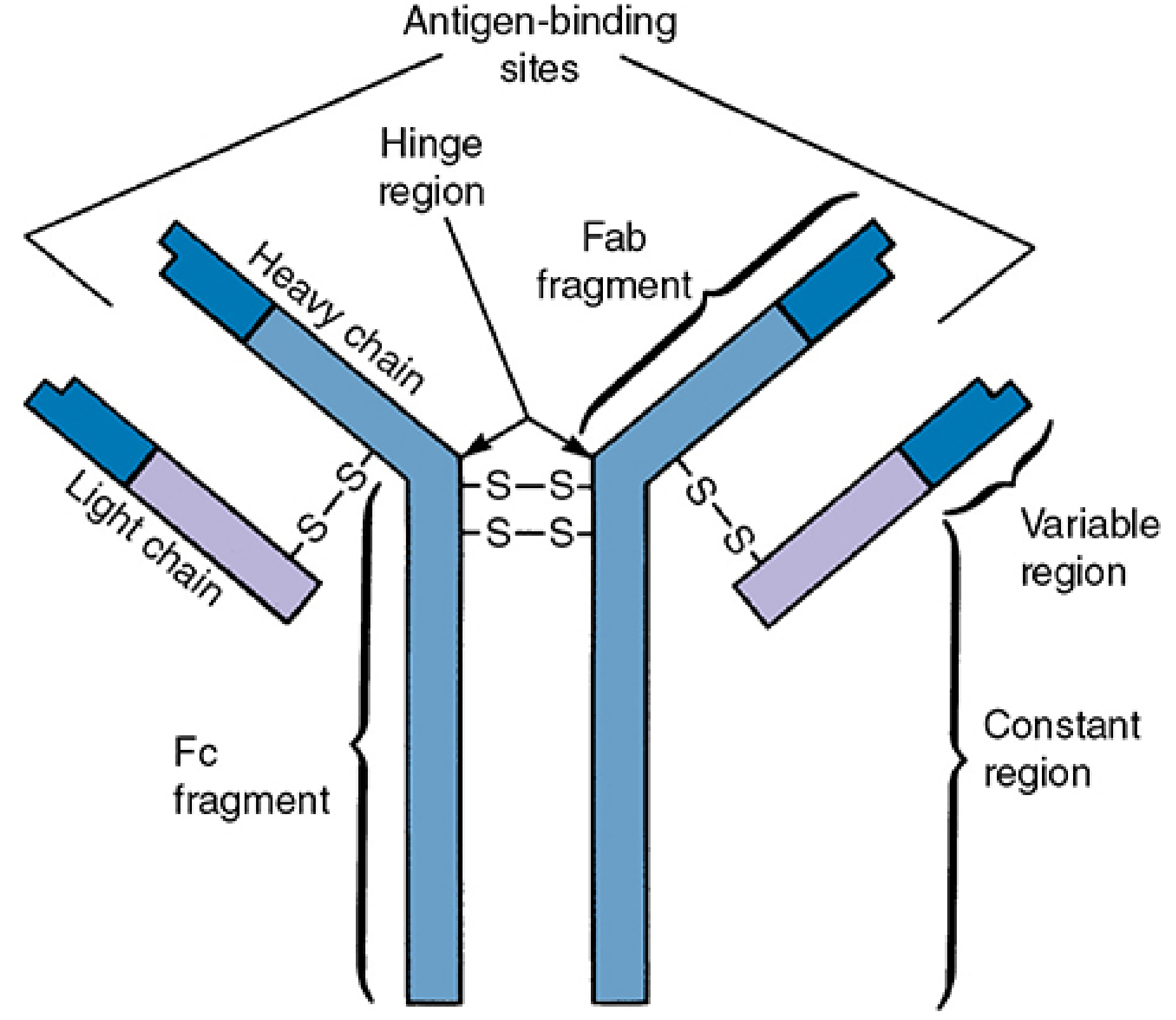
Fig: IgG molecule - the prototype immunoglobulin structure showing heavy chains (dark blue), light chains (light blue), Fab and Fc fragments, and disulfide bonds. (Sherris & Ryan's Medical Microbiology)
Components:
| Component | Description |
|---|---|
| Heavy chains (H) | 2 identical chains, ~50 kDa each; define Ig class |
| Light chains (L) | 2 identical chains, ~25 kDa each; either κ or λ |
| Variable region (V) | N-terminal; contains antigen-binding site (CDRs) |
| Constant region (C) | C-terminal; mediates effector functions |
| Fab fragment | "Fragment antigen-binding" - VH + VL + CH1 + CL; binds antigen |
| Fc fragment | "Fragment crystallizable" - CH2 + CH3; binds complement, Fc receptors |
| Hinge region | Between Fab and Fc; flexible; allows bivalent binding |
| Disulfide bonds | Link H-H chains (inter-chain) and L-H chains |
| CDR (Complementarity-Determining Regions) | Hypervariable loops within V regions; directly contact antigen |
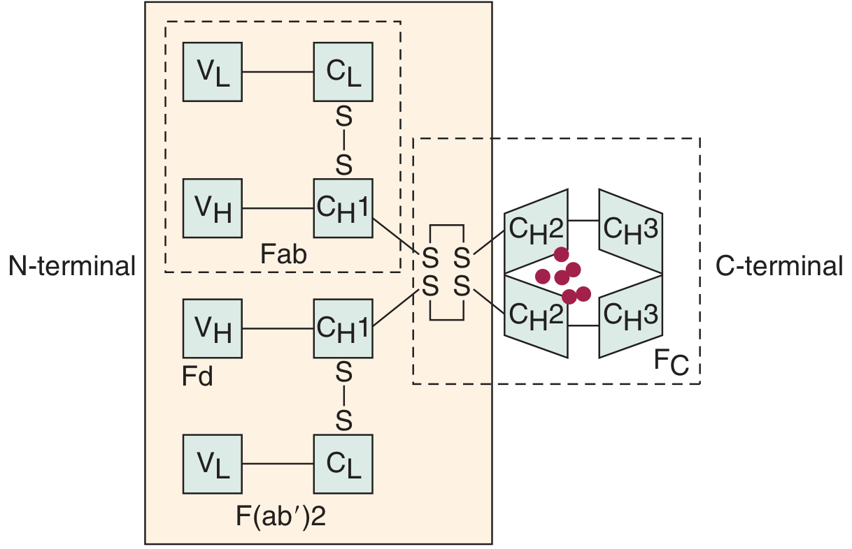
Fig: Domain map of IgG1 showing VL, VH (variable), CL, CH1, CH2, CH3 (constant) domains, Fab, F(ab')2, and Fc regions. Red circles = carbohydrate residues. (Cummings Otolaryngology)
IgG - Immunoglobulin G
a) Diagram
Antigen-binding sites
↙ ↘
[VH-VL] [VH-VL]
| \ / |
CH1 -S-S-S-S- CH1 ← Fab region (×2)
| Hinge |
CH2 — — — — — CH2 ← Fc region begins
| |
CH3 — — — — — CH3
(Fc receptor & complement binding)
(Y-shaped monomer, MW 150,000 Da, 2 antigen-binding sites)
b) Structure
- Form: Monomer (H₂L₂)
- Heavy chain: γ (gamma) - 4 domains: VH, CH1, CH2, CH3
- Light chain: κ or λ - 2 domains: VL, CL
- Subclasses: IgG1, IgG2, IgG3, IgG4 (each with distinct γ chains)
- MW: 150,000 Da
- Half-life: ~21-23 days (longest of all Ig classes)
- Valence: 2 (bivalent)
c) Functions
| Function | Details |
|---|---|
| Most abundant serum Ig | ~75% of total serum immunoglobulins |
| Secondary immune response | Characteristic antibody of anamnestic (memory) response; follows IgM |
| Opsonization | Fc receptors on macrophages, monocytes, neutrophils enhance phagocytosis |
| Complement activation | IgG1, IgG3 fix complement via classical pathway; IgG4 does NOT |
| Neutralization | Neutralizes bacterial exotoxins and viruses by blocking receptor attachment |
| Placental transfer | Only Ig class that crosses the placenta - provides passive immunity to neonate (maternal IgG protects newborn for first 3-6 months) |
| ADCC | Antibody-dependent cellular cytotoxicity via NK cells |
| IgG2 | Directed against polysaccharide antigens - defense against encapsulated bacteria |
IgA - Immunoglobulin A
a) Diagram
Serum IgA (monomer):
[VH-VL] — [VH-VL]
↓ ↓
H-L - S-S - H-L (MW 160,000 Da)
Secretory IgA (dimer):
Monomer1 — J chain — Monomer2
↕
Secretory Component
(protects from proteolytic degradation)
b) Structure
- Serum form: Monomer (MW 160,000 Da)
- Secretory form: Dimer - two monomers joined by J chain + secretory component (SC)
- Heavy chain: α (alpha)
- Subclasses: IgA1 (mainly monomer in serum), IgA2 (mainly polymeric at mucosal surfaces)
- Secretory component: Poly-Ig receptor fragment; added during transcytosis across epithelium; protects sIgA from proteolytic digestion in secretions
- Valence: 2 (monomer), 4 (dimer)
- Humans produce more IgA by total mass than any other Ig class
c) Functions
| Function | Details |
|---|---|
| Mucosal immunity | Primary defense at mucosal surfaces (GI tract, respiratory tract, urogenital tract) |
| Most abundant Ig in secretions | Found in saliva, tears, colostrum, breast milk, nasal secretions, intestinal fluid |
| Neutralization | Neutralizes pathogens and toxins at mucosal surfaces before systemic invasion |
| Prevents adherence | Blocks attachment of bacteria and viruses to epithelial cells |
| Does NOT fix complement | Unlike IgG and IgM |
| Neonatal protection | Present in breast milk and colostrum - protects neonate's GI tract |
| IgA nephropathy | Abnormal IgA deposits cause the most common glomerulonephritis worldwide |
IgM - Immunoglobulin M
a) Diagram
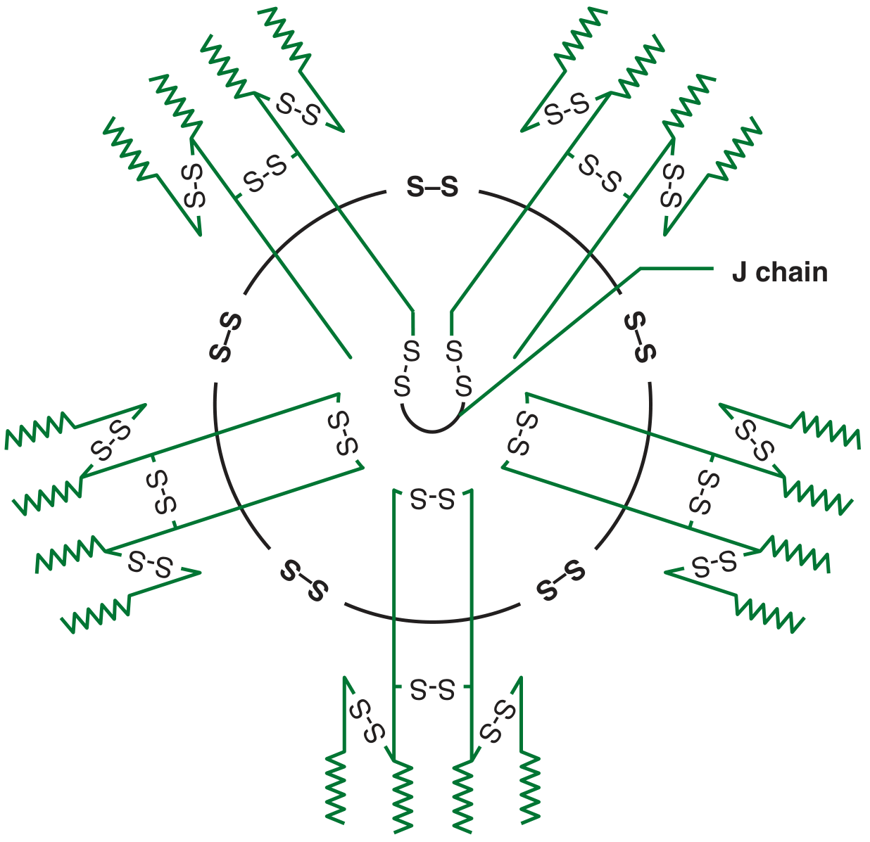
Fig: Pentameric IgM - 5 IgM monomers connected by disulfide bonds and a central J chain. (Jawetz Medical Microbiology)
b) Structure
- Form: Pentamer (5 × H₂L₂ units + 1 J chain)
- Heavy chain: μ (mu) - 5 domains (VH + CH1-CH4, no hinge region)
- J chain: Small glycoprotein that joins the 5 monomers and stabilizes the pentamer
- MW: 900,000 Da (largest Ig)
- Valence: 10 (10 antigen-binding sites) - though functionally only 5 may be used due to steric hindrance
- Serum IgM: Pentamer
- Membrane IgM (mIgM): Monomer - serves as the B-cell antigen receptor (BCR)
c) Functions
| Function | Details |
|---|---|
| First antibody in primary response | Earliest Ig produced after antigen exposure; appears 4-6 days after challenge |
| Best complement activator | Most efficient complement-fixing Ig (classical pathway) - multiple C1q binding sites |
| Agglutination | Most effective agglutinating antibody due to 10 binding sites |
| B-cell receptor | Membrane-bound monomeric IgM is the primary antigen receptor on naive B cells |
| ABO blood groups | Naturally occurring anti-A and anti-B antibodies are IgM |
| Early infection marker | Serum IgM rise indicates acute/recent infection |
| Does NOT cross placenta | Fetal IgM in serum = evidence of intrauterine infection (TORCH) |
| Opsonization | Less effective than IgG as opsonin (Fc not as accessible to phagocytes) |
IgE - Immunoglobulin E
a) Diagram
[VH-VL] [VH-VL]
↓ ↓
H chain H chain
(5 domains:
VH, Cε1, Cε2, Cε3, Cε4)
↕
Fc binds high-affinity FcεRI
on mast cells and basophils
(No hinge region; monomer; MW 200,000 Da)
b) Structure
- Form: Monomer (H₂L₂)
- Heavy chain: ε (epsilon) - 5 constant domains (no hinge region; Cε1-Cε4 + VH)
- MW: 200,000 Da (heaviest Ig monomer - due to extra constant domain)
- Valence: 2
- Serum concentration: Extremely low - 0.004% of total serum Ig (10-400 ng/mL)
- Half-life in serum: ~2 days; but when bound to mast cells/basophils: weeks
- Key receptors:
- FcεRI (high affinity) - on mast cells and basophils
- FcεRII / CD23 (low affinity) - on eosinophils, macrophages, neutrophils, platelets, B cells
c) Functions
| Function | Details |
|---|---|
| Allergic (Type I hypersensitivity) reactions | IgE binds to FcεRI on mast cells/basophils; antigen cross-links IgE → degranulation → histamine, leukotrienes, prostaglandins → allergy, asthma, anaphylaxis |
| Parasitic defense | Elevated IgE in helminth (worm) infections; IgE on eosinophils mediates ADCC against parasites |
| Atopy | Genetic predisposition to produce high IgE → allergic rhinitis, atopic dermatitis, food allergy |
| Local production | IgE can be produced locally in nasal mucosa, tonsils, lymph nodes - "local allergy" even without systemic sensitization |
| ADCC via eosinophils | IgE-coated parasites targeted by eosinophils |
| Does NOT fix complement |
COMPARATIVE SUMMARY TABLE
| Feature | IgG | IgA | IgM | IgE |
|---|---|---|---|---|
| Heavy chain | γ | α | μ | ε |
| Structure | Monomer | Mono/Dimer | Pentamer | Monomer |
| MW (Da) | 150,000 | 160,000 | 900,000 | 200,000 |
| Serum % | 75% | 15% | 10% | 0.004% |
| Valence | 2 | 2 or 4 | 10 | 2 |
| Crosses placenta | ✅ Only one | ❌ | ❌ | ❌ |
| Complement (classical) | ✅ (IgG1,3) | ❌ | ✅ (best) | ❌ |
| Opsonization | ✅ ++ | ❌ | ✅ + | ❌ |
| First in primary response | ❌ | ❌ | ✅ | ❌ |
| Secondary response | ✅ | ✅ | ❌ | ❌ |
| Mucosal immunity | ❌ | ✅ (sIgA) | ❌ | ❌ |
| Allergy/anaphylaxis | ❌ | ❌ | ❌ | ✅ |
| Anti-parasite | ❌ | ❌ | ❌ | ✅ |
| J chain | ❌ | ✅ (dimer) | ✅ (pentamer) | ❌ |
| Secretory component | ❌ | ✅ (sIgA) | ❌ | ❌ |
6. NOTE ON MONOCLONAL ANTIBODIES AND THEIR APPLICATIONS
Definition
Monoclonal antibodies (mAbs) are collections of identical antibody molecules, all produced by the progeny of a single B-cell clone. Every molecule of a given mAb has the same variable region and binds to the same single epitope on an antigen.
"All molecules of a monoclonal antibody are produced by the progeny of a single B cell clone, and all the molecules therefore have the same V region and bind to the same antigen." - Cellular and Molecular Immunology (Abbas)
Production - The Hybridoma Technique (Kohler & Milstein, 1975)
This Nobel Prize-winning technique (1984) is one of the most important advances in biology and medicine.
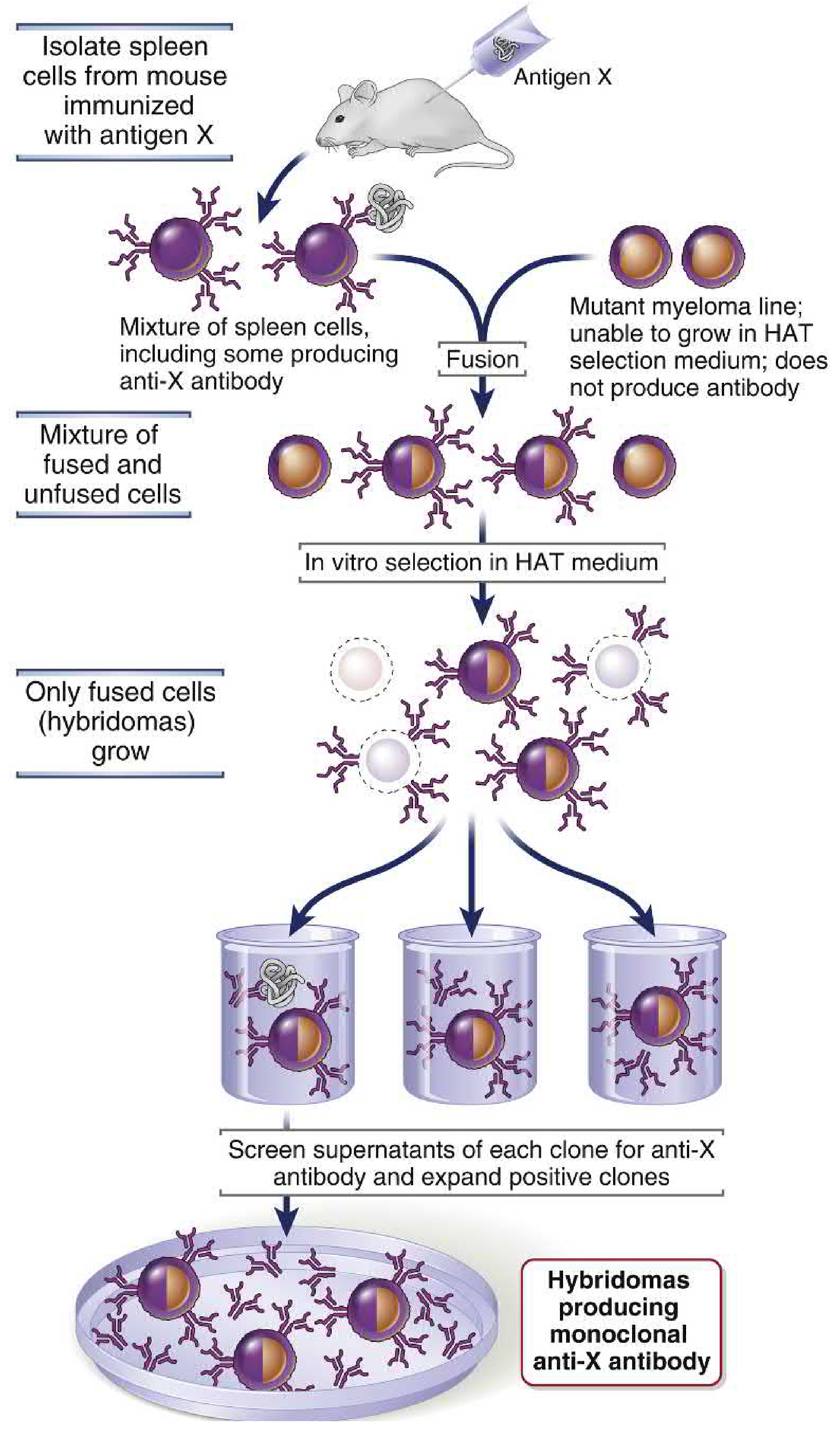
Fig: Generation of monoclonal antibodies by the hybridoma technique. (Cellular and Molecular Immunology, Abbas)
Steps:
- Immunize a mouse with the antigen of interest
- Isolate spleen B cells (produce antibody, but mortal)
- Fuse B cells with myeloma cells (immortal, antibody-negative) using polyethylene glycol
- Select in HAT medium (Hypoxanthine-Aminopterin-Thymidine):
- Unfused myeloma cells die (lack HGPRT for salvage pathway)
- Unfused B cells die naturally
- Only hybridomas survive (get HGPRT from B cell + immortality from myeloma)
- Screen hybridoma clones for desired antibody specificity
- Expand the positive clone → indefinite production of mAb
Types of Therapeutic Monoclonal Antibodies
| Type | Description | Example |
|---|---|---|
| Murine (-omab) | Fully mouse-derived | Muromonab |
| Chimeric (-ximab) | Mouse V region + human Fc | Rituximab, Infliximab |
| Humanized (-zumab) | Mouse CDRs + human framework | Trastuzumab, Bevacizumab |
| Fully human (-umab) | Entirely human | Adalimumab, Pembrolizumab |
(Suffix -mab = monoclonal antibody)
Applications of Monoclonal Antibodies
1. Therapeutic Applications
| Target | Drug | Disease |
|---|---|---|
| TNF-α | Infliximab, Adalimumab | Rheumatoid arthritis, Crohn's disease, psoriasis |
| CD20 (B cells) | Rituximab | B-cell lymphomas, rheumatoid arthritis, multiple sclerosis |
| HER2 | Trastuzumab (Herceptin) | HER2+ breast cancer |
| VEGF | Bevacizumab | Colorectal cancer, macular degeneration |
| PD-1 | Pembrolizumab, Nivolumab | Melanoma, lung cancer, other tumors (checkpoint inhibitors) |
| CTLA-4 | Ipilimumab | Melanoma (checkpoint inhibitor) |
| IgE | Omalizumab | Allergy-related asthma |
| IL-17 | Secukinumab | Psoriasis |
| EGFR | Cetuximab | Colorectal/head & neck cancer |
| Integrin α4 | Natalizumab | Multiple sclerosis, Crohn's disease |
| RANK ligand | Denosumab | Postmenopausal osteoporosis, bone metastases |
| Complement C5 | Eculizumab | Paroxysmal nocturnal hemoglobinuria |
| SARS-CoV-2 | Various | COVID-19 treatment |
| Platelet GPIIb/IIIa | Abciximab | Cardiovascular disease (anti-platelet) |
2. Diagnostic Applications
- Pregnancy tests - detection of hCG using mAbs
- Immunoassays (ELISA, RIA) - detection of antigens/antibodies in blood and urine for infectious disease, cancer markers, hormone levels
- Flow cytometry - CD marker identification on lymphocytes (e.g., CD4:CD8 ratio in HIV)
- Immunohistochemistry - tumor typing from biopsy sections (identify tissue of origin)
- Lateral flow assays - rapid point-of-care tests (e.g., COVID-19 antigen tests)
3. Research Applications
- Cell phenotyping - identification of CD markers (CD4, CD8, CD19, etc.) defining cell populations
- Functional blocking - mAbs that block receptor function help define molecule roles
- Western blotting / flow cytometry / confocal microscopy - standard research tools
- Purification - affinity chromatography using mAbs to purify specific proteins
4. Other Applications
- Radioimmunotherapy - mAbs conjugated with radioisotopes (e.g., I-131) target cancer cells
- Antibody-drug conjugates (ADC) - mAbs carry cytotoxic drugs directly to tumor cells (e.g., Ado-trastuzumab emtansine)
- Bispecific antibodies - engineered to bind two different antigens simultaneously (e.g., Blinatumomab: anti-CD19 × anti-CD3 for B-cell leukemia)
Advantages and Limitations
| Advantages | Limitations |
|---|---|
| Exquisite specificity for single epitope | Expensive to produce |
| Reproducible, unlimited supply | Mouse-derived mAbs cause human anti-mouse antibody (HAMA) response |
| Can be engineered (chimeric, humanized) | Immunogenicity even with humanized versions |
| Wide range of applications | May affect normal physiology of target molecule |
| Can carry payloads (drugs, toxins, radiation) | High cost of development and therapy |
Sources: Cellular and Molecular Immunology (Abbas) | Sherris & Ryan's Medical Microbiology 8th Ed | Cummings Otolaryngology | Jawetz Medical Microbiology 28th Ed | Goldman-Cecil Medicine
what does ig d do
IgD is the least understood immunoglobulin. Its primary known role is as a membrane-bound antigen receptor on mature naive B cells, where it co-expresses with IgM. It makes up only about 0.2% of serum immunoglobulins and has no known major effector function in serum.
Searching sources...
Finding Sources
Finding Sources
Reading File
Reading File
Reading File
Here is a complete note on IgD drawn from multiple authoritative textbooks:
IgD (Immunoglobulin D) - What Does It Do?
Basic Properties
| Feature | Value |
|---|---|
| Heavy chain | δ (delta) |
| Structure | Monomer |
| MW | ~180-184 kDa (contains ~12% carbohydrate) |
| Serum concentration | <1% of total serum Ig (very low) |
| Valence | 2 |
| Half-life in serum | Very short (~2-3 days; highly sensitive to proteolysis) |
| First detectable in serum | ~6 months of age |
The Honest Answer: Its Role Is Largely Mysterious
"To date, IgD has not been assigned a specific biological role as a humoral antibody... the role as a receptor is generally accepted, even though the nature and purpose of the signal it transmits remains controversial." - Henry's Clinical Diagnosis & Management by Laboratory Methods
IgD is the least understood of all immunoglobulin classes. Most of what is known comes from its role on the B-cell surface, not from any circulating effector function.
Known and Proposed Functions
1. B-Cell Antigen Receptor (Main Known Role)
IgD is expressed co-expressed with IgM on the surface of mature naive B cells as a membrane-bound receptor. This is its best-established role.
- Naive mature B cells express both mIgM and mIgD simultaneously on their surface
- The antigen-binding sites (variable regions) and light chains of both are identical - only the constant (heavy chain) regions differ
- When antigen binds to mIgD (or mIgM) on the B-cell surface, it initiates signaling for B-cell activation and clonal proliferation
"The membrane-bound IgD may serve as one of the receptors with which B cells bind antigen and are stimulated to undergo clonal proliferation." - Henry's Clinical Diagnosis
2. Regulation of B-Cell Activation and Tolerance
- Evidence suggests that IgM-bearing B cells can respond to T-independent antigens, but acquisition of IgD is required for B cells to respond to T-dependent antigens (those needing T-cell help)
- If IgD is selectively removed from IgD+IgM+ B cells, those cells become susceptible to tolerance induction rather than activation
- IgD is therefore thought to "turn on, turn off, or modulate" B-cell division or differentiation - essentially a rheostat for B-cell responsiveness
3. Mucosal / Respiratory Immune Defense (Recently Discovered)
- IgD-producing plasma cells are found in tonsils and upper respiratory tract tissues - not just in blood
- At these mucosal sites, secreted IgD binds to galectin-9 on basophils and mast cells
- This binding appears to enhance protective humoral responses against respiratory pathogens AND inhibit IgE-induced allergic reactions at those sites
"IgD-producing plasma cells are found in tonsils and tissues associated with the respiratory tract, where IgD binds to galectin-9 on basophils and mast cells to enhance protective humoral responses and inhibit IgE-induced allergic reactions." - Firestein & Kelley's Textbook of Rheumatology
4. B-Cell Maturation Marker
- IgD surface expression marks the transition from immature to mature naive B cell
- Newborns have a high proportion of IgD-bearing B cells
- As B cells differentiate into plasma cells or memory cells after antigen stimulation, surface IgD is downregulated and lost
What IgD Does NOT Do
| Function | IgD |
|---|---|
| Fix complement | ❌ No |
| Cross the placenta | ❌ No |
| Bind cells via Fc receptor | ❌ No |
| Opsonization | ❌ No |
| Neutralization (serum) | Not established |
| Mucosal secretory form | ❌ No secretory component |
IgD in Disease
Hyper-IgD Syndrome (HIDS)
- Rare autosomal recessive autoinflammatory disorder
- Caused by mutations in the mevalonate kinase gene (MVK) - an enzyme in the cholesterol/isoprenoid biosynthesis pathway
- Features: recurrent episodes of high fever + lymphadenopathy + abdominal pain starting in infancy
- Serum IgD is elevated (>14 mg/dL or >100 IU/mL) - but this is now considered a secondary phenomenon to the underlying inflammatory process, not the cause
- Present equally in males and females
IgD Multiple Myeloma
- Rare (1-2% of myelomas)
- Monoclonal IgD spike in serum
- Generally has worse prognosis than other myeloma types
- Often associated with λ light chains and Bence Jones proteinuria
Chronic Infections
- Serum IgD levels rise non-specifically during chronic infection, along with other immunoglobulins
- No specific disease has been linked uniquely to IgD elevation
Summary
IgD is mostly a:
┌─────────────────────────────────────────────────┐
│ MEMBRANE RECEPTOR on mature naive B cells │
│ Co-expressed with IgM (same V region, diff. C) │
│ Required for T-dependent antigen responsiveness│
│ Modulates B-cell activation/tolerance │
└─────────────────────────────────────────────────┘
With emerging evidence of a MUCOSAL ROLE:
→ Tonsils / upper airway IgD plasma cells
→ Binds galectin-9 on basophils/mast cells
→ Enhances antibacterial responses, inhibits IgE allergy
Sources: Henry's Clinical Diagnosis and Management by Laboratory Methods | Firestein & Kelley's Textbook of Rheumatology | Cummings Otolaryngology | Tietz Textbook of Laboratory Medicine 7th Ed
whats herditary sperocytosis
Hereditary spherocytosis (HS) is the most common inherited hemolytic anemia in people of Northern European descent. It is caused by defects in red blood cell membrane proteins (spectrin, ankyrin, band 3, protein 4.2), causing RBCs to lose their normal biconcave disc shape and become small spheres that are trapped and destroyed in the spleen.
Searching sources...
Finding Sources
Finding Sources
Reading File
Reading File
Reading File
Loading Image
Loading Image
Here is a complete note on Hereditary Spherocytosis:
Hereditary Spherocytosis (HS)
Definition
Hereditary Spherocytosis (HS) is an inherited hemolytic anemia caused by intrinsic defects in the red blood cell (RBC) membrane skeleton proteins, rendering red cells spheroidal, less deformable, and vulnerable to splenic destruction.
"Hereditary spherocytosis is an inherited disorder caused by intrinsic defects in the red cell membrane skeleton that render red cells spheroid, less deformable, and vulnerable to splenic sequestration and destruction." - Robbins & Cotran Pathologic Basis of Disease
Epidemiology
- Most common inherited hemolytic anemia in Northern Europeans
- Prevalence: 1 in 2000-5000 persons of Northern European ancestry
- Much less common in those of African ancestry
- Found worldwide; affects all races
Genetics / Inheritance
| Pattern | Frequency |
|---|---|
| Autosomal dominant | ~75% of cases |
| Autosomal recessive / compound heterozygous | ~25% - usually more severe disease |
| De novo mutations | Occasional |
- Most mutations are "private" (unique to each family) - no selective evolutionary advantage
- Rare homozygous cases can cause fetal death or very severe neonatal hemolysis
Pathogenesis
The normal RBC membrane has a spectrin-actin skeleton tightly linked to the lipid bilayer via two anchoring complexes:
- Spectrin - Ankyrin - Band 3 (vertical linkage)
- Spectrin tail - Protein 4.1 - Glycophorin A (lateral linkage)
In HS: Mutations in one or more of these proteins weaken the membrane skeleton:
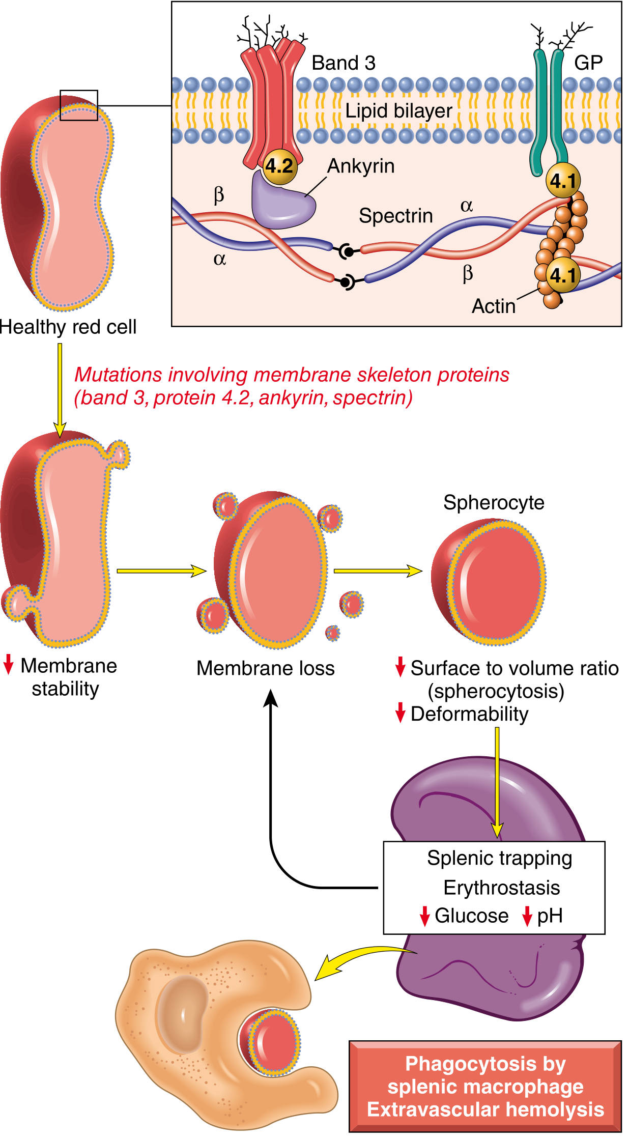
Fig: Pathogenesis of hereditary spherocytosis. Mutations destabilize the membrane skeleton → membrane vesiculation → spherocyte formation → splenic destruction. (Robbins & Cotran Pathologic Basis of Disease)
Defective Proteins in HS:
| Protein | Gene | Frequency |
|---|---|---|
| Ankyrin (ANK1) | ANK1 | Most common (~40-65%) |
| Band 3 (AE1) | SLC4A1 | ~20-25% |
| β-Spectrin | SPTB | ~15-30% |
| α-Spectrin | SPTA1 | ~5% (usually recessive) |
| Protein 4.2 | EPB42 | ~5% (mainly in Japanese) |
The Sequence of Events:
Membrane protein deficiency
↓
Destabilization of lipid bilayer
↓
Membrane fragments shed as RBC ages in circulation
↓
↓ Surface area : Volume ratio
↓
RBC adopts spherical shape (smallest volume for given surface)
↓
Spherocyte - rigid, non-deformable
↓
Trapped in splenic cords (narrow sinusoids)
↓
Hostile splenic environment: ↓ glucose, ↓ pH, ↓ ATP, ↑ free radicals
↓
Further membrane damage → more vesiculation
↓
Phagocytosis by splenic macrophages
↓
EXTRAVASCULAR HEMOLYSIS
Normal RBC lifespan = 120 days. In HS = reduced to 10-20 days
Clinical Features
The classic triad of HS:
Anemia + Jaundice + Splenomegaly
| Feature | Details |
|---|---|
| Anemia | Mild to severe; normocytic/normochromic; compensated by ↑ erythropoiesis |
| Jaundice | Due to unconjugated (indirect) hyperbilirubinemia from hemolysis |
| Splenomegaly | Spleen enlarges due to RBC trapping and hyperplasia of phagocytic cells |
Severity Spectrum:
| Grade | Hb (g/dL) | Bilirubin | Reticulocytes | Features |
|---|---|---|---|---|
| Mild (~20%) | 11-15 | Normal/slight ↑ | 3-6% | Often asymptomatic; detected incidentally |
| Moderate (~60%) | 8-12 | ↑ | 6-10% | Classic triad |
| Severe (~15%) | <8 | ↑↑ | >10% | Transfusion-dependent; may need early splenectomy |
Complications
1. Gallstones (Cholelithiasis) - Most Common Complication
- Pigment stones (bilirubinate) due to chronic ↑ unconjugated bilirubin
- Usually appear in adolescents and young adults
- Can cause biliary colic, cholecystitis, obstructive jaundice
2. Hemolytic Crises
- Triggered by viral illnesses (usually before age 6)
- Sudden worsening of jaundice + ↑ splenomegaly + ↓ hematocrit
- Generally mild and self-limiting
3. Aplastic Crises - Most Dangerous
- Caused by Parvovirus B19 (B19 infects erythroid progenitors)
- Bone marrow suppression → sudden severe drop in Hb + reticulocyte count
- Presents with: fever, vomiting, pallor, severe anemia
- May require transfusion
4. Megaloblastic Crisis
- In states of high folate demand: pregnancy, growing children, recovering from aplastic crisis
- Folate depletion → megaloblastic change on top of hemolysis
5. Uncommon Complications
- Skin ulceration, gout, chronic leg dermatitis
- Cardiomyopathy, spinal cord dysfunction, movement disorders
- Extramedullary hematopoiesis (in severe untreated cases) → hand/skull deformities
Peripheral Blood Smear - Key Finding
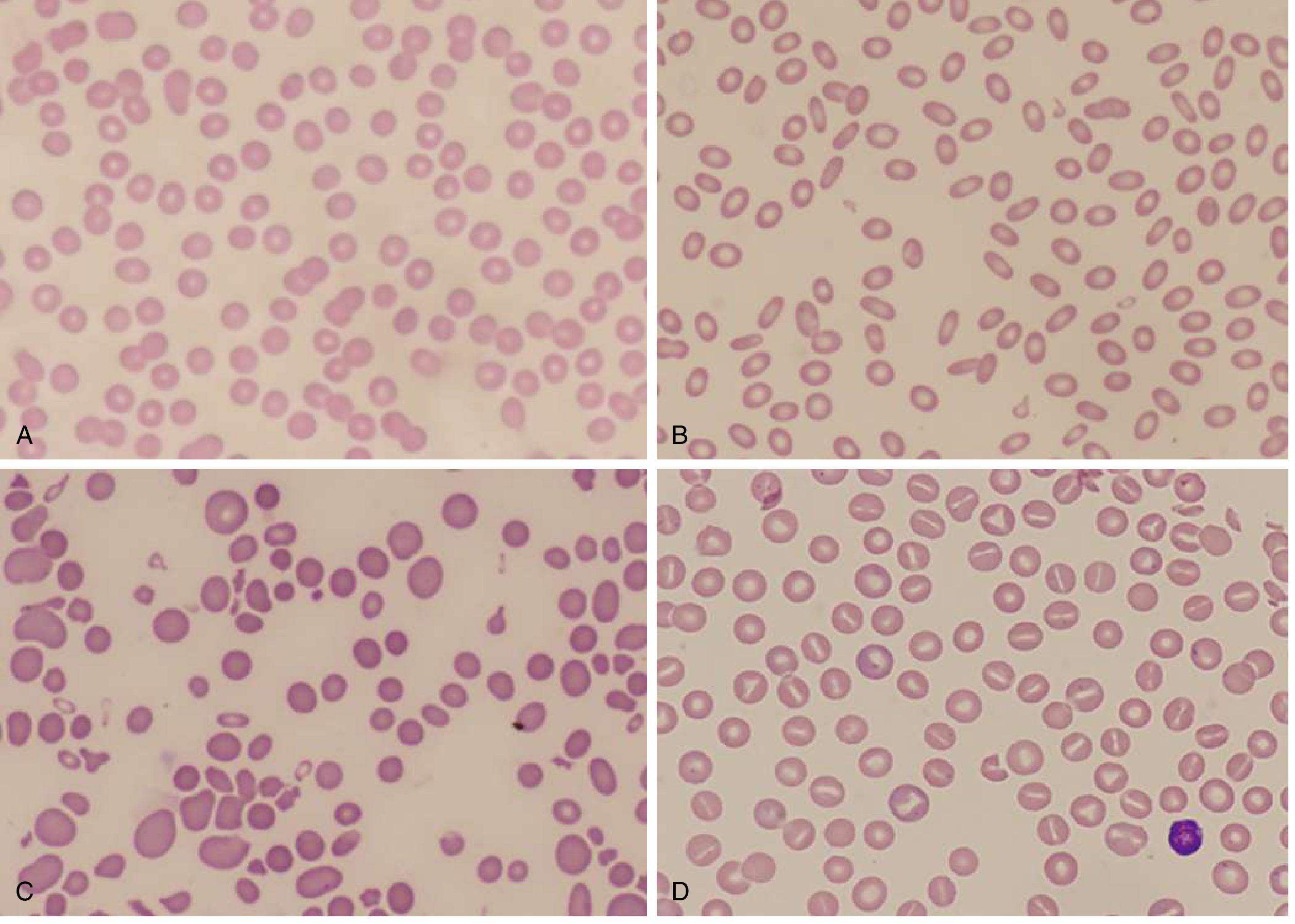
Fig A (top left): HS peripheral smear - small, dense spherocytes uniformly lacking the central pallor seen in normal RBCs. (Goldman-Cecil Medicine)
Key smear features:
- Spherocytes: Small, dense, round cells with no central pallor
- Polychromasia: ↑ reticulocytes (large bluish cells)
- MCHC elevated (>36 g/dL) - hallmark finding (dense cells)
- MCV normal or slightly low
Laboratory Diagnosis
| Test | Finding in HS |
|---|---|
| Peripheral smear | Spherocytes, polychromasia |
| Hb | Low (variable) |
| MCV | Normal or mildly low |
| MCHC | ↑ (>36 g/dL) - key finding |
| Reticulocyte count | ↑ (3-15%) |
| Serum bilirubin (indirect) | ↑ |
| LDH | ↑ |
| Haptoglobin | ↓ |
| Direct Coombs (DAT) | Negative (distinguishes HS from AIHA) |
| EMA binding test | ↓ fluorescence (best screening test; flow cytometry) |
| Osmotic fragility test | ↑ fragility (spherocytes lyse in hypotonic solutions more easily) |
| Cryohemolysis test | ↑ lysis at low temperature |
The EMA (eosin-5'-maleimide) binding test by flow cytometry is now the preferred diagnostic test - sensitive and specific.
Conditions with Spherocytes on Blood Smear (Differential Diagnosis)
- Autoimmune hemolytic anemia (Coombs positive - key distinguisher)
- Thermal injury (burns)
- ABO incompatibility in newborns
- Clostridial septicemia
- Microangiopathic hemolytic anemia
Treatment
1. Splenectomy - Definitive Treatment
- Removes the main site of RBC destruction
- Does NOT correct the membrane defect, but eliminates hemolysis in most patients
- RBC lifespan normalizes after splenectomy
- Timing: Usually deferred until age 6+ (to preserve immune function against encapsulated bacteria)
- Post-splenectomy: Vaccinate against Streptococcus pneumoniae, H. influenzae type b, Neisseria meningitidis before surgery; lifelong penicillin prophylaxis in children
2. Folic Acid Supplementation
- All patients with ongoing hemolysis should take folic acid 1 mg/day to prevent megaloblastic crisis
3. Blood Transfusions
- For severe anemia, aplastic crises, or severe neonatal HS
4. Cholecystectomy
- If gallstones develop; often done simultaneously with splenectomy
5. Partial Splenectomy
- Considered in young children where immune protection of the spleen is important
Summary
HEREDITARY SPHEROCYTOSIS
Cause: Mutation in RBC membrane proteins
(Ankyrin > Band 3 > β-Spectrin > Protein 4.2 > α-Spectrin)
Genetics: 75% autosomal dominant
Result: Membrane vesiculation → spherocytes
→ trapped in spleen → extravascular hemolysis
Triad: ANEMIA + JAUNDICE + SPLENOMEGALY
Key lab: ↑ MCHC, spherocytes on smear, negative Coombs
Test: EMA binding test (flow cytometry) - best screening
Treat: Splenectomy (curative for hemolysis) + folic acid
+ vaccinate before splenectomy
Sources: Robbins & Cotran Pathologic Basis of Disease | Goldman-Cecil Medicine | Robbins & Kumar Basic Pathology | Henry's Clinical Diagnosis and Management by Laboratory Methods
This is a shared conversation. Sign in to Orris to start your own chat.