
Circle of wills
Searching the Web
Circle of Willis anatomy brain
Reading File
Finding Sources
Reading File
Reading File
Reading File
Loading Image
Loading Image
Circle of Willis
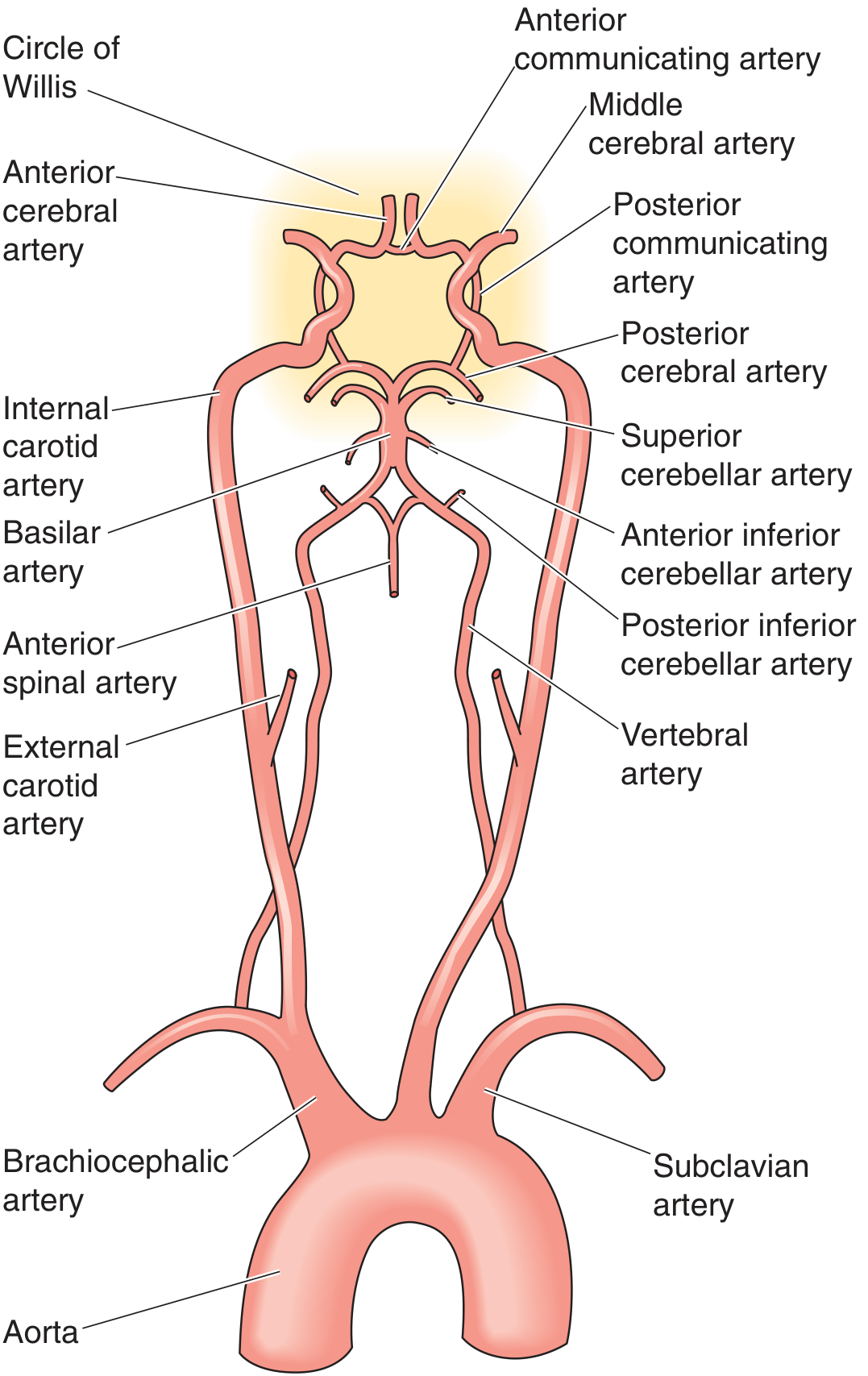
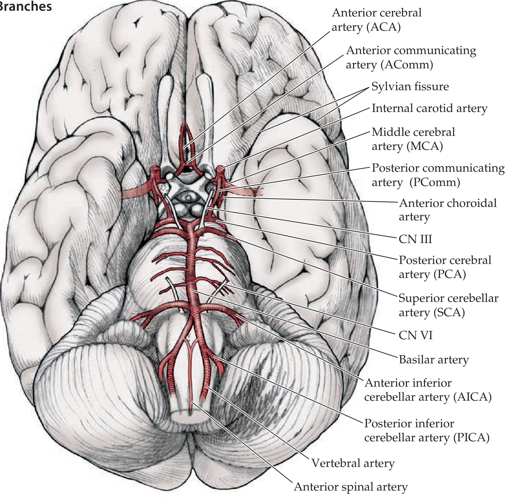
The Circle of Willis (circulus arteriosus cerebri) is an anastomotic arterial ring located at the base of the brain, in the subarachnoid space near the optic chiasm. It connects the anterior (carotid) and posterior (vertebrobasilar) circulations, providing collateral blood flow if one vessel is compromised.
Structure
Here is the full vascular supply diagram from the aorta up:
And the view from the base of the brain showing the circle and its branches:
Component Arteries
Anterior Circulation (from Internal Carotid Arteries)
| Vessel | Notes |
|---|---|
| Internal carotid arteries (ICA) | Arise from common carotids; enter the cranium and bifurcate into ACA + MCA |
| Anterior cerebral arteries (ACA) | Terminal branches of the ICA; supply medial frontal and parietal lobes |
| Middle cerebral arteries (MCA) | Terminal branches of the ICA; supply lateral frontal, parietal, temporal lobes |
| Anterior communicating artery (AComm) | Bridges the two ACAs anteriorly; closes the anterior arch of the ring |
| Posterior communicating arteries (PComm) | Connect each ICA to the ipsilateral PCA; link anterior and posterior circulations |
Posterior Circulation (from Vertebrobasilar System)
| Vessel | Notes |
|---|---|
| Vertebral arteries | Arise from subclavian arteries; ascend through cervical foramina transversaria |
| Basilar artery | Formed by the union of both vertebral arteries; gives off AICA, SCA, and perforators |
| Posterior cerebral arteries (PCA) | Terminal branches of the basilar; supply occipital lobes and medial temporal lobes |
Function
The circle's main role is collateral blood flow: if one feeding artery is occluded or stenosed, blood can be redistributed around the ring to preserve perfusion of at-risk brain territory. This protects against ischemia following vessel disease or injury.
Key Clinical Points
- A complete, full-caliber ring is present in only ~34% of individuals - anatomical variants are the rule, not the exception. - Neuroanatomy through Clinical Cases, 3rd Ed.
- The most common site for saccular (berry) aneurysms is at the junctions of the circle - especially the AComm (most common overall), PComm-ICA junction, and MCA bifurcation. Rupture causes subarachnoid hemorrhage.
- A fetal PCA variant (where the PCA fills primarily from the ICA via PComm rather than from the basilar) is one of the most frequently encountered normal variants.
- The posterior communicating arteries run adjacent to CN III (oculomotor nerve). An aneurysm of the PComm can compress CN III, causing a painful, complete third nerve palsy with a "blown pupil."
Branches to Cerebellum and Brainstem (off the basilar/vertebrals)
These lie outside the ring itself but are part of the posterior circuit:
- PICA (posterior inferior cerebellar artery) - from vertebral artery; occlusion causes Wallenberg (lateral medullary) syndrome
- AICA (anterior inferior cerebellar artery) - from basilar
- SCA (superior cerebellar artery) - from basilar, just before it bifurcates into PCAs
Sources: Neuroanatomy through Clinical Cases, 3rd Ed. | Medical Physiology (Boron & Boulpaep) | Goldman-Cecil Medicine
This is a shared conversation. Sign in to Orris to start your own chat.