
Facial nerve , from embryology to pathology Involving all the anatomy from scratch , syndromes associated , pathway ,everything .
facial nerve anatomy pathway segments diagram
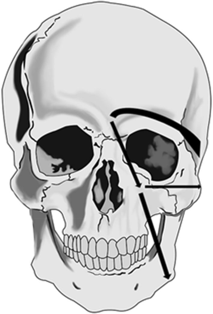
This anatomical diagram presents an anterior view of a human skull overlaid with schematic arrows illustrating facial lymphatic drainage pathways. The illustration focuses on the primary drainage routes from two specific injection sites on the left side of the face (viewer's right). The supra-ocular pathway is shown as a superiorly arching arrow originating above the orbit and directing toward the pre-auricular lymph nodes. The para-nasal pathway originates near the nasal bridge and divides into three distinct directions: inferiorly toward the sub-mandibular lymph nodes, laterally toward the parotid lymph nodes, and superior-laterally toward the pre-auricular lymph nodes. This diagram is utilized in nuclear medicine and clinical anatomy to demonstrate typical patterns of lymphoscintigraphy tracer movement and to highlight the anatomical landmarks (orbit, nasal cavity, mandible) used to map lymphatic flow in patients with facial edema or lymphedema.
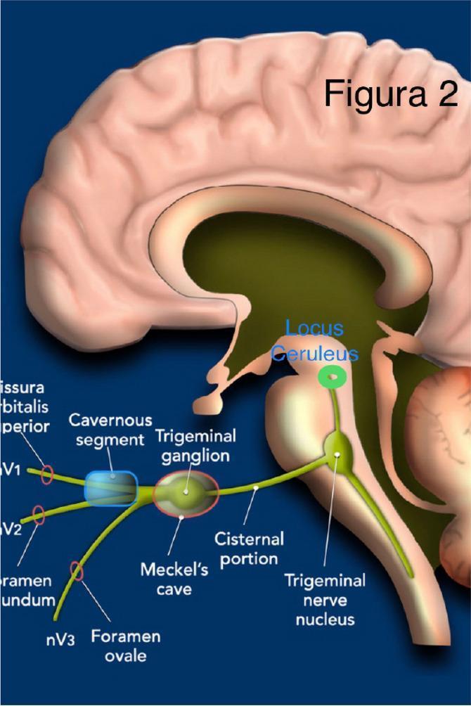
Anatomical diagram of the brain in a sagittal view illustrating the pathway of the trigeminal nerve (CN V). The illustration highlights key anatomical segments and landmarks relevant to neuroanatomy and clinical neurology. Centrally, the trigeminal nerve is traced from its distal branches—V1 (ophthalmic), V2 (maxillary), and V3 (mandibular)—as they exit via the superior orbital fissure, foramen rotundum, and foramen ovale, respectively. Moving proximally, the diagram identifies the cavernous segment (highlighted by a blue oval), the trigeminal ganglion situated within Meckel's cave, and the cisternal portion of the nerve. The nerve is shown entering the brainstem to reach the trigeminal nerve nucleus in the pons. Superior to the nucleus, a green dot marks the locus ceruleus. The diagram provides a clear educational visualization of the transition from peripheral branches to central nuclei, facilitating the study of cranial nerve distribution and relevant neurosurgical corridors.
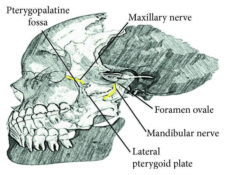
Anatomical diagram of a lateral view of the human skull, specifically focusing on the deep facial nerves and related osteological landmarks. The illustration highlights the trigeminal nerve (CN V) divisions using yellow color-coding. The maxillary nerve (V2) is shown exiting the cranial cavity through the foramen rotundum to enter the pterygopalatine fossa, located anterior to the lateral pterygoid plate. The mandibular nerve (V3) is depicted exiting via the foramen ovale, descending posterior to the lateral pterygoid plate. Key labels include the Pterygopalatine fossa, Maxillary nerve, Foramen ovale, Mandibular nerve, and Lateral pterygoid plate. The diagram serves as an educational resource for understanding the regional anatomy required for performing nerve blocks and identifying neural pathways within the infratemporal and pterygopalatine regions.
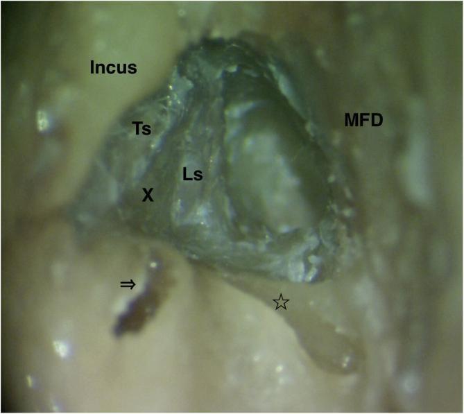
This endoscopic clinical photograph provides a high-magnification view of a surgical dissection of the human left temporal bone, specifically focusing on the facial nerve anatomy. The image demonstrates the labyrinthine segment (Ls) of the facial nerve following the removal of overlying bone. Key anatomical landmarks are labeled: the tympanic segment (Ts) of the facial nerve, the incus, and the middle fossa dura (MFD). The vertical crest (Bill's bar), located at the fundus of the internal acoustic canal, is marked with an 'X'. Additionally, the ampullary ends of the lateral semicircular canal (indicated by an arrow) and the superior semicircular canal (indicated by a star) are visible. The membranous labyrinths have been removed to facilitate clear visualization of these bony boundaries. This view is clinically significant for understanding surgical approaches for facial nerve decompression, particularly at the meatal foramen and the junction of the labyrinthine and tympanic segments.
facial nerve branches parotid gland terminal branches face
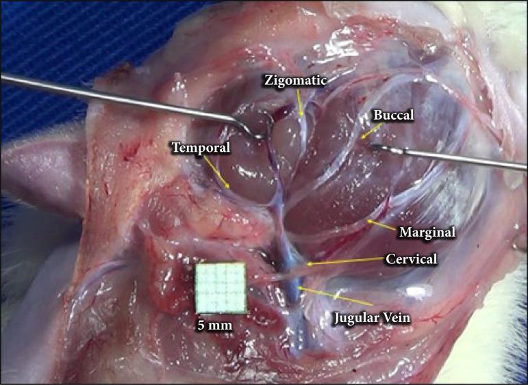
A clinical intraoperative photograph showing the anatomical arrangement of the facial nerve branches in a surgical dissection, following parotidectomy. The image provides a lateral view of the cervicofacial region where the parotid gland has been removed to expose the extratemporal facial nerve plexus. Labeled branches include the temporal branch extending superiorly, the zygomatic and buccal branches traveling toward the mid-face, and the marginal mandibular and cervical branches descending inferiorly. The cervical branch is depicted in close proximity to the jugular vein, which is visible in the lower surgical field. Surgical instruments (retractors) are positioned to maintain exposure of the deep tissues. A 5mm grid scale is included for anatomical measurement. This visual serves as an educational guide for surgical anatomy, identifying key landmarks for nerve preservation during parotid gland surgery or facial nerve decompression.
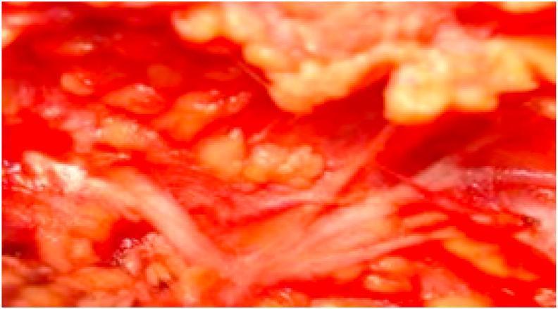
This intraoperative clinical photograph displays an anatomical dissection of the facial nerve (Cranial Nerve VII) within the parotid region. The image illustrates a 'Type II' branching pattern characterized by a single facial nerve trunk (FNT) that divides directly into multiple terminal branches. Notably, this variant lacks the typical primary bifurcation into temporofacial and cervicofacial divisions. The nerve branches appear as thin, pale, string-like structures radiating through the reddish, fleshy tissue of the surgical field. The surrounding area shows exposed glandular and soft tissue relevant to a parotidectomy procedure. This visual serves as an educational reference for surgical anatomy, highlighting normal anatomical variations of the extracranial facial nerve that are critical for surgeons to identify during parotid gland surgery to avoid nerve injury and post-operative facial paralysis.
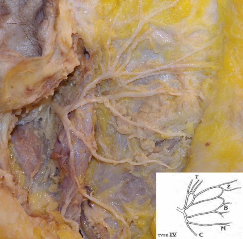
This clinical photograph displays a gross anatomical dissection of the right facial nerve (Cranial Nerve VII) within the parotid region. The image demonstrates a single facial nerve trunk that undergoes bifurcation into primary upper and lower divisions. Following this initial split, the nerve exhibits a complex, interconnected branching pattern identified as Davis Type IV. Multiple secondary branches are visible radiating across a bed of yellowish adipose tissue and the masseter muscle fascia. These branches eventually form the terminal ramifications: temporal (T), zygomatic (Z), buccal (B), marginal mandibular (M), and cervical (C) branches. The parotid gland tissue has been partially reflected to expose the pes anserinus. This specimen serves as an educational example of neuroanatomical variation, specifically highlighting the Davis classification system for surgical planning in parotidectomy and facial reconstructive procedures.
Bell's palsy facial nerve palsy lower motor neuron upper motor neuron
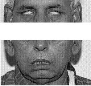
This clinical photograph displays a patient with bilateral lower motor neuron (LMN) facial nerve palsy, likely associated with Ramsay Hunt syndrome or a similar polyneuropathy. The upper panel illustrates bilateral lagophthalmos and Bell's phenomenon: when the patient attempts to close his eyes, the globes roll upward, leaving the sclera visible due to incomplete eyelid closure. The lower panel demonstrates significant facial muscle weakness and hypotonia, characterized by bilateral drooping of the oral commissures and a slack, open-mouthed appearance with visible teeth. There is a notable absence of nasolabial folds and forehead wrinkling, consistent with a complete bilateral facial nerve deficit. The educational focus is on identifying the physical signs of LMN facial paralysis, specifically the loss of motor control in the orbicularis oculi and orbicularis oris muscles, which differentiates it from upper motor neuron lesions.
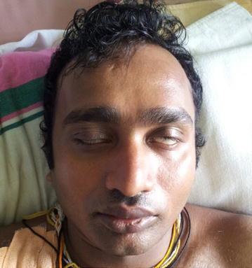
Clinical photograph of a patient's face demonstrating bilateral lower motor neuron facial nerve palsy (CN VII). The image highlights lagophthalmos, where both eyelids are only partially closed despite an attempt at full closure. A positive Bell's phenomenon (Bell's sign) is clearly visible bilaterally, characterized by the upward and outward rotation of the globes, exposing the white sclera beneath the incompletely closed eyelids. The mid-to-lower face shows relative symmetry at rest with neutral oral commissures, suggesting the deficit primarily impacts upper facial musculature or is uniformly bilateral. This visual is typical for clinical presentations of bilateral facial paralysis, which can be seen in systemic neurological conditions such as Guillain-Barré syndrome (specifically the Miller Fisher variant or facial diplegia subtype). The photograph serves as an educational resource for identifying cranial nerve pathology and distinguishing between upper and lower motor neuron signs.
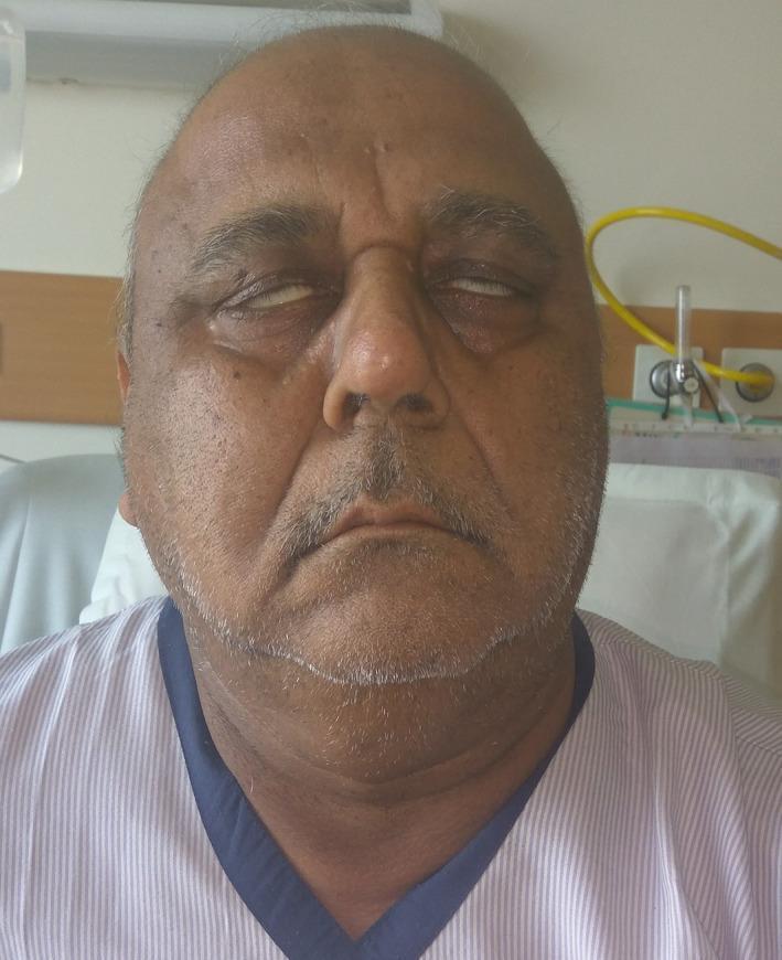
This clinical photograph shows the frontal view of an elderly male patient exhibiting signs of bilateral lower motor neuron facial nerve palsy (Bell's palsy). A prominent finding is Bell's phenomenon, where the patient's eyeballs rotate upward and outward when attempting to close the eyes, leaving the sclera visible due to incomplete eyelid closure (lagophthalmos). The facial features appear relatively symmetrical but mask-like, with a loss of normal expression lines. The mouth is slightly open with evidence of drooling at the commissures, consistent with orbicularis oris muscle weakness. This presentation is highly suggestive of cranial nerve VII involvement, often associated with systemic conditions such as Guillain-Barr syndrome, sarcoidosis, or leptomeningeal metastasis. The image is an educational resource for demonstrating neurological deficits and the physical examination findings of bilateral facial paralysis.
Ramsay Hunt syndrome herpes zoster ear vesicles facial palsy
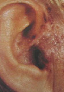
This clinical photograph shows a close-up of the right external ear and preauricular region exhibiting characteristic features of Ramsay Hunt syndrome (herpes zoster oticus). The image displays a cluster of small, fluid-filled vesicles on an erythematous base. These herpetic lesions are localized to the pinna and the external auditory canal entrance, which corresponds to the sensory distribution of the facial nerve (cranial nerve VII). Some vesicles appear intact with clear fluid, while others show signs of early crusting or rupture. The surrounding skin exhibits diffuse erythema and mild edema. This presentation is medically significant for medical students and clinicians as it illustrates the typical cutaneous manifestations of varicella-zoster virus reactivation involving the geniculate ganglion. Key educational points include the recognition of dermatomal vesicular rashes and their association with otalgia and potential facial nerve palsy.
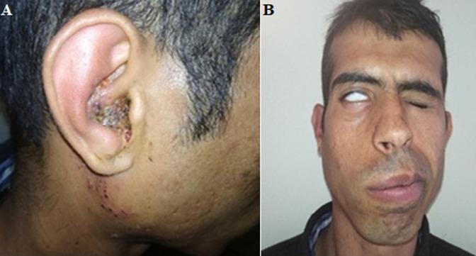
This composite clinical photograph displays the hallmark signs of Ramsay Hunt syndrome (herpes zoster oticus). Image A (left) shows the right ear auricle with inflammatory swelling and a characteristic vesicular eruption within the concha and external auditory meatus. Multiple clustered vesicles and hemorrhagic crusting are visible, extending down the preauricular area and neck. Image B (right) demonstrates an associated ipsilateral peripheral facial nerve palsy (CN VII). Key findings include significant facial asymmetry with right-sided drooping, loss of nasolabial fold depth, and deviation of the mouth to the contralateral (left) side. The patient displays Bell's phenomenon on the right, where the globe rotates upward during an attempt to close the eye, leaving only the white sclera visible due to incomplete eyelid closure (lagophthalmos). These images illustrate the triad of otalgia, auricular vesicles, and facial paralysis typical of varicella-zoster virus reactivation in the geniculate ganglion, targeting medical students and clinicians in neurology, otolaryngology, and dermatology.
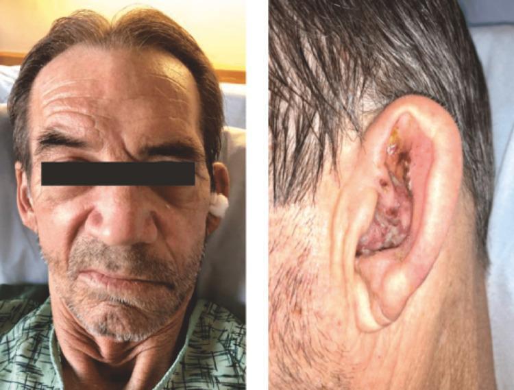
Two-panel clinical photograph illustrating the classic presentation of Ramsay Hunt syndrome (herpes zoster oticus). The left panel displays a frontal view of a patient's face exhibiting signs of peripheral facial nerve (CN VII) palsy. Notable features include ipsilateral facial drooping, flattening of the nasolabial fold, and a downward turn of the mouth corner on the affected side. The right panel provides a close-up of the auricular region, showing a characteristic zoster rash. Multiple erythematous vesicles and areas of crusting are visible within the concha and on the antihelix of the pinna. The image demonstrates the diagnostic association between acute facial paralysis and a painful vesicular eruption in the ear canal or auricle, highlighting the reactivation of the varicella-zoster virus in the geniculate ganglion. This material is suitable for medical education focusing on neurology, dermatology, and infectious diseases.
facial nerve intratemporal course segments labyrinthine tympanic mastoid geniculate ganglion
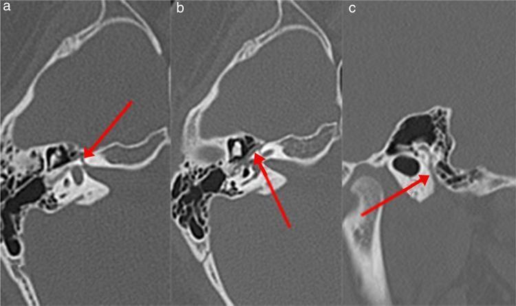
High-resolution axial computed tomography (CT) images of the left temporal bone, presented in three sections (A, B, and C) to demonstrate the course of the facial nerve (Fallopian) canal. Red arrows highlight different segments of the canal. Image A shows the labyrinthine segment, appearing as a narrow radiolucent channel within the dense petrous bone, traveling anterolaterally towards the geniculate ganglion. Image B depicts the tympanic (horizontal) segment, visualized as a thin lucency running along the medial wall of the tympanic cavity, inferior to the lateral semicircular canal. Image C illustrates the mastoid (descending) segment, where the canal assumes a vertical orientation within the mastoid portion of the temporal bone, posterior to the external auditory canal. These images are clinically relevant for evaluating anatomical variations or pathologies, such as Bell's palsy, by assessing the diameter and integrity of the bony canal across its various intratemporal segments.
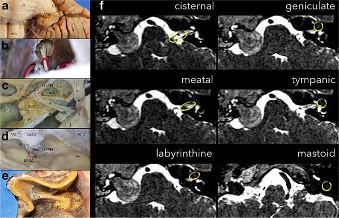
Educational medical graphic combining anatomical dissections (a-e) and diagnostic imaging (f) focused on the segments of the facial nerve (CN VII). Panels (a-b) illustrate the cisternal segment within the cerebellopontine angle and internal acoustic meatus (IAM) via a retrosigmoid approach. Panel (c) shows a superior view of the petrous bone exposing meatal, labyrinthine, and tympanic segments relative to the cochlea and semicircular canals. Panel (d) displays a middle fossa view highlighting the greater superficial petrosal nerve (GSPN) and Gasserian ganglion. Panel (e) depicts the mastoid segment after mastoidectomy. Panel (f) presents a series of axial T2-weighted MRI slices. On the left (normal side), yellow circles track the facial nerve through its cisternal, meatal, labyrinthine, geniculate, tympanic, and mastoid segments. On the contralateral right side, a large, heterogeneous, hyperintense facial nerve schwannoma is visible, causing significant expansion of the IAM and displacement of the surrounding brainstem and cerebellar structures. The content serves to teach the complex three-dimensional course of CN VII and its pathological appearance in schwannomatosis.
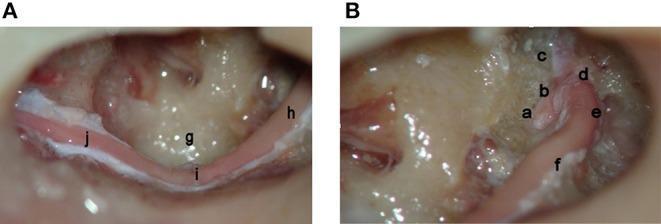
Two intraoperative endoscopic photographs (A and B) demonstrate a complete surgical decompression of the left facial nerve in a cadaveric specimen, tracing the nerve from the labyrinthine meatus to the stylomastoid foramen. Image B shows the proximal segments, including the meatal foramen (a), labyrinthine portion (b), greater superficial petrosal nerve (c), the first genu (d), the geniculate ganglion (e), and the proximal horizontal (tympanic) segment (f). Image A details the distal course, illustrating the continuation of the horizontal segment (h) as it passes the sinus tympani (g), reaching the second genu (i). From this point, the nerve transitions into its vertical (mastoid) segment (j), extending toward the stylomastoid foramen. This anatomical display highlights the relationship between the facial nerve and middle ear landmarks, such as the sinus tympani, and is relevant for otologic surgery, temporal bone fracture management, and facial nerve decompression procedures.
The Facial Nerve (CN VII) - Complete Guide
1. EMBRYOLOGY
Origins and Early Development
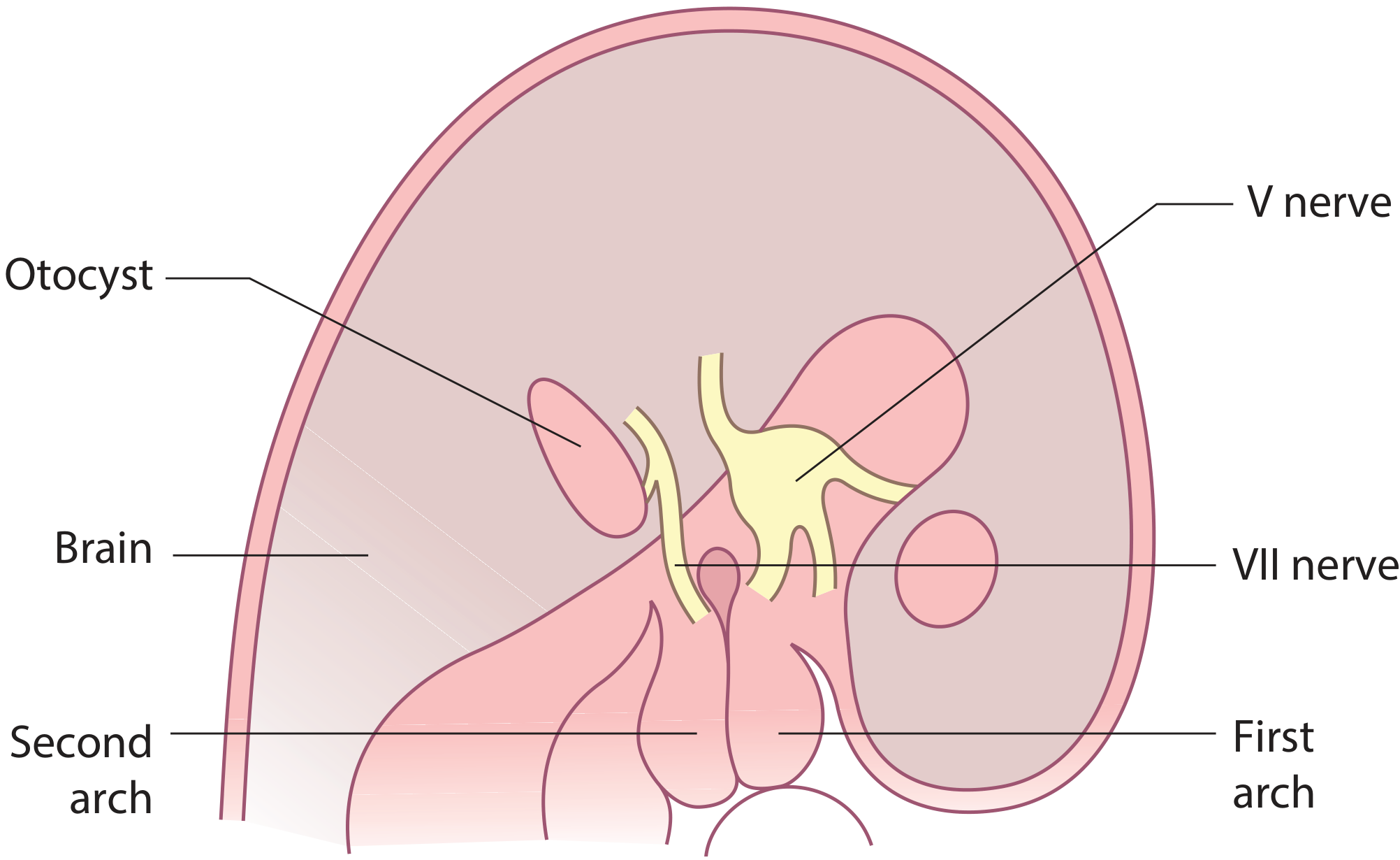
- 5th week: The geniculate ganglion has formed. The facial nerve divides into its main trunk (descending into the 2nd branchial arch) and the chorda tympani (the pretrematic branch, curving cranially into the 1st arch). At this stage, the chorda tympani and main trunk are equal in size.
- Facial nucleus: Neuroblasts in the pons form the facial nucleus, with the 6th nerve nucleus in close proximity. As the pons expands, the 6th nucleus ascends, so facial nerve fibers wrap around it, forming the internal genu. This explains why a single lesion (e.g., Moebius syndrome, pontine stroke) can involve both CN VI and CN VII simultaneously.
- 7th week: The geniculate ganglion (which has a separate origin from the main facial nerve) is well defined and gives rise to the sensory roots forming the nervus intermedius.
Second Branchial Arch Derivatives
-
Muscles of facial expression
-
Stapedius
-
Stylohyoid
-
Posterior belly of digastric
-
Scott-Brown's Otorhinolaryngology Head & Neck Surgery Vol 2, p.275
-
Shambaugh Surgery of the Ear, p.68
2. FIBER COMPOSITION
| Fiber Type | Modality | Function |
|---|---|---|
| Special Visceral Efferent (SVE) | Motor | Muscles of facial expression, stapedius, stylohyoid, digastric (post. belly) |
| General Visceral Efferent (GVE) | Parasympathetic (preganglionic) | Lacrimal gland, nasal/palatal seromucinous glands (via GSPN + pterygopalatine ganglion); submandibular & sublingual glands (via chorda tympani + submandibular ganglion) |
| Special Sensory (SVA) | Taste | Anterior 2/3 of tongue (via chorda tympani); tonsillar fossa & palate (via GSPN) |
| General Somatic Afferent (GSA) | Touch/proprioception | External auditory canal, conchal skin of auricle; proprioception from facial muscles |
| Visceral Afferent (GVA) | Sensation | Mucosa of nose, pharynx, palate |
- Motor nucleus (caudal pons) - SVE fibers
- Superior salivatory nucleus (dorsal to motor nucleus) - GVE fibers
- Nucleus of the solitary tract (medulla oblongata) - taste (SVA) fibers
Key point: The superior portion of the motor nucleus (innervating frontalis and orbicularis oculi) receives both crossed and uncrossed cortical input. The inferior portion receives only ipsilateral cortical input. This is why UMN lesions (cortical/capsular) spare forehead movement.
- Cummings Otolaryngology, p.2451
- Shambaugh Surgery of the Ear, p.68
3. COURSE AND PATHWAY
3A. Intracranial (Cisternal) Segment
- Length: ~24 mm
- Emerges from the brainstem between the pons and olive (the main motor root)
- The nervus intermedius (carrying sensory and parasympathetic fibers) emerges between the pons and inferior cerebellar peduncle
- Both join in the cerebellopontine angle (CPA) and travel together to the internal auditory meatus (IAM) alongside CN VIII
3B. Intracanalicular Segment (IAC)
- Within the IAC, the facial nerve occupies the anterosuperior quadrant, above the cochlear nerve; superior and inferior vestibular nerves are posterior
- At the fundus of the IAC, the nerve is separated from the superior vestibular nerve by a vertical crest of bone (Bill's bar)
- At this segment, the nerve lacks a fibrous sheath or endoneurium and is surrounded only by a thin layer of arachnoid - making it vulnerable to injury
3C. Labyrinthine Segment
- Shortest (~4 mm) and narrowest segment of the fallopian canal
- Runs superior to the cochlea, between the cochlea and vestibule
- Exits to form the geniculate fossa (just deep to the squamous temporal bone)
- The geniculate ganglion is separated from the middle fossa floor by a very thin layer of bone, which is dehiscent in ~25% of ears
- The facial nerve acquires its fibrous sheath at and distal to the geniculate ganglion
3D. Geniculate Ganglion - The Key Branching Point
- Carries preganglionic parasympathetics to the lacrimal gland (via pterygopalatine ganglion)
- Also carries taste fibers from palate
- Travels anteriorly along the middle fossa floor between two layers of dura
- Intraoperative traction on this nerve can cause facial palsy via hemorrhage or edema
3E. Tympanic (Horizontal) Segment
- Length: ~13 mm
- Begins at the first genu where the main trunk turns acutely posterior and slightly inferior
- Occupies the medial wall of the anterior attic (tympanic cavity)
- Passes superior to the cochleariform process, then forms the superior wall of the oval window niche
- Key landmarks: cochleariform process, oval window, pyramidal eminence
3F. Mastoid (Vertical) Segment
- Length: ~20 mm
- Begins at the second genu, just anteroinferior to the lateral semicircular canal, at the pyramidal eminence
- Runs vertically down to the stylomastoid foramen
- Branch given off here:
- Nerve to Stapedius - via a tiny canaliculus at the pyramidal eminence
- Carries taste from anterior 2/3 of tongue
- Carries preganglionic parasympathetics to submandibular and sublingual glands (via submandibular ganglion)
- The space between the mastoid facial nerve and chorda tympani forms the facial recess used in posterior tympanotomy surgery
3G. Extracranial Segment
- Posterior auricular nerve - occipital belly of occipitofrontalis, auricular muscles
- Branch to posterior belly of digastric and stylohyoid
- Upper (temporofacial) division
- Lower (cervicofacial) division
| Branch | Muscles Supplied |
|---|---|
| Temporal | Frontalis, orbicularis oculi (upper), corrugator supercilii |
| Zygomatic | Orbicularis oculi (lower), zygomaticus |
| Buccal | Buccinator, zygomaticus major, levator labii, nasalis |
| Marginal mandibular | Depressor anguli oris, depressor labii, mentalis |
| Cervical | Platysma |
- Scott-Brown's ORL Vol 2, p.1293
- Cummings Otolaryngology, p.2451
- Shambaugh Surgery of the Ear, p.68
4. UPPER MOTOR NEURON vs. LOWER MOTOR NEURON LESIONS
| Feature | UMN Lesion (above motor nucleus) | LMN Lesion (nucleus or peripheral) |
|---|---|---|
| Forehead | Spared (bilateral cortical representation) | Involved (drooping brow, absent forehead wrinkles) |
| Eye closure | Intact | Impaired - lagophthalmos + Bell's phenomenon |
| Mouth | Contralateral lower face weakness | Ipsilateral, all branches |
| Taste | Normal | May be lost (chorda tympani) |
| Lacrimation | Normal | May be reduced (GSPN) |
| Hyperacusis | Absent | Present (stapedius branch) |
| Cause | Stroke, tumor (cortex/internal capsule) | Bell's palsy, RHS, trauma, tumor |
5. SYNDROMES AND ASSOCIATED CONDITIONS
5A. Bell's Palsy (Idiopathic Facial Paralysis) - 70% of acute cases
- Most common cause of acute unilateral facial paralysis (LMN type)
- Diagnosis of exclusion
- Believed to result from reactivation of Herpes Simplex Virus (HSV-1) causing inflammation and edema within the narrow labyrinthine segment of the fallopian canal
- Onset: Rapid (< 48 hours), may involve CN V through XII
- Imaging: MRI shows linear enhancement from distal IAC through labyrinthine segment and geniculate ganglion; HRCT is normal
- Treatment:
- Prednisolone 1 mg/kg for 10 days with 10-day taper (evidence-based first-line)
- Acyclovir/Valacyclovir - considered if seen within first 3 days
- Eye protection (lagophthalmos risk of corneal damage)
- Prognosis: Most recover; ~10% have poor recovery with risk of sequelae
5B. Ramsay Hunt Syndrome (Herpes Zoster Oticus) - 15% of cases
- Reactivation of Varicella-Zoster Virus (VZV) in the geniculate ganglion
- Classic triad:
- Otalgia (often severe)
- Vesicular eruption of the external auditory canal (EAC) and conchal bowl/pinna
- Facial nerve palsy (LMN)
- Higher incidence of associated cochlear (sensorineural hearing loss) and vestibular disturbance than Bell's palsy
- Prognosis is worse than Bell's palsy
- Treatment: Valacyclovir + corticosteroids (Valacyclovir may be more effective here than acyclovir)
5C. Moebius Syndrome
- Congenital agenesis of the facial nucleus AND the 6th nerve nucleus
- Results in bilateral facial palsy + bilateral abducens palsy
- Other cranial nerves may be involved
- FN grafting is not an option (nuclear agenesis)
- Explainable embryologically: as the facial fibers wrap around the 6th nucleus during development, both structures are affected by the same teratogenic insult
5D. Melkersson-Rosenthal Syndrome
- Recurrent unilateral facial palsy
- Orofacial edema (particularly lip swelling - cheilitis granulomatosa)
- Fissured tongue (lingua plicata)
- Granulomatous inflammation on biopsy
- Treatment: corticosteroids + decompression surgery for recurrent cases
5E. Lyme Disease (Borrelia burgdorferi)
- Can cause unilateral or bilateral facial palsy (ratio 3:1 unilateral)
- Associated "bull's eye" (erythema migrans) rash
- Treatment: tetracycline (adults), penicillin (children)
5F. Other Causes of Facial Nerve Palsy
| Cause | Notes |
|---|---|
| Guillain-Barré syndrome | Most common cause of bilateral facial palsy |
| Diabetes mellitus | Microvascular ischemia |
| Sarcoidosis | Heerfordt syndrome (uveoparotid fever) |
| Acoustic neuroma (vestibular schwannoma) | Gradual onset; hearing loss prominent |
| Parotid malignancy | Perineural invasion |
| Cholesteatoma | Erosion of facial canal |
| Otitis media (acute/chronic) | Direct spread |
| Multiple sclerosis | Demyelination |
| Mononucleosis (EBV) | |
| Mumps | |
| Myasthenia gravis | (neuromuscular junction, not truly nerve) |
| Temporal bone fracture | Longitudinal (more common) vs. transverse |
| Birth trauma / forceps delivery | Neonatal facial palsy |
6. SEQUELAE OF FACIAL NERVE INJURY (Aberrant Regeneration)
Synkinesis
- Simultaneous movement of muscles supplied by different branches when only one should move
- Example: corner of mouth contracts when eye closes ("jaw winking")
- Mechanism: axons regenerate into the wrong fascicles (aberrant misdirection)
Crocodile Tears (Bogorad Syndrome)
- Involuntary lacrimation when eating
- Mechanism: Parasympathetic fibers meant for submandibular gland (chorda tympani) aberrantly regenerate into the lacrimal gland pathway (GSPN)
- Gustatory stimulus triggers lacrimation instead of salivation
Frey Syndrome (Gustatory Sweating)
- Sweating and flushing of the preauricular/temporal region while eating
- Occurs after parotid surgery or parotid region trauma
- Mechanism: parasympathetic fibers of the auriculotemporal nerve (originally to parotid) aberrantly reinnervate sweat glands of the overlying skin
- Note: this is a trigeminal-autonomic misdirection, not strictly facial nerve
Hemifacial Spasm
- Involuntary twitching/contractions of one side of the face
- Can follow Bell's palsy recovery; also occurs with vascular compression at the root entry zone of CN VII
- Mechanism: ephaptic (cross-talk) transmission between adjacent nerve fibers
Contracture
-
Permanent shortening of facial muscles following long-standing palsy
-
Adams & Victor's Principles of Neurology, 12th Ed, p.2100
-
Scott-Brown's ORL Vol 2, p.1436
7. ASSESSMENT AND GRADING
House-Brackmann Grading Scale (most widely used)
| Grade | Degree of Dysfunction | Description |
|---|---|---|
| I | Normal | Normal symmetrical function in all areas |
| II | Mild | Slight weakness on close inspection; complete eye closure with minimum effort; slight asymmetry of smile |
| III | Moderate | Obvious but not disfiguring weakness; may not lift eyebrow; complete eye closure with strong effort; obvious but not disfiguring synkinesis |
| IV | Moderately severe | Obvious disfiguring weakness; cannot lift eyebrow; incomplete eye closure; mouth asymmetry |
| V | Severe | Only barely perceptible motion; incomplete eye closure; slight movement at corner of mouth |
| VI | Total paralysis | No movement |
Electrodiagnostic Tests
- Electroneuronography (ENoG): Measures compound motor action potential; >90% degeneration predicts poor prognosis
- Electromyography (EMG): Fibrillation potentials indicate degeneration; voluntary polyphasic potentials indicate early reinnervation
- Nerve excitability test / Maximum stimulation test
8. IMAGING IN FACIAL NERVE DISEASE
MRI
- Gold standard for soft tissue and nerve pathology
- Bell's palsy: Linear enhancement from distal IAC through labyrinthine segment and geniculate ganglion (do not use as a diagnostic tool in typical presentation)
- Facial nerve schwannoma: T1 - isointense expansile mass with smooth margins; T2 - homogeneously high signal; enhances with contrast; can occur anywhere along the course
- Perineural invasion from parotid malignancy: requires imaging from brainstem to parotid
- Hemangioma of geniculate ganglion: Presents with early disproportionate facial weakness; "honeycomb" or irregular calcification on CT
CT (HRCT)
- Best for bony canal evaluation
- Shows fallopian canal dehiscence, fractures, cholesteatoma erosion
- Normal in Bell's palsy
9. SURGICAL CONSIDERATIONS
Facial Recess
- Space between mastoid facial nerve and chorda tympani
- Used for posterior tympanotomy to access the middle ear for cochlear implant surgery
Parotid Surgery
- The facial nerve runs through the substance of the parotid gland (it anatomically divides the parotid into superficial and deep lobes)
- The nerve is identified at the stylomastoid foramen using landmarks: tragal pointer, posterior belly of digastric, tympanomastoid suture
- Must be preserved during superficial parotidectomy
Pediatric Considerations - Important Differences
| Feature | Child | Adult |
|---|---|---|
| Mastoid process | Absent in neonates (incomplete tympanic ring) | Present; tympanic ring complete by adolescence |
| Chorda tympani exit | May exit with main trunk in neonates | Exits separately, proximal to stylomastoid foramen |
| Second genu | More acute and lateral | Less acute and more medial |
| Nerve trunk position | More anterior and lateral | Less anterior, deeper |
| Nerve over mandible | Very superficial (surgical risk) | Less superficial |
- Scott-Brown's ORL Vol 2, p.276
Facial Nerve Repair
- Primary neurorrhaphy: Best if done within 3-5 days of injury; tension-free repair required
- Interposition graft: Greater auricular nerve or sural nerve (gap > 1 cm)
- Hypoglossal-facial nerve transfer (XII-VII): For proximal lesions; causes tongue hemiatrophy; near-nerve VII branch anastomosis preferred when available
- Free muscle transfer: For long-standing paralysis (> 2 years); using gracilis or pectoralis minor with vascular and nerve supply
10. KEY CLINICAL PEARLS
| Symptom | Branch Involved | Significance |
|---|---|---|
| Loss of taste (anterior 2/3 tongue) | Chorda tympani | Lesion distal to geniculate but proximal to chorda tympani takeoff |
| Hyperacusis | Nerve to stapedius | Lesion proximal to pyramidal eminence |
| Decreased lacrimation | GSPN | Lesion at or proximal to geniculate ganglion |
| Forehead spared | UMN | Bilateral cortical representation of upper face |
| Forehead involved | LMN | Ipsilateral, complete |
- Lesion at brainstem (e.g., pontine glioma) → CN VI + CN VII palsy ipsilateral + contralateral hemiplegia (Millard-Gubler syndrome)
- Lesion in CPA (e.g., acoustic neuroma) → facial palsy + hearing loss + tinnitus + vertigo
- Lesion in IAC → same as CPA
- Lesion at geniculate ganglion → facial palsy + loss of lacrimation + hyperacusis + loss of taste
- Lesion at pyramidal eminence → facial palsy + hyperacusis + loss of taste
- Lesion between stapedius and chorda tympani → facial palsy + loss of taste only
- Lesion at stylomastoid foramen or below → pure motor facial palsy (no taste, lacrimation, or hearing changes)
- Scott-Brown's Otorhinolaryngology Head & Neck Surgery, Vol 2 (Chapters 21 and 112)
- Cummings Otolaryngology: Head and Neck Surgery (Chapters 126, 135, 138, 171)
- Shambaugh: Surgery of the Ear (Chapter 2)
- KJ Lee's Essential Otolaryngology
- Adams & Victor's Principles of Neurology, 12th Edition
- Localization in Clinical Neurology, 8th Edition