
Give me the photo of a typical rib that address the all the muscle assessments and the ligament adjustments I mean addressing all the main components on the ribs. I mean all the components must be displayed properly.
labeled typical rib anatomy head neck tubercle angle shaft costal groove anterior end articulation ligaments muscles attachments
typical rib anatomy labeled diagram head neck tubercle angle costal groove ligaments muscle attachments
ribs anatomy
https://www.kenhub.com/en/library/anatomy/the-ribs
Educational anatomical illustration of a typical right rib (a mid-thoracic rib, such as the 6th rib), shown as a clear multi-view labeled study plate. Include superior and inferior/internal views plus a small posterior vertebral articulation inset. Accurately show and label: head with superior and inferior articular facets and crest of head, neck, tubercle with articular facet and non-articular rough part, angle, shaft/body, superior border, inferior border, internal surface, external surface, costal groove, anterior costal end and costal cartilage. Show attachment areas and label the main related structures: external intercostal, internal intercostal, innermost intercostal, levator costarum, subcostalis, serratus anterior, serratus posterior superior and inferior, and iliocostalis where appropriate. In the posterior inset, show the costovertebral and costotransverse joints and label radiate ligament of head of rib, intra-articular ligament of head of rib, proper costotransverse ligament, lateral costotransverse ligament, and superior costotransverse ligament. Include the intercostal neurovascular bundle in the costal groove in correct superior-to-inferior order: vein, artery, nerve. Make anatomy accurate, uncluttered, high-resolution, suitable for medical students.
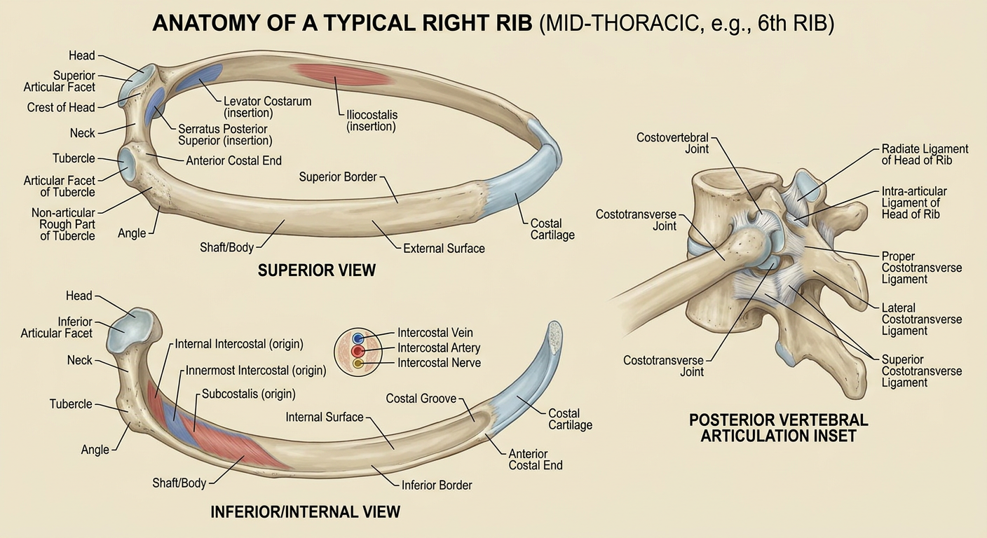
- Head: superior and inferior articular facets, separated by the crest of the head. It articulates with the bodies of its own thoracic vertebra and the vertebra above.
- Neck: between head and tubercle.
- Tubercle: has an articular facet for the transverse process of the corresponding vertebra and a rough non-articular portion.
- Angle: the point where the rib turns anterolaterally.
- Shaft/body: curved, with external and internal surfaces plus superior and inferior borders.
- Costal groove: on the inferior internal border, protecting the intercostal vein, artery, and nerve, in that order from superior to inferior.
- Anterior costal end: joins the costal cartilage.
Also address the cartilages
costal cartilage anatomy costochondral sternocostal interchondral joints ligaments
costal cartilage anatomy
Educational medical anatomy study plate of the thoracic cage centered on one typical right rib and its costal cartilage, with accurate labeled superior/internal and anterior views. Display the complete bony typical rib: head with superior and inferior articular facets and crest, neck, tubercle with articular and non-articular parts, angle, shaft/body, costal groove, and anterior costal end. Continue the rib into its hyaline costal cartilage and show the costochondral junction. Include an anterior sternum inset showing all cartilage relationships: costal cartilages 1 to 7 attach directly to sternum, cartilage 1 to manubrium as a synchondrosis, cartilages 2 to 7 as sternocostal synovial joints, cartilages 8 to 10 joining the cartilage above as false ribs, and ribs 11 and 12 with free anterior ends. Label interchondral joints and interchondral ligaments. Label the sternocostal ligaments, including anterior and posterior radiate sternocostal ligaments, intra-articular sternocostal ligament, and xiphichondral ligaments at the seventh cartilage. Include posterior costovertebral and costotransverse ligament inset with radiate ligament of head of rib, intra-articular ligament, proper, lateral, and superior costotransverse ligaments. Include concise labels for main muscle attachments: external, internal and innermost intercostals, subcostales, levatores costarum, serratus anterior, serratus posterior, and iliocostalis. Show neurovascular bundle in the costal groove as vein, artery, nerve. High-resolution, anatomically accurate, clean labeled instructional plate for medical students.
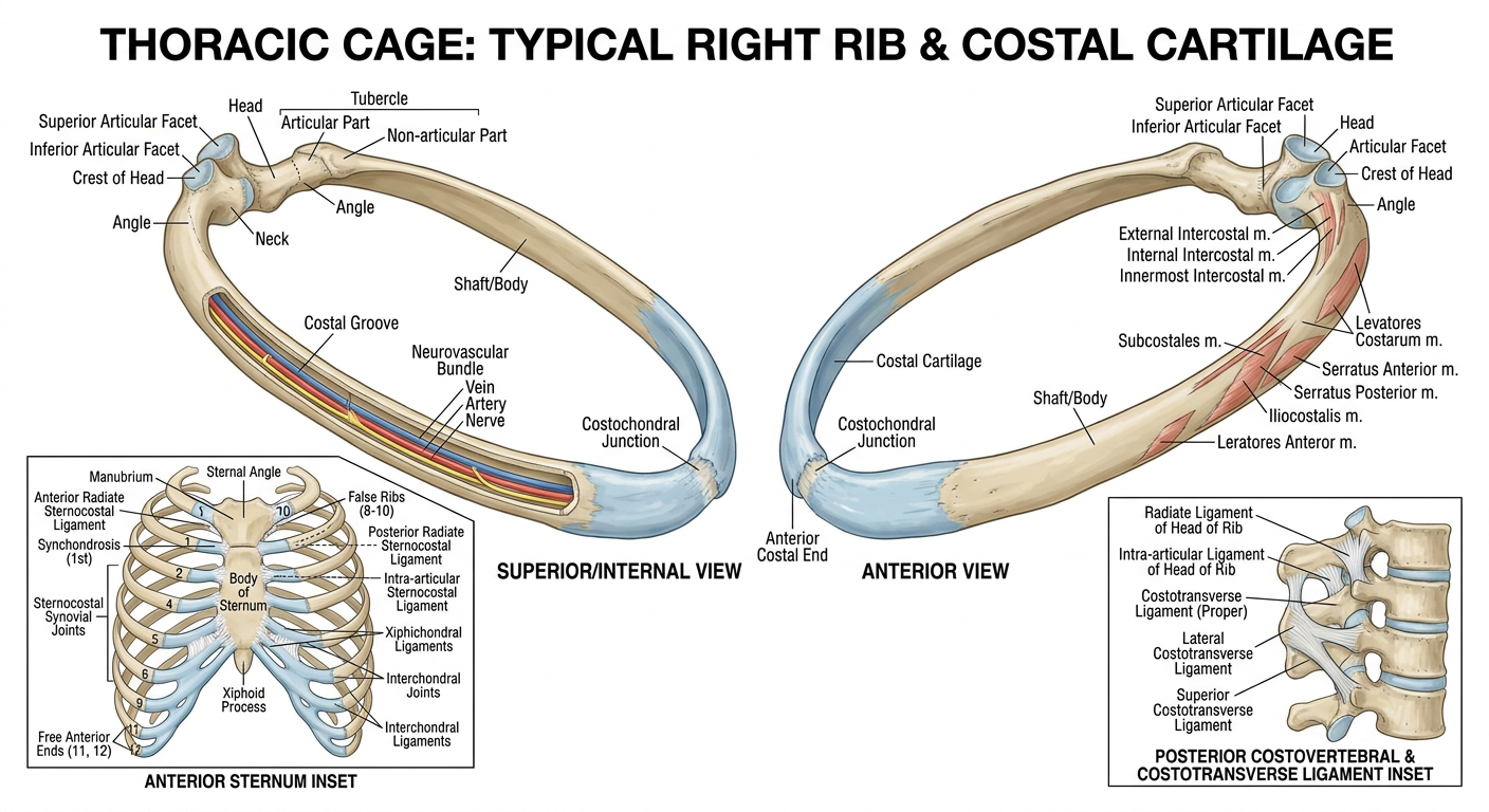
Costal cartilage components included
- Costochondral junction: the continuous transition from the bony anterior end of the rib to its hyaline costal cartilage. It is firmly bound and normally has no movement.
- True ribs, 1-7: each cartilage reaches the sternum directly.
- First costal cartilage joins the manubrium by a primary cartilaginous joint (synchondrosis).
- Costal cartilages 2-7 form sternocostal synovial joints with the sternum.
- False ribs, 8-10: their cartilages join the cartilage immediately above, rather than attaching directly to the sternum.
- Floating ribs, 11-12: do not connect to the sternum. Their anterior ends are free within the abdominal wall.
Cartilage-related ligaments shown
- Anterior and posterior radiate sternocostal ligaments: reinforce the sternocostal joints.
- Intra-articular sternocostal ligament: especially relevant at the second sternocostal joint, where it divides the joint cavity.
- Xiphichondral ligaments: reinforce the seventh costal cartilage's relation to the xiphoid process.
- Interchondral ligaments: stabilize the connections among the lower costal cartilages, especially in the false-rib region.