
dentigerous cyst
Reading File
Now let me search the medical library and PubMed in parallel for dentigerous cyst content.
Finding Sources
Finding Sources
Searching PubMed
"dentigerous cyst"[MeSH Terms]
Good results. Now let me read the detailed textbook sections in parallel.
Reading File
Reading File
Reading File
Now let me get the microscopic features, treatment, and neoplastic potential sections, plus fetch the radiograph image.
Reading File
Loading Image
Loading Image
I now have comprehensive information from multiple textbooks and images. Here is the complete answer:
Dentigerous Cyst (Follicular Cyst)
Definition and Pathogenesis
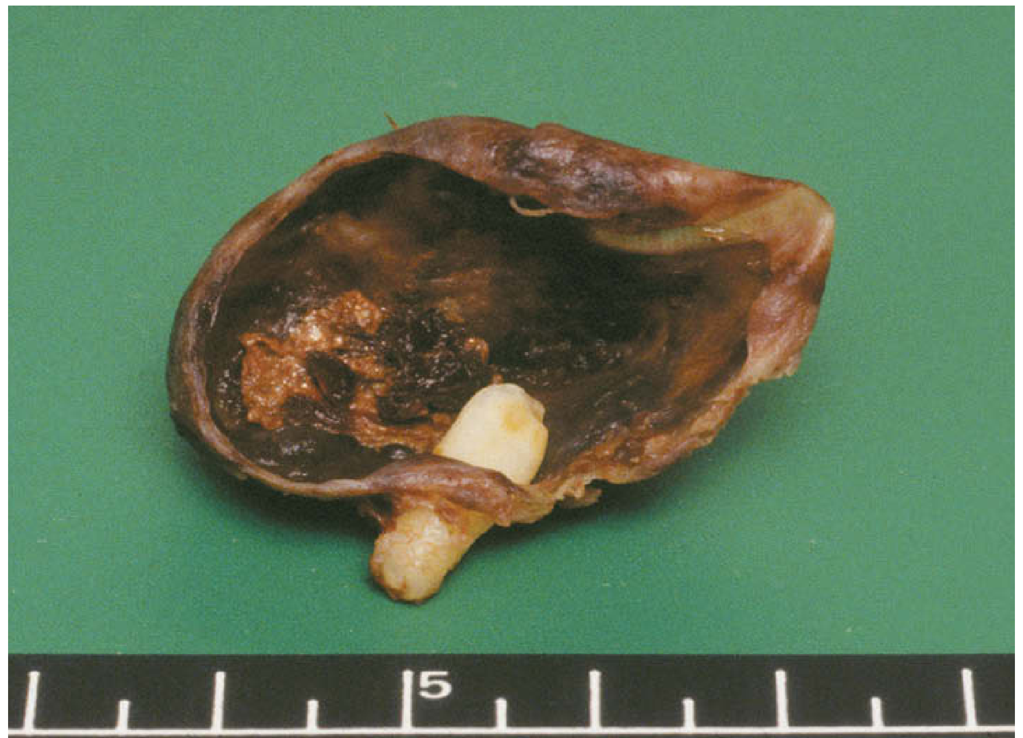
A dentigerous cyst (DC) is, by definition, associated with the crown of an unerupted tooth, a developing tooth, or an odontoma. It forms when fluid accumulates between the reduced enamel epithelium and the tooth crown. The cyst wall attaches at the cementoenamel junction (CEJ), enclosing the crown within the cystic lumen.
- It is the most common developmental odontogenic cyst and the most common cyst of the jaw in the pediatric age group
- It is the second most common odontogenic cyst overall (after the radicular/periapical cyst)
(Cummings Otolaryngology; Scott-Brown's Otorhinolaryngology Head & Neck Surgery)
Epidemiology
- Most common in ages 10-30 years
- More frequent in males
- The longer a tooth remains impacted, the greater the risk of developing a DC
- Predominantly affects the mandibular third molars and maxillary canines (the most commonly impacted teeth), but any impacted tooth is at risk
Clinical Features
- Usually asymptomatic - often an incidental radiographic finding
- May cause painless expansion of bone as it grows
- Can displace the associated tooth considerably
- Aspiration yields straw-colored fluid (18G needle)
- Secondary infection causes pain and swelling
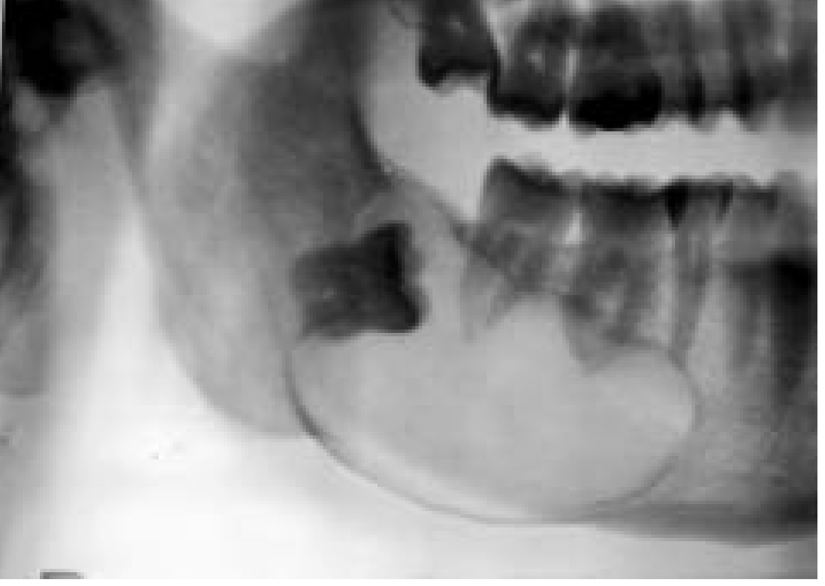
Radiographic Features
A classic dentigerous cyst appears as:
- Well-defined unilocular radiolucency surrounding the crown of an unerupted tooth
- Border is well corticated and well demarcated
- Radiolucency attaches at the cementoenamel junction
- Large cysts can displace the associated tooth over considerable distances
- If secondarily infected: the border may show sclerosis or rarifying osteitis
- Rule of thumb: a dental follicle radiolucency >4 mm from the tooth surface to the outer follicle edge should raise suspicion for cystic change
Macroscopic Appearance
Microscopic (Histopathological) Features
- Fibrous capsule of variably dense fibro-collagenous connective tissue; some areas are loose and myxomatous
- Lining epithelium: nonkeratinized stratified squamous epithelium (thin, 2-3 cell layers unless inflamed)
- Odontogenic epithelial rests scattered within the connective tissue wall
- Mucous prosoplasia within the lumen is not uncommon (should not be overinterpreted)
- Cholesterol slits with associated multinucleated giant cells may appear in inflamed cysts
- Inflammatory infiltrate is present in inflamed cases
(Cummings Otolaryngology)
Neoplastic Potential
Dentigerous cysts retain the ability to transform into true neoplasms - this is clinically important:
| Transformation | Notes |
|---|---|
| Ameloblastoma | ~17% of ameloblastomas are associated with a pre-existing dentigerous cyst |
| Squamous cell carcinoma | Reported in association with DC |
| Mucoepidermoid carcinoma | Also reported - possibly from mucous cells in the cyst lining |
| Odontogenic keratocyst | Can mimic DC radiographically around an unerupted tooth |
This neoplastic potential makes histopathological examination of all enucleated cysts mandatory.
Differential Diagnosis
When a radiolucency is seen around an unerupted tooth, the differential includes:
- Dentigerous cyst (most likely)
- Odontogenic keratocyst (OKC/KCOT) - higher recurrence, thicker parakeratotic lining
- Ameloblastoma - multilocular, "soap bubble" appearance
- Hyperplastic dental follicle - overlaps with small DCs; follicle space normally <4 mm
Eruption Cyst (Subtype)
A variant of the dentigerous cyst confined to the soft tissue overlying an erupting tooth (classified as a DC subtype by WHO 2017):
- Presents as a bluish/purple soft tissue swelling over the alveolar ridge in children under 10 years
- Most require no treatment - resolves spontaneously when the tooth erupts through it
- If eruption is delayed: simple "deroofing" (marsupialisation of the soft tissue)
- Incidence: ~2 per 1,000 births for neonatal eruption cysts
Treatment
| Cyst Size | Approach |
|---|---|
| Small-medium | Tooth extraction + enucleation and curettage in a single stage |
| Large | Two-stage: prolonged decompression/marsupialisation with drain first, then formal enucleation |
- Marsupialization reduces cyst size and allows bone regeneration before definitive surgery
- Large cysts may require allogeneic or xenogeneic bone grafting; autogenous bone grafting reserved for very large cysts where substitutes are unavailable
- Enucleation allows complete histopathological examination of the entire specimen (not just a biopsy)
Prognosis
- Excellent - very low recurrence rate after complete enucleation
- Recurrence is rare if the cyst lining is completely removed
- Follow-up imaging is advisable to rule out residual disease or malignant transformation
Sources: Cummings Otolaryngology Head and Neck Surgery; Scott-Brown's Otorhinolaryngology Head & Neck Surgery; K.J. Lee's Essential Otolaryngology
This is a shared conversation. Sign in to Orris to start your own chat.