


Explain
bilateral undescended testes cryptorchidism ultrasound
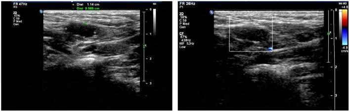
This diagnostic image displays two side-by-side grayscale ultrasonography panels of the inguinal/scrotal region, used to evaluate bilateral cryptorchidism. The left panel shows a longitudinal 2D ultrasound view of an undescended testis, appearing as an oval, hypoechoic, heterogeneous mass measured at approximately 1.14 cm by 0.565 cm. Caliper markers delineate the length and width of the structure within the surrounding soft tissue. The right panel displays the same region using color Doppler imaging, indicated by a rectangular sampling box and a velocity scale (+4.8 to -4.8 cm/s). Small focal areas of blue and red within the sampling box represent blood flow within the testicular parenchyma. Instrument parameters are visible on the left margin, including a frame rate (FR) of 47Hz for the 2D image and 26Hz for the Doppler study. These images demonstrate the classic sonographic appearance of small, extra-scrotal gonads, serving as a primary diagnostic tool for locating non-palpable testes in pediatric patients.
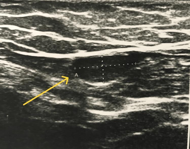
This diagnostic ultrasound image demonstrates an undescended testis (cryptorchidism) located within the inguinal canal. The testis is identified by a yellow arrow pointing to a well-defined, oval, hypoechoic structure relative to the surrounding soft tissue layers. Measurement calipers are positioned over the testicular parenchyma to assess its dimensions. The surrounding anatomical environment shows stratified layers of variable echogenicity, representing subcutaneous fat, fascia, and inguinal musculature. The testicular tissue itself appears relatively homogeneous in texture, a common sonographic finding for an ectopic or undescended testis. This imaging is characteristic of pediatric or adolescent urological evaluations where a testis is non-palpable in the scrotum. The educational focus of this image is the sonographic identification of undescended testes and the differentiation of testicular parenchyma from adjacent inguinal lymph nodes or surrounding soft tissues.
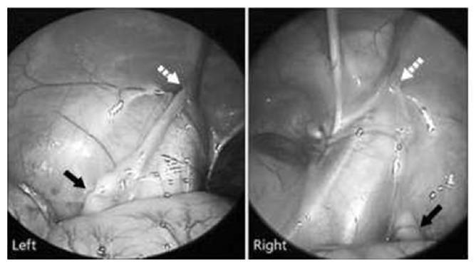
Laparoscopic intra-abdominal view demonstrating bilateral undescended testes (cryptorchidism). The composite clinical image shows two sub-panels, labeled 'Left' and 'Right', illustrating the findings during a diagnostic laparoscopy for impalpable testes. In both images, the intra-abdominal testis (indicated by solid black arrows) is positioned proximal to the internal inguinal ring (indicated by dashed white arrows). The 'Left' panel displays the ovoid morphology of the testis and the associated spermatic vessels. The 'Right' panel shows surgical instrumentation, likely a probe or grasper, used for mobilization or inspection of the cord structures. Key anatomical landmarks visible include the internal inguinal ring, the ductus deferens, and the testicular vessels. This visual is diagnostic for intra-abdominal cryptorchidism and serves as a surgical planning tool for orchidopexy, such as the Fowler-Stevens procedure. The images highlight the proximity of the gonads to the inguinal canal entry point within the retroperitoneal space.
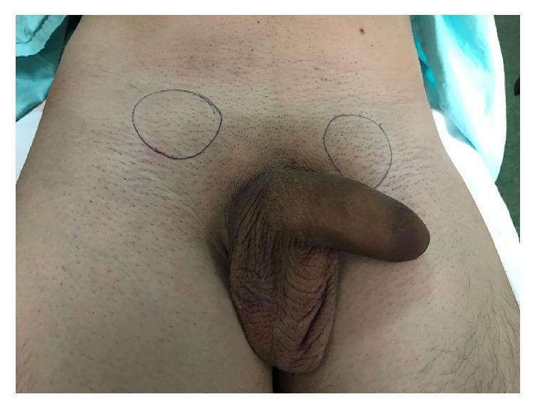
Clinical photograph of the male pelvic and inguinal region demonstrating bilateral cryptorchidism. The image shows a normotrophic but empty scrotal pouch, characterized by wrinkled, hyperpigmented skin without visible or palpable testicular contents. In the bilateral inguinal areas, two circular markings have been drawn with a surgical marker, indicating the location of ectopic or undescended testes identified in the subcutaneous tissue. The surrounding skin of the lower abdomen and groin appears normal with typical fine hair distribution. This visual serves as an educational tool for identifying the physical signs of traumatic testicular dislocation or congenital cryptorchidism, highlighting the contrast between a developed scrotal sac and the absence of its contents, as well as the utility of surface marking for surgical planning in orchiopexy.
bilateral cryptorchidism undescended testes adult management
Explanation of the Two Scan Reports
Report 1: USG Abdomen and KUB (Kidney, Ureter, Bladder)
| Organ | Finding | Meaning |
|---|---|---|
| Liver | 14.2 cm, homogeneous, no abscess/mass | Slightly enlarged (normal upper limit ~13 cm) but no serious lesion |
| Gall Bladder | Normal, no calculi (stones) | No gallstones |
| Common Bile Duct | Normal, no calculi | No blockage |
| Pancreas | Normal | No pancreatitis or tumor |
| Spleen | Normal, 11.8 cm | Within normal limits |
| Aorta | Normal, no para-aortic nodes | No lymph node enlargement around the aorta |
| Peritoneal Cavity | Normal | No free fluid (ascites) |
| Right Kidney | Normal cortex and collecting system, no calculi | No kidney stones or hydronephrosis |
| Left Kidney | Normal cortex and collecting system, no calculi | No kidney stones or hydronephrosis |
| Bladder | Normal | No thickening or stone |
| Prostate | Normal, no intra-vesical enlargement | Normal for age |
| RIF (Right Iliac Fossa) | No inflamed bowel loops | Rules out appendicitis or ileitis |
Report 2: USG Scrotum - THE KEY FINDING
Right Testis:
- Not seen in the scrotum (where it should normally be)
- Found in the right side of the pelvis, near the anterolateral aspect of the urinary bladder (i.e., inside the abdomen/pelvis)
- Size: 3 x 1.4 cm (smaller than normal; a normal adult testis is ~4 x 3 x 2 cm)
- Tiny calcifications in the testicular tissue
- Minimal vascularity on Doppler (reduced blood flow - concerning)
Left Testis:
- Not seen in the scrotum
- Found near the anterior abdominal wall of the pelvis, adjacent to the deep inguinal ring (at the entrance of the inguinal canal, but hasn't passed through into the scrotum)
- Size: 3 x 1.6 cm (also small)
- Multiple tiny calcifications in the testicular tissue
- Normal vascularity on Doppler (blood flow intact)
Impression: Bilateral Undescended Testes (Bilateral Cryptorchidism) - Located Intra-abdominally
What is Bilateral Cryptorchidism?


Why Is This Significant? - Major Risks
- Men with cryptorchidism have a 10 to 40-fold increased risk of developing testicular cancer in the undescended testis - Goldman-Cecil Medicine
- Intra-abdominal testes carry the highest risk for malignancy - Family Medicine Textbook
- The calcifications found in both testes (called testicular microlithiasis) can also be associated with germ cell tumors
- Even the normally descended testis has higher risk; here both are undescended, doubling the concern
- Risk increases with every year of delay after 18 months
- The scrotal temperature (~2°C lower than body temperature) is essential for sperm production
- At body temperature (inside the abdomen), sperm production is severely impaired
- Bilateral cryptorchidism means both testes are affected - virtually complete infertility is expected at this age
- Campbell-Walsh Urology notes that reproductive prognosis worsens significantly when orchidopexy is delayed
- Undescended testes have a higher risk of twisting on their blood supply (torsion), which can cause sudden loss of the testis
- Both testes are atrophic (smaller than normal). The right testis especially shows minimal vascularity, suggesting poor function
What the Calcifications Mean
- Is commonly associated with prolonged undescended testes
- Increases the risk of germ cell tumors (testicular cancer)
- Warrants close surveillance or tumor marker testing (AFP, beta-hCG, LDH)
What Should Happen Next
- Urgent surgical referral (already referred to Dr. Raju, M.S.) - Orchidopexy (if the testes are viable) or Orchidectomy (removal, if testicular function is absent/minimal and cancer risk outweighs preservation)
- Tumor markers: AFP, beta-hCG, LDH to screen for occult germ cell tumor
- Hormone evaluation: Testosterone, LH, FSH to assess testicular function
- Fertility counseling: Sperm analysis, sperm banking if any viable sperm remain
- Genetic testing if there is any suspicion of a syndromic cause (e.g., Prader-Willi, Noonan syndrome)
Sources: Campbell-Walsh Wein Urology; Harrison's Principles of Internal Medicine 22E; Goldman-Cecil Medicine; Family Medicine Textbook 9e
Disclaimer: This is an educational explanation only. Please follow up with Dr. Raju or a urologist/andrologist for clinical management.
Possible of death
Can It Lead to Death?
- Men with intra-abdominal undescended testes (like in this report) have a 10 to 40-fold higher risk of developing testicular germ cell cancer
- At 26 years old, with both testes undescended AND showing calcifications (microlithiasis) in both - the cancer risk is at its highest
- If testicular cancer develops and is left undetected or untreated, it can spread (metastasize) to lymph nodes, lungs, liver, and brain - and that can be fatal
- The reduced vascularity (blood flow) in the right testis is an additional red flag
The Good News
The Real Risk Here
| Risk Factor | Present in This Patient? |
|---|---|
| Bilateral undescended testes | Yes |
| Intra-abdominal location (highest risk zone) | Yes |
| Testicular microlithiasis (calcifications) | Yes - in BOTH testes |
| Minimal vascularity (right testis) | Yes |
| Age 26 - untreated into adulthood | Yes |