


Make a infographic exam answer by this picture for cataract with diagram in picture
A professional veterinary pathology exam infographic titled "CATARACT - Pathology of the Lens" with a dark navy blue background and organized sections. Top header: "CATARACT" in large bold gold text, subtitle "Opacity of the Lens" in white. Small note: "Common in dogs, rare in other animals" Section 1 - DEFINITION box (teal): "Opacity of the lens" Section 2 - ETIOLOGY (two columns side by side): Left box labeled "CONGENITAL" (blue): - Failure of hyaloid artery to regress - Impaired lens translucence - Hereditary predisposition - Deficiency of Vitamin E Right box labeled "ACQUIRED" (orange): - Trauma / Luxation - Degenerative ocular disease - Senility (old stallions) - Diabetes mellitus (dogs) - Nutritional: Vit D, C, cysteine deficiency - Toxins / Ergot poisoning - Absorbed radiation Section 3 - PATHOGENESIS (purple box): Three arrows pointing down: 1. Proliferation of capsular epithelium 2. Necrosis of lens fibres 3. Increased sclerosis of nucleus fibres Section 4 - FOUR TYPES OF CATARACT (four color-coded boxes in a row): Box 1 RED "1. SUBCAPSULAR": Horses, dogs, birds. Proliferation of lens epithelium under capsule. Anterior polar (front) + Posterior polar (back). Disorganized proliferation → opacity Box 2 GREEN "2. CORTICAL": Most common. Lens fibres affected. Follows corneal ulcer. Stellate opacity center to periphery. Progressive. Box 3 BLUE "3. LAMELLAR/ZONULAR": Young animals. Congenital or acquired. After distemper or rickets. Non-progressive. Between nucleus and cortex. Box 4 ORANGE "4. NUCLEAR": Senile changes. Fibres at centre become denser → dull/hazy nucleus. Leads to blindness/vision impairment. Center diagram - Detailed cross-section of EYE AND LENS anatomy with labeled parts: Cornea, Iris, Anterior chamber, Lens, Vitreous humor, Retina, Sclera, Ciliary muscle, Zonular fibres, Lens capsule, Anterior capsule, Posterior capsule, Lens nucleus, Cortex, Epithelial cells. Clean anatomical illustration in blue/teal tones. Bottom - CLASSIFICATION summary box: "By Extent: Partial / Complete | By Origin: Congenital / Acquired | By Location: Subcapsular, Cortical, Lamellar, Nuclear" Clean, modern medical infographic style with clear typography, color-coded sections, white text on dark background, professional veterinary pathology layout.
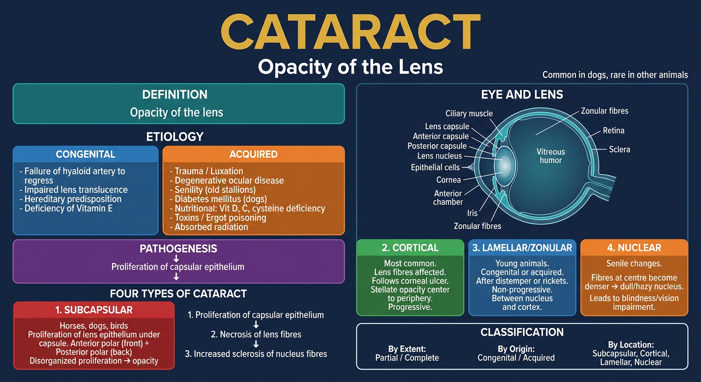
CATARACT - Pathology of the Lens
ETIOLOGY
| Congenital | Acquired |
|---|---|
| Failure of hyaloid artery to regress | Trauma / Luxation |
| Impaired translucence of the lens | Degenerative ocular disease |
| Hereditary predisposition | Senility (common in old stallions) |
| Deficiency of Vit E | Diabetes mellitus (occasionally in dogs) |
| Nutritional: deficiency of Vit D, C, cystein | |
| Toxins / Ergot (cattle & pigs) | |
| Absorbed radiation |
PATHOGENESIS
- Proliferation of the capsular epithelium
- Necrosis of the fibre of the lens
- Increased sclerosis of the fibres forming the nucleus
CLASSIFICATION
- By extent: Partial or complete
- By origin: Congenital or acquired
- By nature/location of lesion: 4 types
4 TYPES OF CATARACT
- Seen in: horses, dogs, birds
- Anterior polar cataract = proliferation at the front surface
- Posterior polar cataract = extends to posterior surface (lens degeneration)
- Disorganized proliferation → thicker layers → opacity via posterior synechia/repeated attacks
- Involves lens fibres - front (anterior cortical) or back (posterior cortical)
- Usually follows a corneal ulcer
- Accumulation of interstitial fluid → fibres disintegrated and disorganized
- Stellate opacity spreading from centre to periphery
- Progressive condition
- Occurs in young animals
- Congenital or acquired (puppies after distemper or rickets)
- Located between the nucleus and cortex
- Non-progressive
- Caused by senile changes - fibres at centre become denser → dull/hazy nucleus
- Sequelae: Blindness / impairment of vision
EYE & LENS ANATOMY (Diagram Key)
Sequale
Simple sequeale
Sequelae of Cataract
From your notes (Nuclear Cataract section):
"Blindness / Impairment of vision develops"
Full Sequelae (Veterinary Pathology):
| # | Sequela | Notes |
|---|---|---|
| 1 | Blindness | Complete loss of vision |
| 2 | Impairment of vision | Partial or progressive visual loss |
| 3 | Lens-induced uveitis | Leaking lens proteins → inflammatory reaction |
| 4 | Lens luxation | Lens slips from normal position |
| 5 | Secondary glaucoma | Increased intraocular pressure |
| 6 | Lens liquefaction | Lens contents liquefy (hypermature cataract) |
| 7 | Posterior synechia | Adhesion between iris and lens capsule |
| 8 | Rupture of lens capsule | Releases lens material → severe uveitis |
Simple Memory Point:
Cataract → Opacity → Blindness (the core sequela in exams)
- Blindness and impairment of vision - as stated directly in your notes under Nuclear Cataract sequelae.