
Bullos disorders
Reading File
Bullous disorders are a group of conditions characterized by fluid-filled blisters (bullae) on the skin and/or mucous membranes. The main types include pemphigus vulgaris, bullous pemphigoid, dermatitis herpetiformis, and epidermolysis bullosa, among others.
Searching sources...
Finding Sources
Finding Sources
Reading File
Reading File
Reading File
Finding Sources
Reading File
Reading File
Loading Image
Loading Image
Loading Image
Now I have comprehensive information. Let me compile a thorough answer.
Bullous (Blistering) Disorders
Bullous disorders are a group of skin diseases in which fluid-filled blisters (bullae/vesicles) are the primary and most distinctive feature. The level at which the blister forms within the skin is a critical diagnostic clue.
Level of Blister Formation
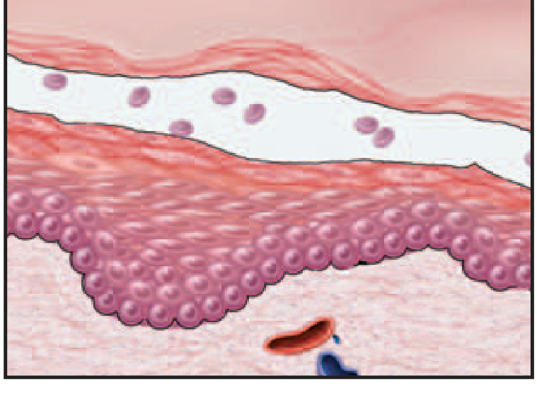
Fig. 22.8 - Levels of blister formation: (A) Subcorneal, (B) Suprabasal, (C) Subepidermal
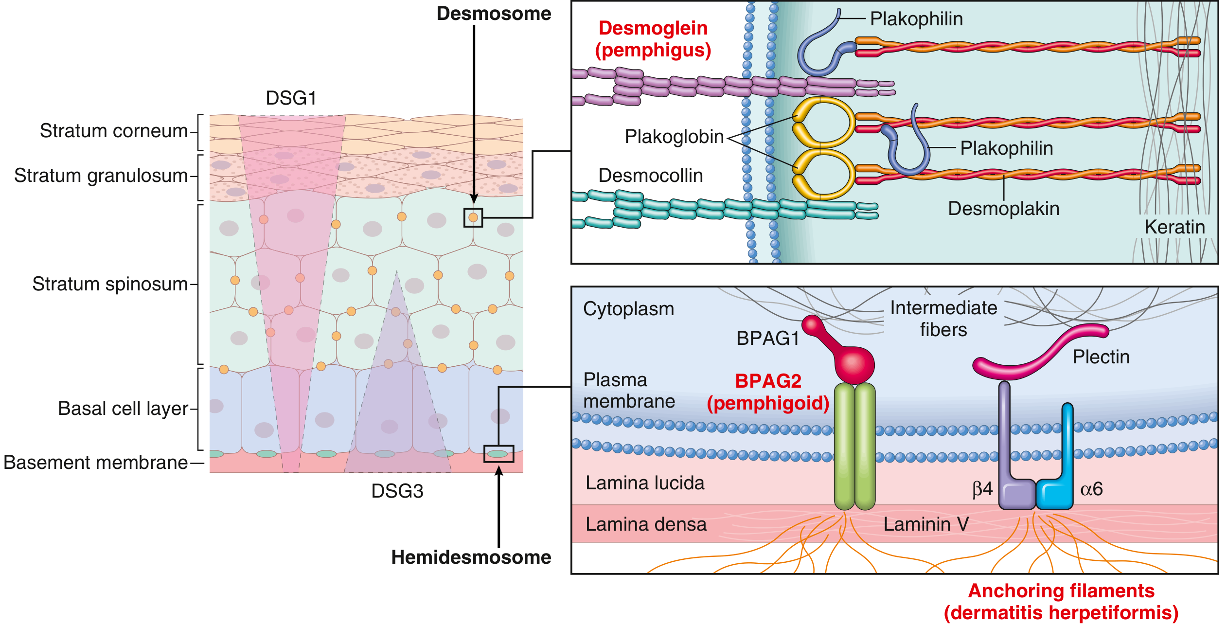
The diagram below shows the molecular targets in each disorder:
1. Pemphigus Vulgaris
| Feature | Detail |
|---|---|
| Type | Autoimmune (IgG - Type II hypersensitivity) |
| Target antigen | Desmoglein-1 (DSG1) + Desmoglein-3 (DSG3) |
| Blister level | Suprabasal (acantholytic) |
| Immunofluorescence | Fish-net / chicken-wire intercellular IgG deposits |
| Sites | Scalp, face, axillae, groin, trunk, mucosa |
| Appearance | Flaccid bullae that rupture easily, leaving painful erosions with serum crust |
| Treatment | Systemic immunosuppression (steroids + steroid-sparing agents), often lifelong |
Pathogenesis: IgG autoantibodies bind to desmosomal proteins DSG1 and DSG3, disrupting intercellular adhesion (acantholysis) and activating intracellular proteases. Associated with specific HLA alleles.
Key hallmark: Nikolsky sign positive (lateral pressure causes skin to slide/blister).
2. Pemphigus Foliaceus
| Feature | Detail |
|---|---|
| Target antigen | Desmoglein-1 (DSG1) only |
| Blister level | Subcorneal (superficial - at stratum granulosum) |
| Sites | Skin only - mucous membranes spared |
| Appearance | Superficial blisters; erythema and crusting; milder than vulgaris |
| Note | DSG3 compensates in mucosa, explaining mucosal sparing |
3. Paraneoplastic Pemphigus
| Feature | Detail |
|---|---|
| Association | Underlying malignancy (especially lymphoproliferative disease) |
| Common tumors | Non-Hodgkin lymphoma, CLL, Castleman disease, thymoma |
| Presentation | Painful hemorrhagic oral erosions + polymorphous skin eruption (may mimic BP, EM, GVHD, lichen planus) |
| Organ involvement | Lung, thyroid, kidney, GI tract |
| Pathogenesis | Both cellular and humoral immunity |
| Prognosis | Poor with malignancy-associated disease; better with benign tumors |
4. Bullous Pemphigoid (BP)
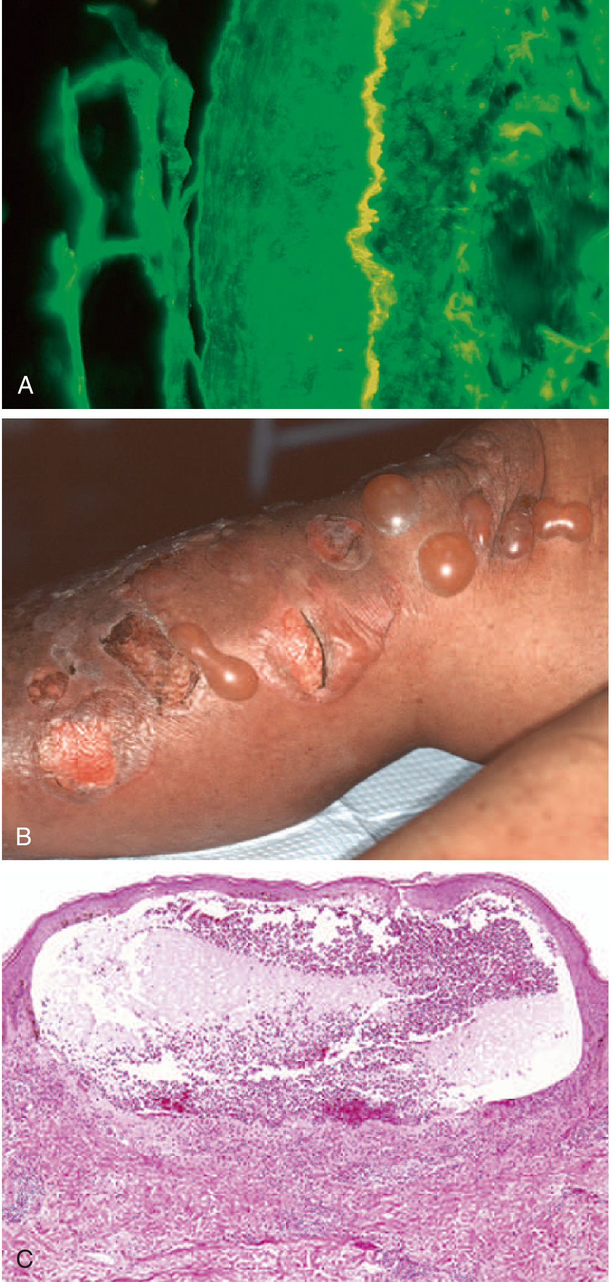
Fig. 22.12 - Bullous pemphigoid: (A) Linear IgG deposits at BMZ on immunofluorescence, (B) Tense, intact blisters, (C) Subepidermal blister with eosinophil infiltrate
| Feature | Detail |
|---|---|
| Type | Autoimmune (IgG - Type II hypersensitivity) |
| Target antigen | BPAG2 (BP180, type XVII collagen) + BPAG1 - hemidesmosome proteins |
| Blister level | Subepidermal (no acantholysis) |
| Immunofluorescence | Linear IgG + C3 at the dermoepidermal junction (BMZ) |
| Epidemiology | Elderly patients |
| Appearance | Tense, intact bullae (do not rupture as easily as pemphigus); pruritus prominent |
| Histology | Subepidermal blister with eosinophil-rich infiltrate; intact epidermis with no acantholysis |
| Course | Remitting/relapsing; responds to topical or systemic immunosuppressives |
Key distinction from pemphigus: Blister roof = full-thickness epidermis with intact intercellular junctions; blisters are tense not flaccid.
Gestational Pemphigoid (Herpes Gestationis): Subtype appearing in 2nd-3rd trimester of pregnancy; resolves after delivery but may recur in subsequent pregnancies. No viral etiology despite the misnomer.
5. Dermatitis Herpetiformis (DH)
| Feature | Detail |
|---|---|
| Type | Autoimmune IgA-mediated |
| Target antigen | IgA to epidermal transglutaminase (cross-reacts with tissue transglutaminase / endomysium) |
| Blister level | Subepidermal (neutrophil microabscesses at dermal papillae tips) |
| Immunofluorescence | Granular IgA deposits at tips of dermal papillae (distinctive!) |
| Association | Gluten sensitivity / celiac disease (up to 80% of cases) |
| Epidemiology | Males > females; 3rd-4th decade |
| Appearance | Extremely pruritic, bilateral symmetric grouped vesicles/papules - elbows, knees, buttocks |
| Treatment | Gluten-free diet + dapsone |
| Malignancy risk | Increased risk of non-Hodgkin lymphoma (both B-cell and T-cell, including enteropathy-associated T-cell lymphoma) |
Comparison Table
| Feature | Pemphigus Vulgaris | Pemphigus Foliaceus | Bullous Pemphigoid | Dermatitis Herpetiformis |
|---|---|---|---|---|
| Antibody | IgG | IgG | IgG | IgA |
| Antigen | DSG1 + DSG3 | DSG1 | BPAG1/BPAG2 | Epidermal TGase |
| Blister level | Suprabasal | Subcorneal | Subepidermal | Subepidermal |
| Mucosa involved | Yes | No | Rarely | No |
| Blister type | Flaccid | Superficial/crusted | Tense | Vesicles (often scratched) |
| IF pattern | Intercellular fish-net | Intercellular | Linear BMZ | Granular papillae tips |
| Key association | HLA alleles | - | Elderly | Celiac disease/gluten |
| Histology | Acantholysis, suprabasal split | Acantholysis, subcorneal split | Eosinophils, no acantholysis | Neutrophil microabscesses |
Key Concepts to Remember
- Acantholysis (loss of intercellular adhesion) = hallmark of pemphigus group; absent in bullous pemphigoid and dermatitis herpetiformis.
- Blister level predicts pathomechanism: desmosomes (upper epidermis) → pemphigus; hemidesmosomes/BMZ → pemphigoid and DH.
- IgA + granular papillary tips on DIF = pathognomonic of dermatitis herpetiformis.
- Linear IgG at BMZ on DIF = bullous pemphigoid.
- Gluten-free diet helps both celiac disease and dermatitis herpetiformis, and may reduce lymphoma risk in DH.
Sources: Robbins & Kumar Basic Pathology, pp. 884-890; Fitzpatrick's Dermatology, Vol. 1-2
Sle
Searching sources...
Finding Sources
Finding Sources
Finding Sources
Finding Sources
Reading File
Reading File
Reading File
Excellent - I now have very rich content. Let me read the morphology/organ involvement section which was truncated.
Reading File
Loading Image
Reading File
Reading File
Reading File
Finding Sources
Reading File
Loading Image
I now have all the information needed for a comprehensive SLE review. Let me compile the full answer.
Systemic Lupus Erythematosus (SLE)
SLE is a chronic, multisystem autoimmune disease characterized by the production of a vast array of autoantibodies (particularly antinuclear antibodies), with injury caused mainly by immune complex deposition and direct antibody binding to cells and tissues. It follows a remitting and relapsing course and can affect virtually any organ.
Epidemiology
- Prevalence: up to 1 in 2500 in certain populations
- Female predominance: 9:1 (F:M) in the reproductive age group (17-55 years); ratio drops to 2:1 in childhood-onset or >65 years
- Peak onset: 20s and 30s
- More prevalent and severe in individuals of African, Hispanic, and Asian descent in the United States
Pathogenesis
SLE is fundamentally driven by loss of self-tolerance to nuclear antigens and excessive autoantibody production.
Genetic Factors
- HLA associations (HLA-DR2, HLA-DR3)
- Deficiency of complement components (C1q, C2, C4) - impairs clearance of immune complexes and apoptotic debris
- Variants in TLR signaling and IFN-regulatory genes
- X-chromosome gene effects (partly explain female predominance, independent of hormones)
Immunologic Abnormalities
- Failure of apoptotic clearance: UV irradiation and other insults cause cell apoptosis; nuclear antigens (DNA, histones, Ro, La) are not cleared and accumulate
- TLR activation: Self nucleic acids mimic microbial patterns and activate TLRs on B cells and dendritic cells, driving production of type I interferons (IFN-α)
- Defective B- and T-cell tolerance: Self-reactive lymphocytes survive and are stimulated by nuclear antigens
- Positive feedback cycle: Autoantibodies form immune complexes → Fc receptor binding on B cells and DCs → internalization of nucleic acids → more TLR engagement → more autoantibodies and interferons
Environmental Factors
- UV light: Induces apoptosis, enhances TLR recognition of altered DNA, stimulates keratinocyte IL-1
- Sex hormones: Estrogen promotes immune activation
- Drugs: Hydralazine, procainamide, D-penicillamine can induce a drug-induced lupus-like syndrome
Autoantibodies in SLE
| Antibody | Specificity / Clinical Significance |
|---|---|
| ANA (anti-nuclear antibody) | Present in ~100% of SLE patients; screener but not specific |
| Anti-dsDNA | Highly specific for SLE; titers correlate with disease activity and nephritis |
| Anti-Sm (Smith) | Highly specific for SLE; targets small nuclear ribonucleoproteins |
| Anti-histone | Characteristic of drug-induced lupus |
| Anti-Ro (SS-A) / Anti-La (SS-B) | Neonatal lupus, congenital heart block; also in Sjogren's |
| Antiphospholipid antibodies | Thrombosis, recurrent miscarriages, false-positive VDRL |
| Anti-RBC antibodies | Hemolytic anemia (Coombs positive) |
| Anti-platelet antibodies | Thrombocytopenia |
Anti-dsDNA and Anti-Sm are the most specific antibodies for SLE.
Mechanisms of Tissue Injury
- Type III hypersensitivity (immune complex deposition) - responsible for most systemic lesions (nephritis, vasculitis, skin, serositis)
- Type II hypersensitivity (direct antibody binding) - hemolytic anemia, thrombocytopenia
- Antiphospholipid antibodies - thrombosis, miscarriages (secondary antiphospholipid syndrome)
- Neuropsychiatric - antibodies crossing blood-brain barrier binding neurons/neurotransmitter receptors; cytokine effects
1997 ACR Diagnostic Criteria (≥4 of 11 for diagnosis)
Clinical:
- Acute cutaneous lupus - malar (butterfly) rash; photosensitivity
- Chronic cutaneous lupus - discoid rash (erythematous raised patches with keratotic scaling + follicular plugging)
- Nonscarring alopecia
- Oral/nasal ulcers (usually painless)
- Nonerosive arthritis (≥2 peripheral joints)
- Serositis - pleuritis or pericarditis
- Renal disorder - proteinuria >0.5 g/24h or red cell casts
- Neurologic disorder - seizures, psychosis, myelitis, neuropathy
Hematologic:
9. Hemolytic anemia
10. Leukopenia (<4000/mm³) or lymphopenia (<1500/mm³)
11. Thrombocytopenia (<100,000/mm³)
Immunologic:
12. ANA (positive)
13. Anti-dsDNA (abnormal titer)
14. Anti-Sm
15. Antiphospholipid antibodies
16. Low complement (C3, C4, CH50)
17. Direct Coombs test
Organ Involvement and Morphology
Prevalence of Manifestations
| Clinical Manifestation | Prevalence (%) |
|---|---|
| Hematologic abnormalities | 100 |
| Fatigue | 80-100 |
| Arthritis/arthralgia/myalgia | 80-90 |
| Skin | 85 |
| Fever | 55-85 |
| Weight loss | 60 |
| Renal | 50-70 |
| Pleuritis | 45 |
| Raynaud phenomenon | 15-40 |
| Neuropsychiatric | 25-35 |
| Pericarditis | 25 |
| GI | 20 |
| Peripheral neuropathy | 15 |
| Ocular | 5-15 |
Skin
- Butterfly (malar) rash: Fixed erythema over nose and cheeks (~50% of patients); photosensitive
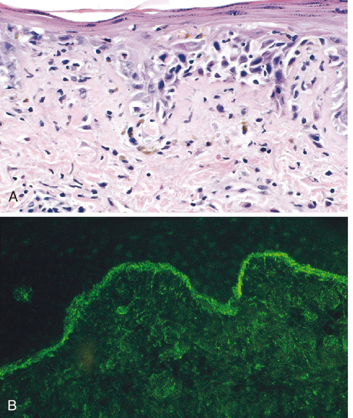
- Histology: vacuolar degeneration of basal layer, dermal edema, perivascular inflammation, fibrinoid necrotizing vasculitis
- Immunofluorescence: IgG + complement deposits along the dermoepidermal junction ("lupus band test")
Kidney - Lupus Nephritis (ISN/RPS Classification)
| Class | Name | Description |
|---|---|---|
| I | Minimal mesangial | Normal histology; mesangial deposits only by EM/IF |
| II | Mesangial proliferative | Mesangial hypercellularity + matrix expansion |
| III | Focal nephritis | <50% glomeruli involved; subendothelial deposits |
| IV | Diffuse nephritis | >50% glomeruli; subendothelial deposits ± crescents; worst prognosis untreated |
| V | Membranous nephritis | Subepithelial deposits; thickened BM; nephrotic syndrome |
| VI | Sclerotic nephritis | Global sclerosis of almost all glomeruli; end-stage |
- Class I & II: Mild; excellent prognosis; minimal or no therapy needed
- Class III & IV: Active proliferative; treat with high-dose steroids + cyclophosphamide or mycophenolate mofetil
- Class IV most severe - high anti-dsDNA, low complement, hematuria, RBC casts, proteinuria, hypertension
Cardiovascular
- Libman-Sacks endocarditis: Non-bacterial vegetations on both surfaces of mitral/aortic valves (pathognomonic); caused by immune complex deposition
- Pericarditis: up to 50% of patients
- Myocarditis: less common; causes tachycardia + ECG changes
- Accelerated coronary atherosclerosis: young patients with long-standing disease (multifactorial - immune complex + antiphospholipid antibody-mediated endothelial damage + corticosteroid effects)
Joints
- Nonerosive synovitis - contrasts with rheumatoid arthritis (which is erosive)
- No joint deformity despite significant pain
Blood Vessels
- Acute necrotizing vasculitis (fibrinoid necrosis) of capillaries, arterioles, small arteries in any tissue
- Chronic: fibrotic thickening with luminal narrowing
Spleen
- Splenomegaly, follicular hyperplasia
- "Onion-skin" lesions: concentric intimal smooth muscle hyperplasia of penicillary arteries (characteristic but not pathognomonic)
Serosal Cavities
- Pleuritis, pleural/pericardial effusions (up to 50%)
- Fibrinous exudate in acute phase → fibrous thickening and obliteration in chronic phase
Lungs
- Pleuritis, pleural effusions
- Chronic interstitial fibrosis, secondary pulmonary hypertension
CNS
- Neuropsychiatric lupus: psychosis, seizures, cognitive dysfunction
- Non-inflammatory occlusion of small vessels (intimal proliferation)
- No clear histologic abnormality in most cases
Hematologic
- Hemolytic anemia (Coombs positive)
- Leukopenia/lymphopenia
- Thrombocytopenia
- LE bodies (hematoxylin bodies) in bone marrow - strongly indicative of SLE
LE Cell (Historical)
- A phagocytic leukocyte (neutrophil/monocyte) that has engulfed the denatured nucleus of an injured cell (coated with ANAs)
- LE cell test: now largely of historical interest, replaced by ANA immunofluorescence assay
Variants of Lupus
| Type | Key Features |
|---|---|
| Drug-induced lupus | Anti-histone antibodies; caused by hydralazine, procainamide, D-penicillamine; resolves on stopping drug; anti-dsDNA usually absent |
| Discoid lupus erythematosus (DLE) | Skin-limited; erythematous patches with scarring; 5-10% progress to SLE |
| Subacute cutaneous LE (SCLE) | Anti-Ro antibodies; widespread non-scarring rash; intermediate between DLE and SLE |
| Neonatal lupus | Maternal anti-Ro/La cross-placenta; congenital heart block; rash; transient |
| Antiphospholipid syndrome (secondary) | Thrombosis, miscarriages, thrombocytopenia in SLE context |
Clinical Course and Prognosis
- Highly variable - from indolent (mild skin and urinary changes for years) to rapidly fatal
- ~90% 5-year survival, ~80% 10-year survival with treatment
- Disease flares correlate with rising anti-dsDNA titers and falling complement (hypocomplementemia)
- Main causes of death: renal failure, intercurrent infections, cardiovascular disease
Treatment Overview
- Hydroxychloroquine (antimalarial): cornerstone for mild-moderate disease; reduces flares and mortality
- NSAIDs: for arthritis and serositis
- Corticosteroids: for moderate-severe flares
- Immunosuppressives: cyclophosphamide or mycophenolate mofetil for severe nephritis/CNS disease
- Belimumab (anti-BLyS/BAFF): approved biologic for active SLE
- Sun protection (UV avoidance)
Sources: Robbins, Cotran & Kumar Pathologic Basis of Disease, pp. 210-219; Harrison's Principles of Internal Medicine 22E
Sle derma
Searching sources...
Finding Sources
Finding Sources
Reading File
Reading File
Reading File
Finding Sources
Reading File
Reading File
Reading File
Loading Image
Loading Image
Loading Image
Loading Image
Finding Sources
Reading File
Finding Sources
Loading Image
I now have comprehensive data. Here is the full answer on SLE dermatology.
SLE - Dermatological Manifestations
The skin and mucous membranes are affected in ~85% of SLE patients, making cutaneous involvement one of the most common features of the disease. Skin lesions are divided into lupus-specific (directly caused by lupus pathology) and lupus-nonspecific (reflect systemic immune activation or vasculopathy but are not unique to lupus).
Classification of Lupus-Specific Skin Lesions
The Gilliam classification divides lupus-specific lesions into three major categories based on chronicity, depth of inflammation, and scarring potential:
| Category | Types |
|---|---|
| Acute Cutaneous LE (ACLE) | Localized (malar rash), Generalized |
| Subacute Cutaneous LE (SCLE) | Annular-polycyclic, Papulosquamous/psoriasiform |
| Chronic Cutaneous LE (CCLE) | Discoid LE, Tumid LE, Lupus panniculitis (LEP) |
The morphology of each lesion depends on: depth/intensity of inflammatory infiltrate, presence/absence of epidermal basal cell damage, follicular involvement, dermal mucin, and scarring tendency.
1. Acute Cutaneous Lupus Erythematosus (ACLE)
Malar (Butterfly) Rash - Classic ACLE
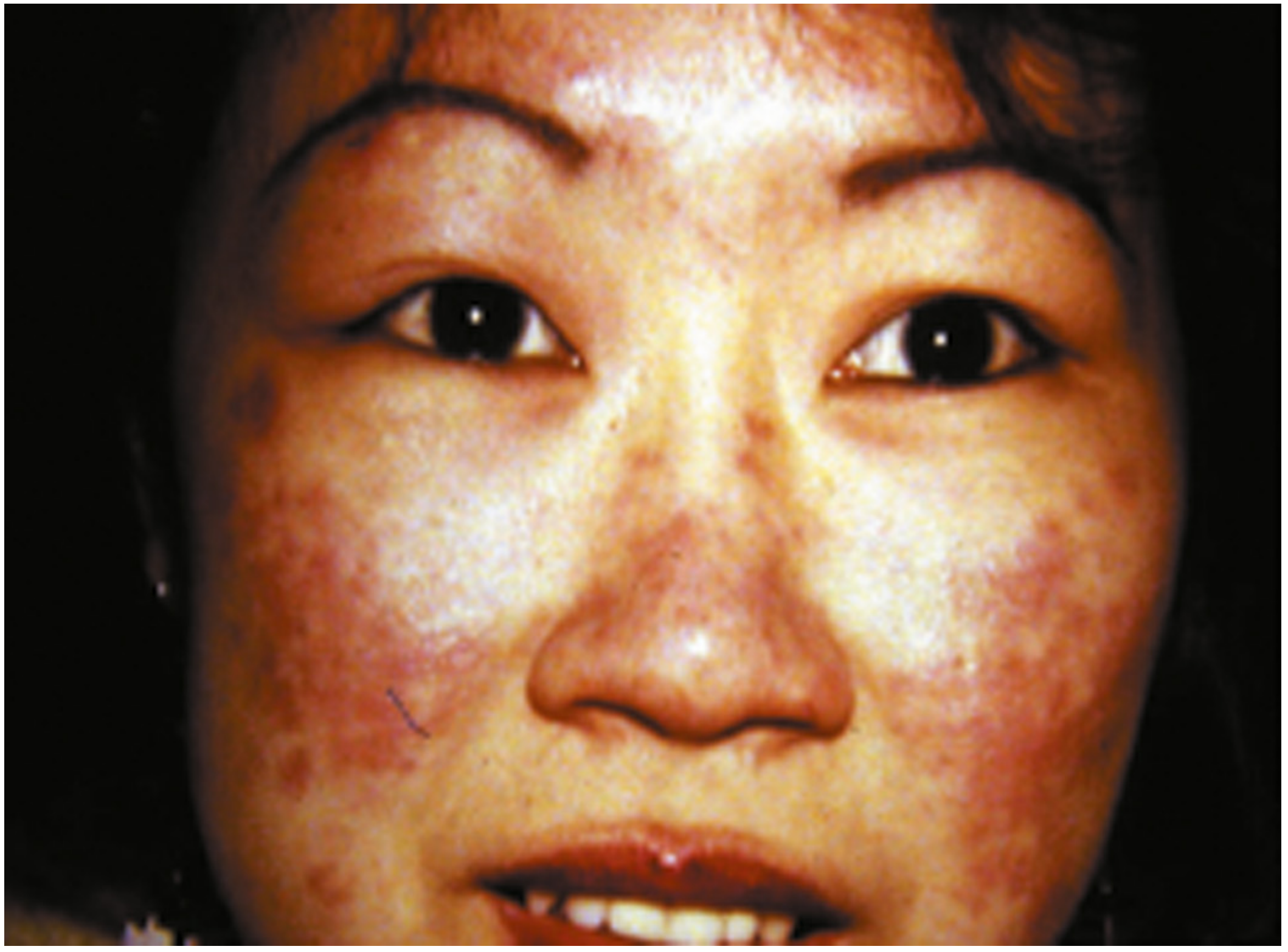
- Appearance: Fixed erythematous rash over the malar eminences and nasal bridge, characteristically sparing the nasolabial folds
- Seen in 30-60% of SLE patients
- Often triggered by UV/sun exposure (photosensitive)
- Distribution: sun-exposed face, neck, extensor arms, dorsal hands; skin over knuckles relatively spared
- Lesions may be transient (hours to days) or persistent
- When severe: facial edema may be prominent; oral lesions often concurrent
- Superficial inflammation - little propensity to scar
Key characteristics of ACLE:
- Strong association with systemic disease activity - the appearance/worsening of ACLE often heralds a flare
- Skin biopsy usually NOT performed on malar rash (transient, biopsy scar, other diagnostic means available)
Differential diagnosis of Malar Rash:
| Condition | Distinguishing Feature |
|---|---|
| Rosacea | Papulopustules, telangiectasias; no systemic features |
| Seborrheic dermatitis | Greasy yellow scale; involves nasolabial folds |
| Dermatomyositis | More violaceous; Gottron's papules on knuckles |
| Polymorphous light eruption | Papules/vesicles; no systemic symptoms |
| Atopic dermatitis | Itch predominant; not photosensitive |
2. Subacute Cutaneous Lupus Erythematosus (SCLE)
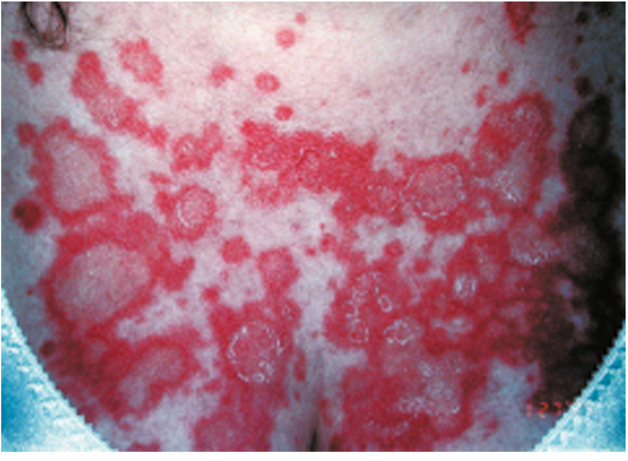
- Morphology: Two main types:
- Annular-polycyclic: Ring-shaped erythematous plaques with central clearing and scaly edge
- Papulosquamous/psoriasiform: Scaly erythematous plaques resembling psoriasis
- Distribution: Sun-exposed areas - arms, upper trunk, neck, sides of face; midfacial area usually spared (unlike ACLE)
- Predominantly affects fair-skinned individuals
- Strongly associated with anti-Ro/SSA antibodies
- Lesions resolve with hypopigmentation or depigmentation - rarely scar (intermediate between ACLE and DLE)
- ~15% of patients with SCLE have or will develop significant systemic disease (SLE, Sjogren's, or overlap)
Drug-induced SCLE:
- Causes: hydrochlorothiazide, proton pump inhibitors, terbinafine (seen in ~1/3 of SCLE cases)
- Resolves after stopping the offending drug
Differential: Psoriasis, tinea corporis, polymorphous light eruption, erythema multiforme
3. Chronic Cutaneous LE (CCLE)
A. Discoid Lupus Erythematosus (DLE)
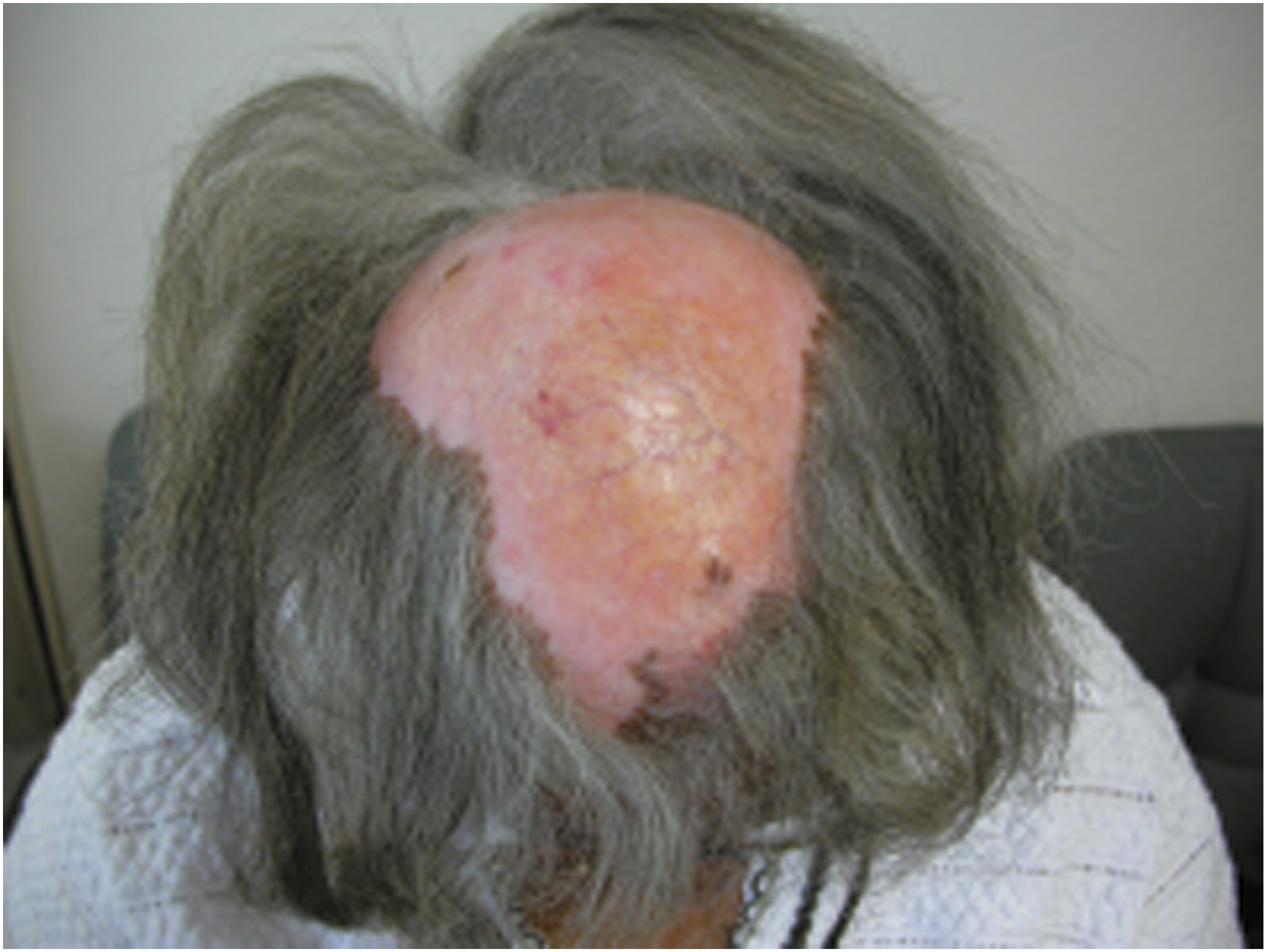
- Most common of the persistent lupus-specific skin lesions
- Active lesions: Erythematous indurated papules/plaques; palpably firm due to dense dermal lymphocytic infiltrate
- Established lesions: Dyspigmentation (central hypopigmentation/depigmentation + peripheral hyperpigmentation); follicular plugging; scarring
- Sites: Scalp, ears, face - usually above the neck; mucosal surfaces occasionally involved
- Scarring alopecia when scalp involved (permanent hair loss)
- Hypertrophic DLE: variant with thickened epidermis
- Sun exposure may exacerbate (but lesions also occur in sun-protected areas of scalp/ears)
- Risk of SLE: ~5-10% progress to systemic SLE; up to 15-20% may meet ACR criteria (often based on mucocutaneous findings)
- ~35% have positive generic ANA; anti-dsDNA rarely present
- Complications: Squamous cell carcinoma can develop in long-standing DLE lesions
- Blacks have 3-5x higher incidence than whites
Histology of DLE:
- Interface (vacuolar/lichenoid) dermatitis: vacuolar degeneration of basal keratinocytes
- Dense perivascular and periappendageal lymphocytic infiltrate (deep, involving follicles)
- Follicular plugging, epidermal atrophy
- Dermal mucin deposition
- Scarring in chronic lesions
Immunofluorescence (DIF):
- Lupus Band Test: Granular IgG deposits at the dermal-epidermal junction of lesional skin only (unlike SLE where non-lesional skin also positive)
B. Tumid Lupus Erythematosus (TLE)
- Erythematous, indurated, boggy papules/plaques with substantial lymphocytic infiltrate and abundant dermal mucin
- No epidermal abnormalities, follicular involvement, or scarring (key distinction from DLE)
- Most common on the face; may be reproduced by phototesting
- Low risk for SLE
- DIF: Ig deposits NOT generally present
- Controversial - some argue TLE is the same as Jessner lymphocytic infiltrate
C. Lupus Panniculitis (LEP) / Lupus Profundus
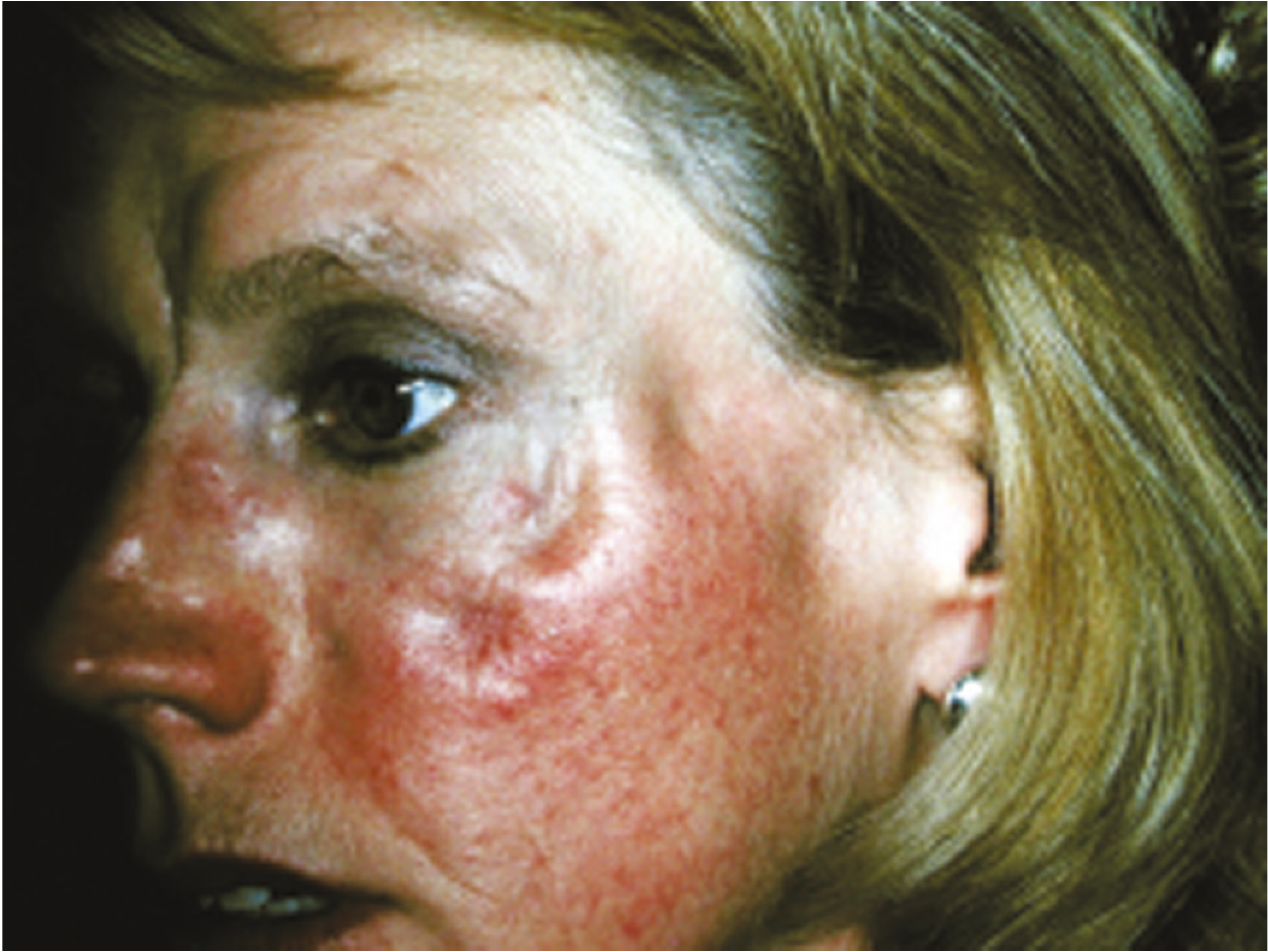
- Inflammation in subcutaneous fat → deep, firm, indurated plaques → disfiguring depressed atrophic areas after resolution
- Sites: face, scalp, upper trunk, breasts, upper arms, buttocks, thighs
- Some patients with LEP have or will develop SLE
- Must be differentiated from panniculitic lymphoma (clinical mimicker)
4. Lupus-Nonspecific Skin Lesions
These reflect immune activation or vasculopathy but are not unique to lupus:
| Lesion | Description |
|---|---|
| Livedo reticularis | Diffuse, lacy net-like violet pattern on skin; associated with antiphospholipid antibodies |
| Nail fold infarcts | Arteriolar spasm/infarcts in nail folds due to vasculopathy |
| Petechiae/purpura | From thrombocytopenia or vasculitis |
| Urticarial vasculitis | Hive-like lesions that last >24 hours, may leave purpura |
| Raynaud phenomenon | 15-40% of SLE patients; vasospasm of fingers/toes |
| Mucosal ulcers | Buccal mucosa and upper palate; usually painless |
| Alopecia (non-scarring) | Diffuse hair thinning; marker of active disease; reversible |
| Bullae | From intensive basal cell damage in ACLE/SCLE; or autoantibodies to type VII collagen |
Histology of SLE Skin Lesions
Common to all subtypes (varying degrees):
- Vacuolar/interface dermatitis: vacuolar degeneration of basal layer keratinocytes
- Perivascular lymphocytic infiltrate (superficial in ACLE; deep/periappendageal in DLE)
- Dermal edema and mucin deposition
- Epidermal atrophy (in chronic lesions)
- Follicular plugging (in DLE)
Lupus Band Test (Direct Immunofluorescence)
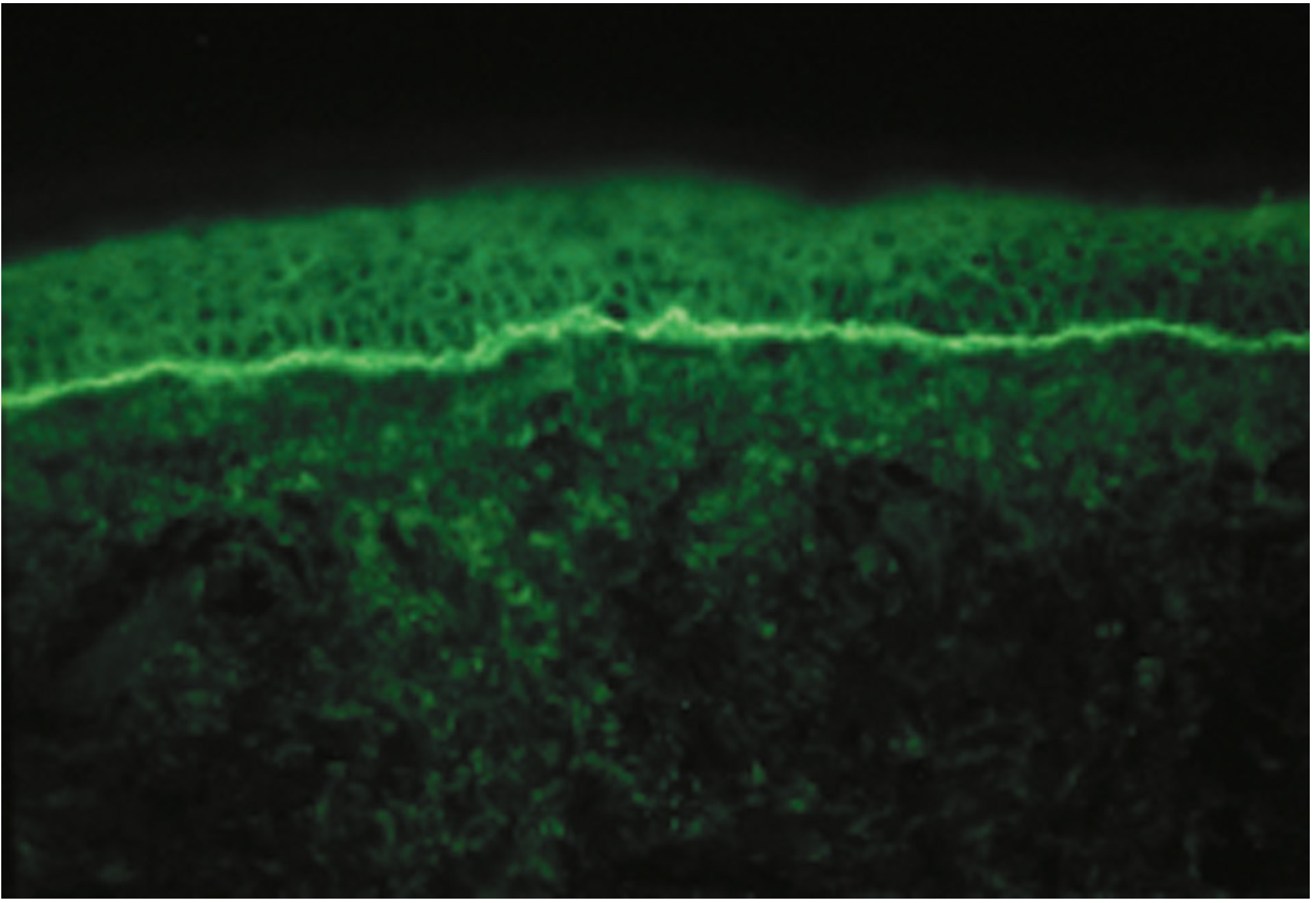
- Granular IgG (± C3, IgM, IgA) deposits at the dermal-epidermal junction
- Lesional skin: Positive in DLE, SCLE, SLE - diagnostically useful
- Non-lesional (normal) skin:
- Positive → strongly indicative of SLE (indicates systemic immune complex deposition)
- Negative → suggests localized/cutaneous-only disease
Comparison of Major Cutaneous Subtypes
| Feature | ACLE (Malar) | SCLE | DLE | TLE | LEP |
|---|---|---|---|---|---|
| Depth of inflammation | Superficial | Superficial-mid | Deep (follicular) | Deep (no epidermal) | Subcutaneous |
| Scarring | No | Rarely | Yes | No | Yes (atrophy) |
| Alopecia | Non-scarring | No | Scarring | No | Possible |
| Photosensitivity | Prominent | Prominent | Variable | Possible | No |
| Key antibody | Anti-dsDNA | Anti-Ro/SSA | Generic ANA | None specific | None specific |
| SLE risk | Very high | ~15% | 5-10% | Low | Variable |
| DIF | Positive (lesional) | Positive (lesional) | Positive (lesional) | Usually negative | Positive (lesional) |
| Distribution | Malar/face | Trunk/arms | Scalp, ears, face | Face | Trunk/limbs |
Treatment of Cutaneous Lupus
- Sun protection: Broad-spectrum SPF, protective clothing - critical for all photosensitive subtypes; tobacco cessation (exacerbates disease, reduces drug response)
- Topical therapy (first-line for localized disease):
- Topical corticosteroids (most commonly used)
- Intralesional corticosteroids (for DLE)
- Topical calcineurin inhibitors (tacrolimus, pimecrolimus)
- Topical retinoids
- Systemic - first line: Hydroxychloroquine (antimalarial) - backbone of cutaneous lupus treatment
- Antimalarial-resistant: Chloroquine, add quinacrine; baricitinib (JAK inhibitor) for familial chilblain lupus
- Refractory disease: Methotrexate, mycophenolate mofetil, dapsone (especially for bullous LE), thalidomide/lenalidomide
- Systemic corticosteroids: for severe/widespread acute flares
Sources: Firestein & Kelley's Textbook of Rheumatology Vol. 2; Goldman-Cecil Medicine; Robbins & Kumar Basic Pathology
Leprosy
Searching sources...
Finding Sources
Finding Sources
Reading File
Reading File
Reading File
Reading File
Reading File
Loading Image
Reading File
Finding Sources
Reading File
Reading File
Loading Image
Finding Sources
I have rich, comprehensive content from multiple authoritative textbooks. Here is the complete answer.
Leprosy (Hansen Disease)
Leprosy is a chronic, slowly progressive infectious disease caused by Mycobacterium leprae (and, less commonly, the recently described M. lepromatosis) that primarily affects the skin and peripheral nerves. Despite its low communicability, it remains a WHO-classified neglected tropical disease, with ~130,000-150,000 new cases annually, predominantly in Southeast Asia, East Africa, and Brazil.
The Pathogen
| Feature | Detail |
|---|---|
| Organism | Mycobacterium leprae (rarely M. lepromatosis) |
| Type | Obligate intracellular, acid-fast bacillus |
| Culture | Cannot be cultured in vitro |
| Optimal temperature | 32-34°C (human skin surface) - explains distribution |
| Virulence factor | Lipid PGL-1 (phenolic glycolipid-1) critical for host cell invasion |
| Immune evasion | Inhibits mitochondrial energy metabolism; replication in cool peripheral tissues |
| Transmission | Nasal/respiratory secretions (prolonged close contact); rare zoonotic from armadillos |
| BCG | Confers some cross-protection (shared antigens with M. tuberculosis) |
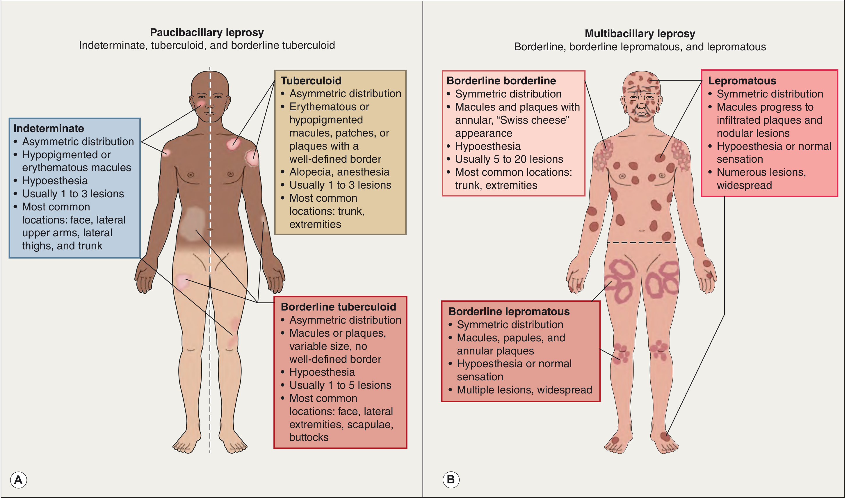
The Spectrum of Disease - Ridley-Jopling Classification
The type and severity of disease is determined entirely by the host T-helper cell (Th1 vs Th2) immune response to M. leprae, not by the organism itself.
Immunologic Poles
| Feature | Tuberculoid (TT) | Lepromatous (LL) |
|---|---|---|
| Immune response | Strong Th1 (IL-2, IFN-γ, Th17) | Weak Th1; relative Th2/regulatory T cell dominance |
| Cell-mediated immunity | Strong | Weak/absent |
| Bacterial load | Low (paucibacillary) | High (multibacillary) |
| Antibody production | Low | High (but non-protective; form immune complexes) |
| Lepromin (Mitsuda) test | Positive | Negative |
| Contagiousness | Low | Higher |
| Prognosis | Self-limiting tendency | Progressive without treatment |
WHO Classification (Practical/Treatment-Based)
| Type | Skin Lesions | Skin Smear |
|---|---|---|
| Paucibacillary (PB) | ≤5 lesions | Negative |
| Multibacillary (MB) | >5 lesions | Positive or negative |
Clinical Presentations by Type
1. Tuberculoid Leprosy (TT) - Paucibacillary
- Few (1-3) well-defined lesions with sharply demarcated, raised, hyperpigmented borders and central hypopigmentation/atrophy
- Anesthesia/hypoesthesia within lesions (loss of pain, temperature, touch) - hallmark
- Alopecia within plaques (loss of hair follicles)
- Anhidrosis within lesions
- Asymmetric distribution - face and extremities common
- Peripheral nerve involvement: asymmetric, single or few nerves enlarged, painful
- Nerve damage causes skin anesthesia, muscle atrophy, contractures, paralysis
- Facial nerve involvement → lagophthalmos → corneal ulceration
- Palpably enlarged nerves: most commonly affected - ulnar (claw hand), common peroneal (foot drop), great auricular, radial cutaneous, posterior tibial
- Bacilli almost never found on smear or biopsy
2. Lepromatous Leprosy (LL) - Multibacillary
- Multiple, widespread, symmetric lesions
- Initially poorly defined erythematous macules, papules, nodules, plaques
- Progression to massive skin infiltration:
- Leonine facies: thickened, nodular facial skin (lion-face)
- Madarosis: loss of eyebrows and eyelashes
- Saddle nose deformity: destruction of nasal septum cartilage
- Infiltration of earlobes (thickened, nodular)
- Acquired ichthyosis on lower extremities
- Anesthesia: glove-and-stocking distribution (symmetric)
- Peripheral nerves: symmetrically invaded with bacteria but minimal inflammation; loss of sensation and trophic changes
- Nasal involvement: persistent inflammation, bacilli-laden discharge
- Ocular: lagophthalmos (facial/trigeminal nerve involvement), corneal anesthesia
- Testes: extensive destruction → sterility
- Systemic: lymph nodes (foamy macrophage aggregates), liver, spleen in advanced disease
- CNS and vital organs: NOT affected (too warm for M. leprae replication)
3. Borderline Leprosy (BT / BB / BL)
- Intermediate features between tuberculoid and lepromatous poles
- Clinically and immunologically unstable - can shift toward either pole
- BT (Borderline Tuberculoid): asymmetric; larger plaques with less defined borders than TT; hypoesthetic; 1-5 lesions
- BB (Mid-borderline): symmetric; annular plaques with "Swiss cheese" appearance (central clearing); ill-defined outer borders; 5-20 lesions
- BL (Borderline Lepromatous): symmetric; macules, papules, annular plaques; multiple widespread lesions; may have normal sensation
4. Indeterminate Leprosy
- Earliest form - not yet classified into a pole
- 1-3 hypopigmented or erythematous macules, asymmetric
- Face, lateral upper arms, lateral thighs, trunk
- Mild hypoesthesia
- May resolve spontaneously OR progress to any type on the spectrum
Histopathology
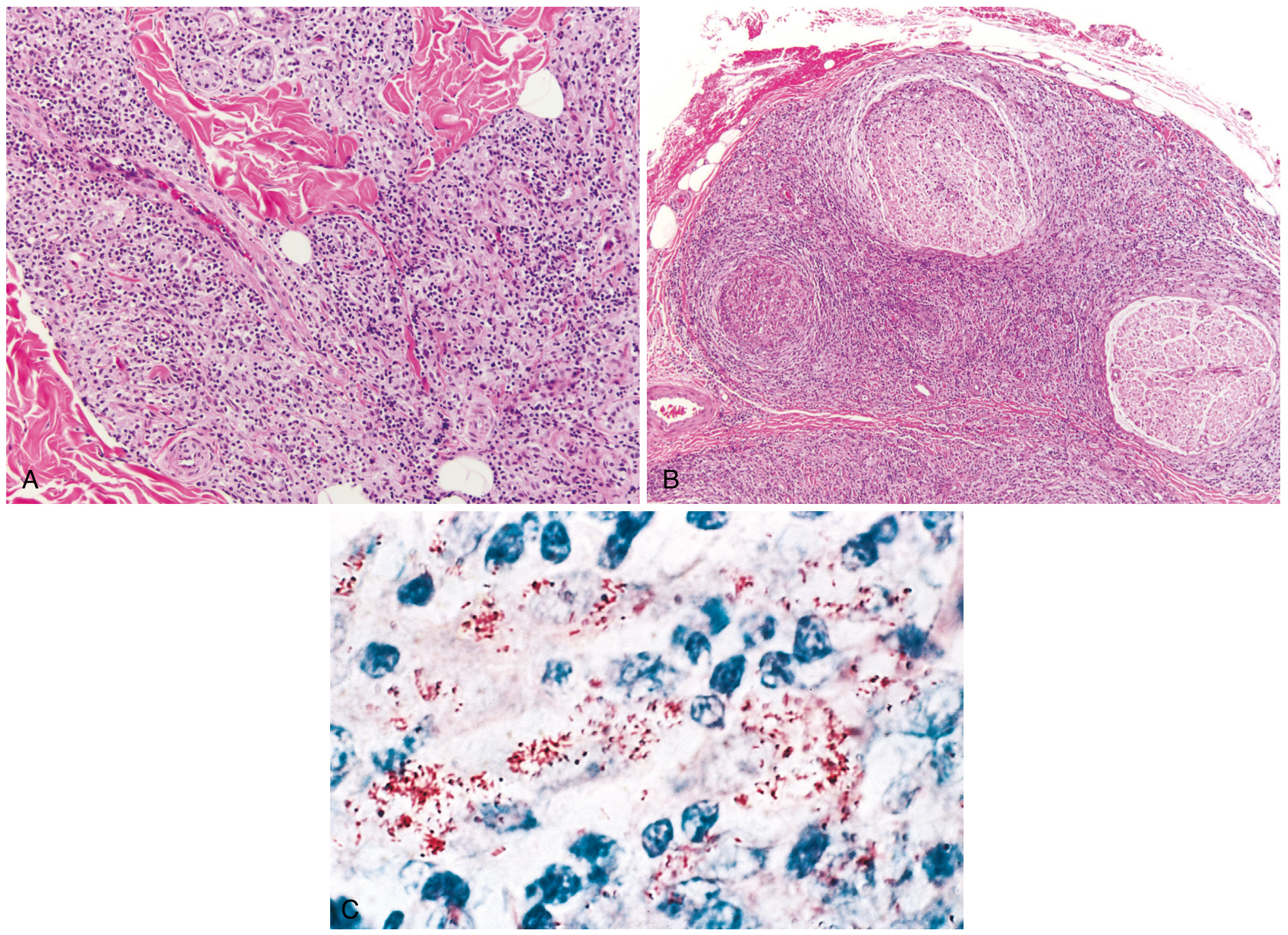
Tuberculoid Pattern
- Epithelioid granulomas closely resembling tuberculosis (non-caseating)
- Granulomas surround and invade dermal nerves, adnexa, and vessels
- Dense lymphocytic infiltrate (CD4+ Th1)
- Bacilli absent (or extremely rare)
- Strong T-cell immunity = paucibacillary
Lepromatous Pattern
- Virchow cells (lepra cells): foamy, lipid-laden macrophages packed with masses of bacilli ("globi")
- Grenz zone (Unna band): band of normal-appearing dermis separating epidermis from the infiltrate - characteristic
- Plasma cells, few lymphocytes
- Minimal inflammation around nerves despite heavy bacillary load
- Staining: Fite-Faraco stain (preferred, more sensitive) or Ziehl-Neelsen stain → bacilli stain bright red
- Multibacillary - readily visualized in tissue
Borderline Pattern
- Intermediate between the two; number of granulomas and bacilli inversely proportional to immunity
Leprosy Reactions (Complications)
Reactions occur in 30-50% of patients, especially during/after treatment. They are acute inflammatory episodes.
Type 1 - Reversal Reaction
| Feature | Detail |
|---|---|
| Mechanism | Delayed-type hypersensitivity; sudden increase in cell-mediated (Th1) immunity |
| Occurs in | Borderline (BT, BB, BL); also TT with "upgrading" |
| Clinical | Pre-existing lesions become acutely inflamed, edematous, erythematous |
| Neuritis | Acute neuritis - nerve pain, swelling, sensory and motor loss |
| No systemic symptoms | |
| Treatment | Oral prednisone |
Type 2 - Erythema Nodosum Leprosum (ENL)
| Feature | Detail |
|---|---|
| Mechanism | Immune complex-mediated; Th2 pattern + neutrophil recruitment; excessive humoral immunity |
| Occurs in | Lepromatous (LL) and BL - high bacterial index |
| Clinical | Erythematous tender papules and subcutaneous nodules; occasionally pustules, bullae, ulcers |
| Systemic | Fever, arthralgias, malaise, iritis, orchitis, neuritis, nephritis |
| May cause | Vasculitis, glomerulonephritis |
| Treatment | Thalidomide (drug of choice); prednisolone |
Lucio Phenomenon
- Seen in diffuse lepromatous leprosy (Mexico/Central America; M. lepromatosis)
- Necrotic ulcers, retiform purpura → necrotizing vasculitis
- Associated with antiphospholipid antibodies + thrombosis
- Severe, potentially fatal
- On a worldwide basis, leprosy is a common cause of cutaneous vasculitis in low-income countries
Disabilities and Deformities
| Grade | WHO Definition |
|---|---|
| Grade 0 | Normal protective sensation |
| Grade 1 | Loss of protective sensation (hands, feet, eyes) |
| Grade 2 | Visible deformities - ulcers, claw hand, foot drop, bone resorption, lagophthalmos, saddle nose |
Nerve damage mechanisms: granulomatous infiltration (tuberculoid) or direct bacillary invasion of Schwann cells and endoneurial macrophages (lepromatous) → demyelination and axonal loss → anesthesia, muscle atrophy, trophic ulcers, contractures, auto-amputation
Diagnosis
| Test | Use |
|---|---|
| Skin biopsy (H&E + Fite-Faraco) | Gold standard; confirms type and bacillary load |
| Slit-skin smear | AFB from earlobe, elbow, forehead; positive in multibacillary only |
| Lepromin (Mitsuda) test | Intradermal M. leprae antigen; positive in tuberculoid (good immunity); not diagnostic per se |
| Nerve conduction studies | Detect subclinical neuropathy |
| Clinical triad | Anesthetic skin lesion + thickened peripheral nerve + AFB in smear |
Treatment: WHO Multidrug Therapy (MDT)
First-line regimen (WHO 2018 guidelines): Rifampicin + Dapsone + Clofazimine for ALL patients
| Drug | Paucibacillary | Multibacillary |
|---|---|---|
| Rifampicin | 600 mg once/month | 600 mg once/month |
| Dapsone | 100 mg daily | 100 mg daily |
| Clofazimine | 300 mg/month + 50 mg/day | 300 mg/month + 50 mg/day |
| Duration | 6 months | 12 months |
After the first dose, the patient is no longer infectious to others.
Key Drug Side Effects
| Drug | Side Effects |
|---|---|
| Rifampicin | Orange-red discoloration of urine/tears; hepatitis; flu-like syndrome; induces CYP450 (reduces OCP efficacy) |
| Clofazimine | Orange-brown skin/mucosa/cornea discoloration (reversible); crystal enteropathy (abdominal pain); cardiac arrhythmias |
| Dapsone | Hemolytic anemia (all patients to some degree); methemoglobinemia; G6PD deficiency - clinically significant hemolysis; Dapsone hypersensitivity syndrome (4-6 weeks): fever, rash, lymphadenopathy, hepatitis |
Treating Reactions
- Type 1 (Reversal): Oral prednisone
- Type 2 (ENL): Thalidomide (drug of choice) + prednisolone; clofazimine can reduce frequency
Alternative Drugs (resistance/intolerance)
- Minocycline, clarithromycin, ofloxacin/moxifloxacin/levofloxacin
- ROM scheme (Rifampicin + Ofloxacin + Minocycline): for PB disease, even single-dose for single-lesion
Key Comparisons Summary
| Feature | Tuberculoid | Lepromatous |
|---|---|---|
| Immunity | Strong Th1 | Weak Th1/Th2 predominance |
| Lesion number | Few (1-3) | Many, widespread |
| Lesion borders | Well-defined | Poorly defined |
| Distribution | Asymmetric | Symmetric |
| Anesthesia | Within lesions | Glove/stocking |
| Nerve damage | Asymmetric, severe | Symmetric, minimal inflammation |
| Bacilli | Absent | Abundant (globi in Virchow cells) |
| Lepromin test | Positive | Negative |
| Grenz zone | Absent | Present |
| Classification | Paucibacillary | Multibacillary |
| Reactions | Type 1 (reversal) | Type 2 (ENL), Lucio |
Sources: Robbins, Cotran & Kumar Pathologic Basis of Disease; Dermatology 5e (Elsevier); Goldman-Cecil Medicine; Firestein & Kelley's Textbook of Rheumatology
Atopic dermatitis
Searching sources...
Finding Sources
Finding Sources
Your credits are exhausted. You can purchase a top-up or subscribe to a plan to continue using Orris. If you're already subscribed, your daily credits will reset tomorrow.
This is a shared conversation. Sign in to Orris to start your own chat.