
Explain me Hives patho physiology symptoms and treatment
urticaria hives skin wheals clinical appearance
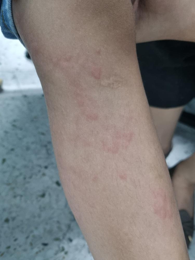
A clinical photograph showing a close-up of a child's lower extremity demonstrating urticaria. The image displays multiple raised, edematous skin lesions known as wheals or hives. These lesions are characterized by pale-to-skin-colored central swellings of varying sizes and irregular shapes, surrounded by a distinct zone of erythema (redness). The distribution is scattered across the leg, illustrating the typical appearance of a hypersensitivity reaction or allergic response. This image serves as an educational reference for identifying cutaneous manifestations of urticaria, focusing on the morphology of superficial dermal edema and associated inflammatory flare.
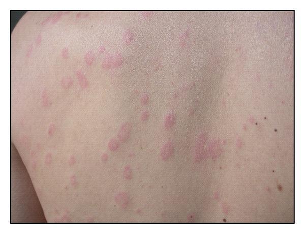
This clinical photograph displays a human patient's back presenting with multiple pink-to-red edematous lesions characteristic of urticaria (hives). The lesions are primarily wheals of varying sizes, ranging from small papules to larger, confluent plaques. A distinct morphological feature of these lesions is their annular or targetoid appearance, characterized by raised, erythematous margins with central pallor. The distribution is scattered and non-contiguous across the dorsal trunk, with clear skin visible between the affected areas. There are no signs of secondary changes such as scaling or ulceration, though the presentation is typical for transient inflammatory skin reactions. The image serves as an educational example of common dermatological manifestations of urticaria, illustrating the classical morphology of wheals and flares used in clinical diagnosis and primary care dermatology.
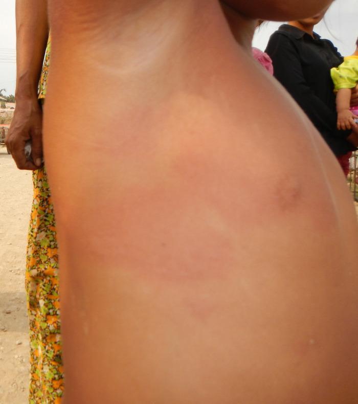
This clinical photograph shows the torso and upper arm of a child, illustrating generalized urticaria (hives). The skin exhibits diffuse erythema and inflammation, characterized by widespread red patches across the lateral chest and abdominal region. Several erythematous wheals and edematous plaques of varying sizes are visible, some of which demonstrate central pallor or darker red centers. The distribution is extensive and lacks a specific dermatomal pattern, consistent with a systemic allergic reaction or hypersensitivity response. The visual presentation focuses on the morphology of skin lesions, specifically the characteristic raised, red, and itchy appearance of acute urticaria in a pediatric patient. This image is relevant for dermatology and pediatrics, demonstrating post-vaccination cutaneous manifestations and the typical appearance of Type I hypersensitivity reactions.
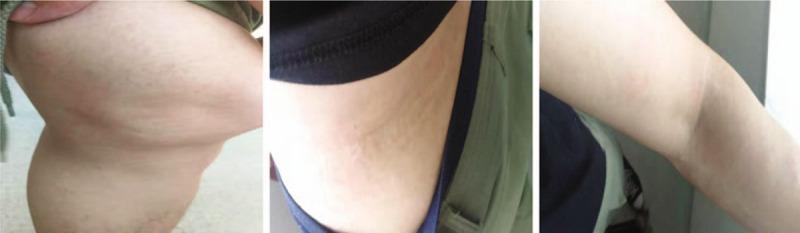
A series of three clinical photographs demonstrating the dermatological resolution of urticaria (hives) across three anatomical regions: the knee joint (A), the lateral waist (B), and the elbow (C). The images show characteristic wheals—edematous, circumscribed, and slightly elevated skin lesions. Panel A displays the knee area with subtle, flesh-colored to slightly erythematous elevations and an uneven skin texture consistent with resolving wheals. Panel B shows the lateral waist with faint, linear, and confluent dermal edema. Panel C depicts the elbow where the lesions have largely flattened, leaving only minimal, faint markings on the skin surface. The progression illustrates the transient nature of urticarial lesions as they gradually diminish in size and elevation. This visual serves as an educational reference for identifying skin morphology in the recovery phase of an acute urticarial reaction, emphasizing the characteristic lack of secondary skin changes (such as scaling or scarring) during the resolution of wheals.
urticaria treatment guidelines
Hives (Urticaria) - Pathophysiology, Symptoms & Treatment
Definition
Classification
| Type | Duration | Key Features |
|---|---|---|
| Acute urticaria | < 6 weeks | Evanescent wheals; individual lesions rarely last > 12 hours |
| Chronic spontaneous urticaria (CSU) | > 6 weeks | > 50% of cases; unknown cause; twice as common in women |
| Inducible (physical) urticaria | Triggered by stimuli | Up to 35% of chronic cases |
- Andrews' Diseases of the Skin, p. 175
Pathophysiology
Core Mechanism
- Antigen exposure (food, drug, insect venom, pollen) leads to cross-linking of IgE antibodies on mast cell surfaces via the high-affinity FcεRI receptor
- Cross-linking triggers mast cell degranulation, releasing:
- Histamine - binds H1 receptors on postcapillary venules causing vasodilation and increased vascular permeability
- Slow-reacting substances of anaphylaxis (leukotrienes C4, D4, E4)
- Prostaglandins, kinins, cytokines (TNF-α, IL-4, IL-5)
- The result: localized dermal edema (wheal) + surrounding flare (axon reflex vasodilation)
- ~33% of chronic urticaria patients have IgG autoantibodies directed against the FcεRI alpha subunit (or less frequently against IgE itself)
- These autoantibodies directly activate mast cells without needing allergen
- Associated with CD4+ lymphocytes, monocyte, and granulocyte infiltration on biopsy
- Often associated with autoimmune thyroiditis
- Goldman-Cecil Medicine, p. 4603
- Certain drugs and chemicals directly trigger mast cell degranulation without IgE involvement:
- Opiates, polymyxin B, tubocurarine, radiocontrast dye
- Aspirin and NSAIDs (also inhibit COX, shifting arachidonic acid toward leukotriene production)
- Vancomycin, tartrazine, benzoates
- Andrews' Diseases of the Skin, p. 175
- Anaphylatoxins C3a and C5a produced by complement activation (e.g., hepatitis B/C, EBV, parasites forming immune complexes) activate mast cells and granulocytes
- Tietz Textbook of Laboratory Medicine
Histopathology
- Sparse superficial perivenular mononuclear infiltrate with rare neutrophils and eosinophils
- Superficial dermal edema causes splaying and wider spacing of collagen bundles
- Mast cells reside around superficial venules and degranulate at these sites
- Robbins & Kumar Basic Pathology, p. 879
Common Triggers / Etiology
| Category | Examples |
|---|---|
| Infections | URIs, viral infections (most common in children), hepatitis B/C, H. pylori, helminths |
| Foods | Shellfish, nuts, strawberries, eggs, milk, chocolate, food additives, azo dyes |
| Drugs | NSAIDs, antibiotics (penicillin), opiates, vancomycin, radiocontrast media |
| Inhalants | Pollen, house dust mites, animal dander, molds |
| Physical stimuli | Dermatographism, cold, heat, pressure, vibration, solar, aquagenic, cholinergic |
| Autoimmune | Thyroid autoantibodies, anti-FcεRI antibodies |
Clinical Features / Symptoms
- Wheals: raised, edematous, pale-to-skin-colored central swellings of varying shapes and sizes, surrounded by a red flare (erythematous halo)
- Pruritus (severe itching) - the hallmark; also stinging or pricking sensations
- Individual lesions resolve within 24 hours without residual marks (no scarring, no scaling)
- Lesions may coalesce producing annular or polycyclic patterns
- Angioedema: deeper subcutaneous swelling, often of lips, eyelids, hands, and genitalia - accompanies wheals in ~40% of cases
- Acute episodes: may present with systemic features if part of anaphylaxis (bronchospasm, hypotension, laryngeal edema)
- Fever + joint/bone pain + urticaria without angioedema → consider autoinflammatory disease
- Lesions lasting > 24 hours + bruising → consider urticarial vasculitis (biopsy required)
- Schnitzler syndrome: chronic non-pruritic urticaria + fever + bone pain + elevated ESR + monoclonal IgM
Treatment
Step 1 - Identify & Avoid Triggers
- Remove the causative agent where possible (drug substitution, dietary avoidance)
- H. pylori eradication if confirmed (can lead to urticaria resolution)
Step 2 - Antihistamines (First-line for all types)
- Oral non-sedating H1 antihistamines are the mainstay
- If unresponsive: short course of systemic corticosteroids
- For severe/anaphylactic reactions: epinephrine (adrenaline) 0.3 mL of 1:1000 IM every 10-20 minutes + IM antihistamines + IV corticosteroids
| Step | Treatment |
|---|---|
| 1st line | 2nd-generation H1 antihistamine (cetirizine 10 mg, loratadine 10 mg, fexofenadine 180 mg, levocetirizine 5 mg, desloratadine 5 mg) once daily |
| If no response at 2 weeks | Increase dose up to 4x standard dose (e.g., cetirizine up to 40 mg/day) |
| Add-on | Classical (sedating) H1 antihistamine at night only (hydroxyzine, chlorphenamine) |
| H2 blocker | Cimetidine or famotidine as adjunct (15% of skin histamine receptors are H2) |
| Omalizumab | Anti-IgE monoclonal antibody - 300 mg SC every 4 weeks; approved for antihistamine-refractory CSU; highly effective |
| Immunosuppressives | Cyclosporine, mycophenolate - reserved for refractory cases due to side effect profile |
| Corticosteroids | Short courses only; avoid long-term use |
- Andrews' Diseases of the Skin; Dermatology 5e, p. 386
Key Drug Dosing Reference (Dermatology 5e)
| Drug (Generation) | Dose |
|---|---|
| Chlorphenamine (1st) | 4 mg three times daily |
| Hydroxyzine (1st) | 10-25 mg three times daily |
| Cetirizine (2nd) | 10 mg once daily |
| Loratadine (2nd) | 10 mg once daily |
| Fexofenadine (2nd) | 180 mg once daily |
| Levocetirizine (newer 2nd) | 5 mg once daily |
| Desloratadine (newer 2nd) | 5 mg once daily |
| Bilastine (newer 2nd) | 20 mg once daily |
Special Situations
- Physical urticaria: may require higher-dose antihistamines; can be refractory
- Pregnancy/lactation: chlorphenamine is preferred based on safety record
- Severe angioedema/anaphylaxis: epinephrine IM is first-line (not effective in hereditary angioedema/HAE)
- HAE (Hereditary Angioedema): epinephrine is NOT effective; use C1-esterase inhibitor, icatibant, or tranexamic acid
Prognosis
- Acute urticaria: > 2/3 of cases are self-limited
- Chronic urticaria: 50% clear within 1 year; ~20% of those with wheals + angioedema continue having symptoms at 20 years; mean disease duration approximately 6 years
- Dermatology 5e, p. 388