
Parathyroid medicine all information
parathyroid gland anatomy location posterior thyroid diagram
"primary hyperparathyroidism" AND management
parathyroid adenoma histology
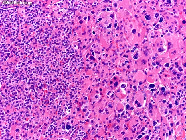
This histopathology image portrays a hematoxylin and eosin (H&E) stained parathyroid tissue section examined under bright-field light microscopy at approximately 400x magnification. The left hemisphere emphasizes small, uniform chief cells arranged in cords and granular cytoplasm with round to ovoid nuclei and inconspicuous nucleoli, consistent with typical parathyroid parenchyma. The right hemisphere shows oxyphil cells characterized by larger size, abundant eosinophilic cytoplasm, and markedly enlarged, hyperchromatic nuclei; these cells can occur as a normal variant in mature parathyroid glands. Adipose stroma is variably present, reflecting age-related involution of parathyroid tissue. There is no cytologic atypia, mitotic index is low, and there is no evidence of capsular or vascular invasion. Mitotic figures may occasionally appear but are not increased, aligning with benign histology. The overall architecture is preserved without destructive growth patterns, supporting a benign interpretation rather than neoplasia such as parathyroid adenoma or carcinoma. This image is suitable for educational comparisons of parathyroid cellular phenotypes (chief vs oxyphil cells) and for teaching differential diagnosis between normal histology and pathology requiring clinical correlation, including primary hyperparathyroidism, parathyroid adenoma and hyperplasia. Clinically, recognizing these normal parathyroid variants avoids misdiagnosis of neoplasia and guides correlation with serum calcium and parathyroid hormone (PTH) levels and imaging findings when indicated.
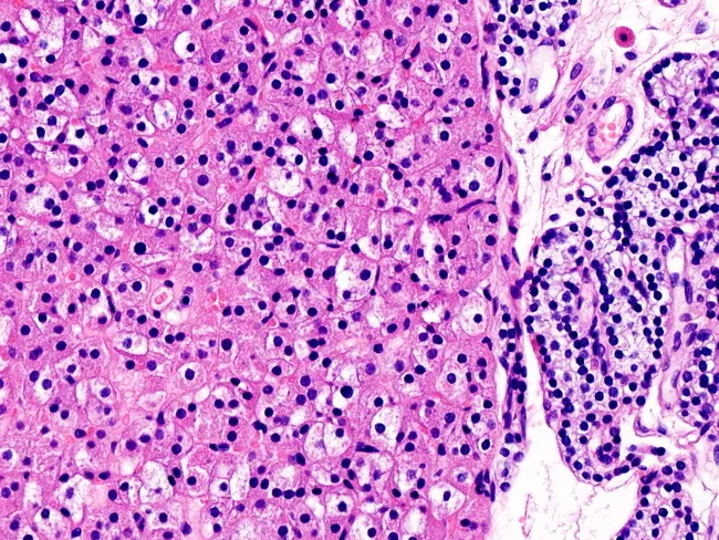
This is a light microscopic histology image of parathyroid tissue stained with hematoxylin and eosin (H&E). The left side shows an oxyphil cell–rich parathyroid adenoma characterized by sheets and cords of large, polygonal cells with abundant eosinophilic cytoplasm and centrally located round to ovoid nuclei. The dense eosinophilic cytoplasm is due to abundant mitochondria, giving the cells a characteristic acidophilic appearance. The stroma is relatively scant, with delicate capillary networks and minimal hematopoietic infiltration. A normal parathyroid gland is visible on the right for comparison, containing smaller, less eosinophilic chief cells arranged in cords with lighter cytoplasm and a richer vascular stroma. The juxtaposition helps distinguish adenomatous parathyroid tissue from normal parenchyma. Clinically, oxyphil cell–predominant adenomas can cause primary hyperparathyroidism, though nonfunctional adenomas exist. Pathology notes include a well-circumscribed lesion lacking significant mitotic activity or atypia, consistent with benign parathyroid adenoma rather than carcinoma. This image is valuable for educational purposes in surgical pathology, endocrine pathology, and histology training, illustrating cellular morphology, dysplastic features, and differential diagnosis with parathyroid hyperplasia. Correlation with serum calcium, parathyroid hormone levels, and radiological localization studies is recommended in clinical workups. It also aids understanding of oxyphilic differentiation and benign behavior in educational settings today.
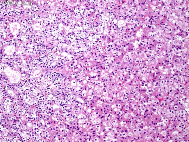
Imaging modality: Light microscopy of Hematoxylin and Eosin stained parathyroid tissue section (parathyroid adenoma). Anatomy: parathyroid gland within the cervical neck, typically near the thyroid; left half features sheets of chief cells; right half shows oxyphil cells; other cell types include water-clear cells and transitional cells. Histology: chief cells with amphophilic to lightly eosinophilic cytoplasm and conspicuous intracellular lipid/fat droplets; oxyphil cells with abundant granular eosinophilic cytoplasm and small pyknotic nuclei; cytoplasmic features reflect numerous mitochondria; cellular arrangement is lobular and highly cellular; vascular stroma is variable; fatty stroma reduced relative to normal parathyroid tissue. Pathology: parathyroid adenoma characterized by clonal proliferation of chief cells with admixture of oxyphil and water-clear cells; cytologic atypia minimal; mitotic activity low; absence of invasive growth in typical cases; indicates benign neoplasm; clinical correlation: hyperparathyroidism due to autonomous parathyroid hormone secretion; diagnostic significance includes correlation with elevated PTH and calcium levels; differential diagnoses include parathyroid hyperplasia, parathyroid carcinoma, and non-neoplastic thyroid tissue; educational use: illustrates cellular heterogeneity and diagnostic features used to differentiate hyperplasia from adenoma by architectural pattern, fat content, and cytoplasmic characteristics; suitable for pathology teaching, exam review, and database annotation. Representative images aid learners in recognizing parathyroid neoplasia and guiding surgical management.
thyroid and parathyroid glands
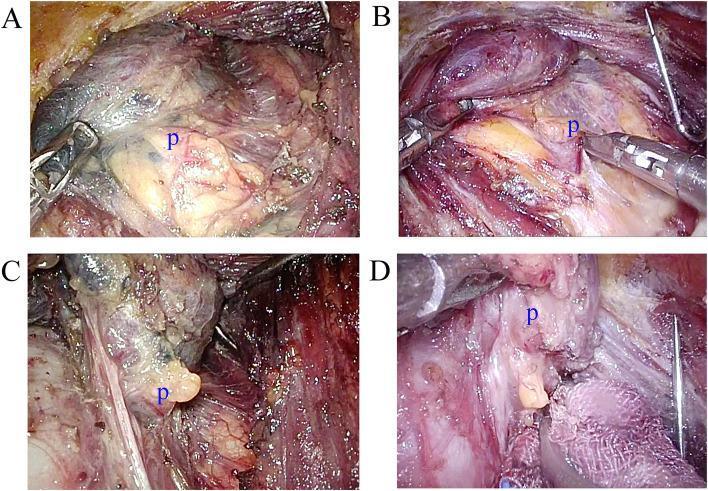
This set of four intraoperative clinical photographs (A-D) demonstrates the surgical exposure and identification of parathyroid glands (labeled 'p') during thyroid surgery, comparing the use of carbon nanoparticles (CN) for tissue contrast. Panels A and C show the inferior and superior parathyroid glands, respectively, after CN injection. In these images, the thyroid gland and surrounding lymphatic tissues exhibit dark grey or black staining due to carbon nanoparticle uptake, which provides visual contrast against the yellowish-tan, lobulated parathyroid glands that remain unstained. Panels B and D serve as control images showing inferior and superior parathyroid glands without the use of CN tracers. In the absence of CN, the parathyroid glands appear as pale yellow or ovoid structures, but the surrounding thyroid and fatty tissues maintain their natural reddish-pink and translucent hues, providing less distinct visual differentiation compared to the CN-stained groups. This educational visual illustrates the clinical application of negative mapping tracers to protect parathyroid glands during endocrine surgery by enhancing the visibility of surrounding structures.
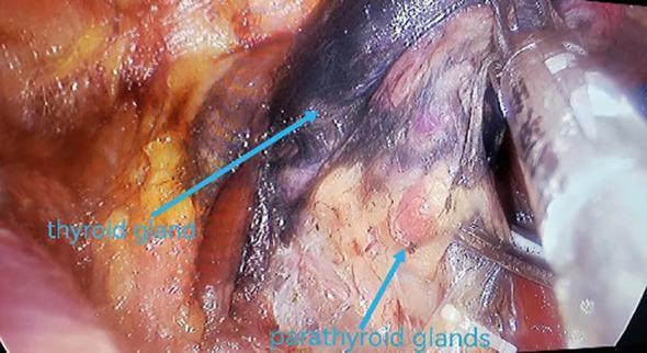
This intraoperative clinical photograph demonstrates the concept of 'negative opacification' during a thyroidectomy and central lymph node dissection. Following the injection of a nanocarbon suspension, the thyroid gland and central compartment lymph nodes exhibit significant black staining as they absorb the lymphatic tracer. In contrast, the parathyroid glands are highlighted by their failure to take up the nanocarbon, appearing as distinct, lighter-colored (yellow-tan) structures against the darkened thyroid tissue. Blue arrows and text labels specifically identify the blackened thyroid gland and the relatively pale parathyroid glands. This technique serves as a surgical aid to facilitate the identification and preservation of parathyroid glands in situ while ensuring the radical clearance of potentially metastatic, stained lymph nodes. The image illustrates a critical step in endocrine surgery for protecting parathyroid function and improving oncological outcomes in thyroid carcinoma cases.
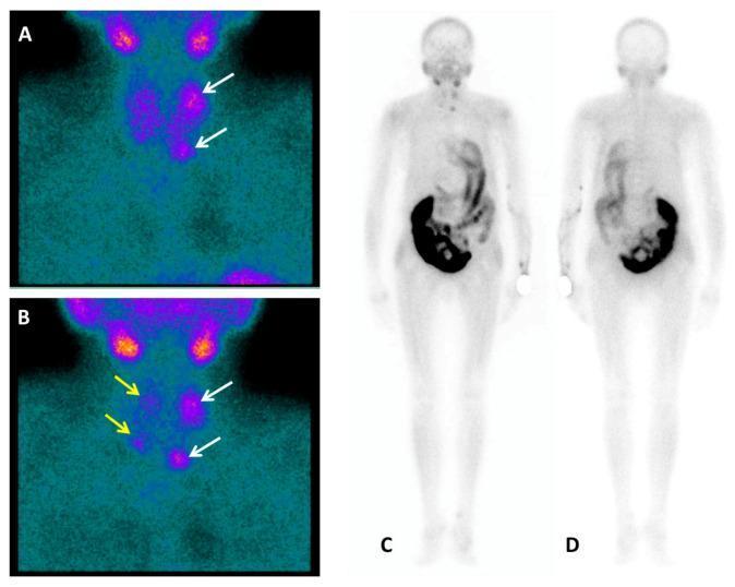
Diagnostic nuclear medicine imaging featuring Tc-99m-Sestamibi (MIBI) planar scintigraphy for the evaluation of tertiary hyperparathyroidism. Panels A and B display dual-phase anterior neck views. Early acquisition (A) shows initial radiotracer uptake in the thyroid and left superior/inferior parathyroid glands (white arrows). Delayed acquisition (B) demonstrates characteristic thyroid washout with persistent focal uptake in the left parathyroid glands and emerging mild concentration in the right parathyroid glands (yellow arrows), suggesting multiglandular hyperplasia or adenomas. Panels C (anterior) and D (posterior) show whole-body planar scans. These images reveal physiological tracer distribution, notably intense uptake in the abdominal digestive organs, while specifically demonstrating an absence of tracer accumulation in the skeletal system. The study highlights the diagnostic use of MIBI scintigraphy to differentiate hyperfunctioning parathyroid tissue from thyroid tissue and to screen for metabolic bone involvement in patients with chronic renal failure and elevated PTH levels.
Parathyroid Glands: Complete Overview
1. Anatomy and Embryology
- Superior parathyroids: derived from the 4th pharyngeal pouch endoderm
- Inferior parathyroids: derived (with the thymus) from the 3rd pharyngeal pouch endoderm
- Supernumerary glands occur in 3-5% of people, often in ectopic locations along the migration path (including within the thymus or thyroid) - this is clinically important during neck surgery.
2. Physiology - Parathyroid Hormone (PTH)
- Bone: stimulates osteoclastic bone resorption (indirectly, via increased RANKL and decreased osteoprotegerin from osteoblasts), releasing calcium and phosphate.
- Kidney: increases renal tubular calcium reabsorption while promoting phosphate excretion (phosphaturic effect); also stimulates 1-alpha-hydroxylase, converting vitamin D to its active form, calcitriol.
- Intestine: increases calcium absorption indirectly via calcitriol.
3. Disorders
A. Hyperparathyroidism
- Causes: adenoma (85-95%), primary hyperplasia (5-10%), parathyroid carcinoma (~1%)
- Molecular drivers: cyclin D1 gene rearrangement (10-20% of adenomas) and MEN1 mutations (30-35% of sporadic tumors)
- Clinical presentation: most cases today are picked up incidentally on routine calcium testing and are asymptomatic. Classic ("stones, bones, groans, and psychiatric overtones") presentation is now uncommon:
- Stones: nephrolithiasis, nephrocalcinosis
- Bones: bone resorption, osteitis fibrosa cystica, "brown tumors," reduced bone density
- Groans: GI symptoms, peptic ulcer, pancreatitis
- Psychiatric overtones: fatigue, irritability, weakness, depression
- May occur as part of MEN1 or MEN2A syndromes.
B. Hypoparathyroidism
4. Diagnostic Work-Up
- Serum calcium (total and ionized), phosphate, intact PTH, 25-OH vitamin D, 24-hour urine calcium
- Imaging for localization prior to surgery: Tc-99m sestamibi scintigraphy, ultrasound, 4D-CT, and increasingly 18F-choline PET (shown by a 2023 meta-analysis to have high detection/cure rates - PMID 36756935)
5. Medicines and Treatment
For Hyperparathyroidism (hypercalcemia)
| Drug/Approach | Mechanism / Role |
|---|---|
| Parathyroidectomy | Definitive treatment for symptomatic primary HPT, parathyroid carcinoma, and most tertiary HPT |
| Cinacalcet (calcimimetic) | Allosteric activator of the CaSR - "mimics" high calcium, lowering PTH secretion. Used for parathyroid carcinoma, primary HPT when surgery isn't feasible, and secondary HPT in dialysis patients (initial dose ~30 mg) |
| Etelcalcetide | Newer IV calcimimetic, similar mechanism, used mainly in secondary HPT/dialysis |
| Bisphosphonates | Reduce bone resorption; adjunct for skeletal protection in nonsurgical candidates |
| Vitamin D analogs / phosphate control | Used early in CKD-related secondary HPT to prevent progression |
For Hypoparathyroidism (hypocalcemia)
| Drug | Role |
|---|---|
| Calcitriol (active vitamin D) | First-line, combined with oral calcium, because impaired 1-alpha-hydroxylation means standard vitamin D is less effective |
| Oral calcium supplementation | Maintains serum calcium alongside calcitriol |
| Recombinant PTH (1-84) / Teriparatide-class analogs | Used in select refractory chronic hypoparathyroidism as hormone replacement |
| Magnesium repletion | Needed if hypomagnesemia is an underlying cause (magnesium is required for PTH secretion/action) |
Recent Evidence (PubMed, last 3 years)
- A 2024 systematic review appraising clinical practice guidelines for primary hyperparathyroidism found variable quality/rigor across existing guidelines, suggesting some published recommendations may lack strong methodological backing (PMID 38551756).
- A 2024 systematic review on primary HPT in pregnancy compared surgical vs non-surgical management, generally favoring surgical management (2nd trimester parathyroidectomy) to reduce maternal/fetal complications when feasible (PMID 38918290).
- A 2023 meta-analysis described "normohormonal primary hyperparathyroidism" (elevated calcium with PTH in the normal range) as a distinct, likely under-recognized variant (PMID 37100740).