
Make notes on sebaceous gland anatomy and physiology along with diagram for md dermatology theory exams
Reading File
Finding Sources
Searching Images
sebaceous gland anatomy diagram skin structure
Searching PubMed
"sebaceous gland" AND physiology
Reading File
query_images_db
Reading File
Reading File
Reading File
Reading File
Finding Sources
Finding Sources
Reading File
Reading File
Sebaceous Gland: Anatomy and Physiology (MD Dermatology Notes)
1. Introduction
Sebaceous glands are holocrine exocrine glands of the skin that secrete sebum. Along with eccrine and apocrine glands, they form the three major skin gland systems - but unlike the merocrine sweat glands, sebaceous glands release their entire cell content (cytoplasm + membranes) into the duct.
2. Embryology (brief)
- Develop from the epithelial placode (bulge) of the developing hair follicle at 13-16 weeks of fetal life, arising in a cephalocaudal sequence.
- The hair follicle bulge stem cell reservoir can regenerate sebaceous glands, though some evidence suggests glands may also be maintained independently of the bulge.
- Key signalling: Sonic hedgehog (Shh) and Myc signalling promote sebocyte differentiation; Wnt/β-catenin signalling promotes hair follicle differentiation instead (reciprocal switch) - a favorite viva question.
3. Anatomy
Structure
- Multilobular, holocrine glands composed of acini (lobules) draining into a common sebaceous duct lined by undifferentiated keratinocytes (stratified squamous epithelium).
- Gland periphery = single layer of small, cuboidal, mitotically active undifferentiated sebocytes (basal layer) → cells migrate centrally, accumulate lipid droplets, terminally differentiate, lose all organelles, then rupture and die, discharging contents into the duct.
- Usually opens into the hair follicle infundibulum, forming the pilosebaceous unit (gland + hair follicle + arrector pili muscle). When the associated hair is vestigial/tiny with a disproportionately large gland, this is called a sebaceous follicle (the site of acne).
Distribution
- Present wherever hair follicles exist (whole body).
- Highest density: nose (~1600 glands/cm²) > face and scalp (400-900 glands/cm²) - "T-zone" seborrhea correlates directly with this density.
- Absent on palms and soles (no hair follicles); sparse on dorsa of hands/feet.
- Free (ectopic) sebaceous glands - not associated with hair, open directly onto epithelial surface:
- Meibomian glands (tarsal glands) - eyelids
- Montgomery glands (areolar glands) - nipple/areola
- Tyson glands - prepuce/genital skin
- Fordyce spots - lips, buccal mucosa, genital mucosa (visible 1-3 mm papules due to oral epithelial transparency)
Sebocyte maturation (histologic stages, often asked as a spotter): Undifferentiated → Early differentiated → Advanced differentiated → Fully differentiated → Mature (about to rupture)
Diagram
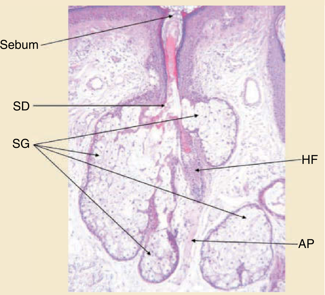
Figure: Cross-section of a pilosebaceous unit - multiacinar sebaceous gland (SG) opening via the sebaceous duct (SD) into the hair follicle (HF), alongside the arrector pili muscle (AP). (Zouboulis CC, Tsatsou F, in Pathogenesis and Treatment of Acne and Rosacea, 2014; reproduced in Fitzpatrick's Dermatology, p. 100)
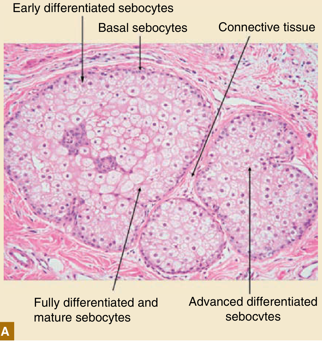
Figure: H&E section of human sebaceous gland - Fitzpatrick's Dermatology, p. 101
For your answer sheet, a simple labelled hand-drawn diagram should show: epidermis → hair shaft → sebaceous duct opening into follicular infundibulum → multilobular acini (with a peripheral basal layer and central disintegrating lipid-filled sebocytes) → arrector pili muscle attached obliquely between the follicle and papillary dermis.
4. Physiology
A. Holocrine Secretion
- Not simple cell swelling; it is a programmed, lysosomal DNase2-mediated cell death pathway distinct from apoptosis, necroptosis, and cornification.
- Sebocytes accumulate lipid droplets as they move centrally → disintegrate near the duct → release neutral lipids (proteins/nucleic acids/membrane phospholipids are recycled, not secreted).
- Transit time: ~7.4 days total (4-7 days undifferentiated stage, 14-25 days differentiated/lipid-producing stage) - a commonly asked numeric fact.
B. Lipid Composition of Sebum
Sebum leaving the gland = mixture of neutral lipids:
| Lipid | Approx. % |
|---|---|
| Triglycerides + diglycerides + free fatty acids | 40-60% |
| Wax esters | 25-30% |
| Squalene | 12-15% |
| Cholesterol esters | 3-6% |
| Cholesterol | 1.5-2.5% |
- Wax esters and squalene are unique to sebum (absent from internal organ lipids) - key differentiating feature in exams.
- Ductal bacterial lipases hydrolyze triglycerides → free fatty acids (implicated in acne/comedogenesis).
- Unique fatty acids: sapienic acid (16:1Δ6, major fatty acid of adult human sebum, from Δ6 desaturation of palmitic acid) and sebaleic acid (18:2Δ5,8) - both essentially unique to human sebum.
C. Regulation of Sebaceous Gland Size and Sebum Production
- Gland size increases with age (mean area rises from ~0.2 to ~0.4 mm²); gland number stays roughly constant through life, but turnover slows in older adults.
- Regulated by ligand-receptor systems on sebocytes:
- Androgen and estrogen receptors (androgens are the principal stimulus for sebum production - clinically central to acne pathogenesis)
- PPAR (peroxisome proliferator-activated receptor) and LXR (liver-X receptor)
- Neuropeptide receptors (e.g., CRH receptors - corticotropin-releasing hormone increases lipogenesis and IL-6/IL-8 release, linking stress to acne flares)
- Retinoid receptors (basis of isotretinoin's mechanism - sebosuppressive)
- Vitamin D receptor
- Sebocytes retain stem-like, bipotential characteristics despite being programmed for terminal differentiation.
D. Functions of the Sebaceous Gland (Fitzpatrick's functional classification - good for a "functions of sebaceous gland" essay)
-
Protection
- Natural UVB photoprotection
- Thermoregulation and water-repelling properties
- Possible role in wound healing
- Production of vernix caseosa (neonatal skin protection)
-
Transportation
- Vehicle for delivering antioxidants to/from the skin surface
- Vehicle for pheromone/fragrance-related compounds
-
Inflammation and Immunity
- Produces both pro- and anti-inflammatory lipids
- TLR2 activation upregulates lipogenesis
- Lipid-induced innate antimicrobial activity
- Secretes antimicrobial peptides: cathelicidin, psoriasin, β-defensin-1, β-defensin-2 (direct antimicrobial activity, relevant to acne and rosacea pathogenesis)
- Promotes Th17 differentiation (contributes to inflammatory skin disease)
-
Endocrine Properties
- Sebaceous gland is a peripheral steroidogenic organ - expresses all steroidogenic enzymes and synthesizes androgens locally
- Expresses vitamin D receptor and vitamin D-metabolizing enzymes
- Expresses retinoid-metabolizing cytochrome P450 enzymes
- Contributes to hormonally-mediated skin aging
5. Clinical Correlation (high-yield linkage for exams)
| Sebaceous gland abnormality | Disorder |
|---|---|
| Increased activity/hyperplasia | Acne vulgaris, rosacea, seborrhea, senile sebaceous hyperplasia |
| Neoplastic | Sebaceoma, sebaceous carcinoma, Muir-Torre syndrome |
| Decreased activity | Xerosis, cicatricial alopecias (lichen planopilaris, pseudopelade) |
| Ectopic gland pathology | Fordyce spots (normal variant), nevus sebaceus |
6. Quick Exam Recall Points
- Sebaceous glands = holocrine, sweat glands = merocrine
- Density highest at nose (1600/cm²); absent on palms/soles
- Transit time ~7.4 days
- Unique lipids: wax esters, squalene, sapienic acid, sebaleic acid
- Chief hormonal driver: androgens (basis for antiandrogen/isotretinoin therapy in acne)
- Free sebaceous glands mnemonic: "MFTM" - Meibomian (eyelid), Fordyce (mucosa), Tyson (prepuce), Montgomery (areola)
Source: Zouboulis CC. "Skin Glands: Sebaceous, Eccrine, and Apocrine Glands" - Fitzpatrick's Dermatology, 9th ed., Vol 1, pp. 99-105.
Recent literature note (for viva awareness, not core physiology): recent systematic reviews have focused on sebaceous gland pathology rather than baseline physiology - e.g., malignant transformation risk in nevus sebaceus (PMID: 38725109), energy-device effects on sebum in acne (PMID: 39129257), and sebaceous neoplasms in Lynch/Muir-Torre syndrome (PMID: 36418753). These don't change the core anatomy/physiology but are useful if asked about clinical correlations or recent advances.
This is a shared conversation. Sign in to Orris to start your own chat.