
Pulmonary embolism
Reading File
Now let me search the medical library for pulmonary embolism content across multiple angles simultaneously.
Finding Sources
Searching PubMed
"pulmonary embolism"[MeSH Terms] AND diagnosis AND treatment
Good - I have strong hits from Murray & Nadel's (pulmonology reference) and Rosen's Emergency Medicine. Let me read the most comprehensive sections in parallel.
Reading File
Reading File
Reading File
Excellent content from Rosen's Emergency Medicine. Let me read more of that block for diagnosis and management, and also grab the main Murray & Nadel section.
Reading File
Reading File
I have rich content from both Rosen's and Murray & Nadel's. Let me read the core management/treatment section from Rosen's which appeared in the truncated part.
Reading File
I went past the main content. Let me read the treatment and diagnosis sections from the Rosen's block at the right offset.
Reading File
I now have very comprehensive content. I have enough to write a detailed, well-organized answer on pulmonary embolism. Let me also fetch the autopsy image that was referenced.
Loading Image
I now have comprehensive content from authoritative sources. Here is a full, detailed overview of pulmonary embolism.
Pulmonary Embolism (PE)
Definition and Overview
Pulmonary embolism is the obstruction of the pulmonary arterial circulation by a thrombus (or, less commonly, air, fat, amniotic fluid, or tumor). It is a life-threatening manifestation of venous thromboembolism (VTE) that most frequently originates from deep venous thrombosis (DVT) of the lower extremities. PE accounts for approximately 25% of sudden cardiac deaths and is a leading cause of maternal mortality in the United States. - Murray & Nadel's Textbook of Respiratory Medicine, p. 960; Rosen's Emergency Medicine, p. 1202
Pathophysiology
Hemodynamic consequences: The obstructing embolus increases right ventricular afterload. If obstruction is large enough, right ventricular (RV) dilation occurs, the interventricular septum bows leftward (D-sign), and left ventricular (LV) filling drops - reducing cardiac output, systemic blood pressure, and coronary perfusion. This spiral of RV failure drives mortality. Hypotension (SBP < 90 mmHg) is the single most important predictor of PE mortality, conferring a 4-fold increase in death risk. - Rosen's Emergency Medicine, p. 1203
Hypoxemia mechanism: PE creates dead space (occluded segments receive no perfusion). Patients mount a compensatory hyperventilation, so most present with hypocarbia, not hypercarbia. Hypoxemia results from:
- Redistribution of blood flow to non-obstructed zones, creating low V/Q areas
- Shunt (from atelectasis and surfactant loss) appearing hours to days later
- Markedly low mixed venous PO2 from depressed cardiac output (amplifies venous admixture)
- Loss of surfactant causing atelectasis in the infarcted zone
Notably, ~12% of patients with PE have PaO2 > 80 mmHg, and the alveolar-arterial gradient can be normal in a small minority. - Murray & Nadel's, p. 960
Risk Factors (Virchow's Triad)
| Category | Examples |
|---|---|
| Hypercoagulability | Malignancy, thrombophilia (Factor V Leiden, prothrombin G20210A, protein C/S/antithrombin deficiency), antiphospholipid syndrome, OCP/HRT, pregnancy |
| Venous stasis | Prolonged immobilization, long-haul travel, paralysis, heart failure, pregnancy (uterine IVC compression) |
| Endothelial injury | Surgery, trauma, pelvic vein injury at delivery, central venous catheters |
Special population - Pregnancy: VTE risk is ~5x higher than age-matched non-pregnant women. The postpartum period (especially post-cesarean section) carries the highest risk. - Murray & Nadel's, p. 3029
Clinical Presentation
Symptoms
- Dyspnea: most common symptom (75-80%), but absent in ~25%
- Chest pain: 2nd most common; can be pleuritic (sharp, respirophasic - seen in ~20%, usually with peripheral/infarction PE), dull, or vague
- Hemoptysis, cough, fever: suggest pulmonary infarction; fever > 38.6°C (101.5°F) suggests infection rather than PE
- Syncope: PE in <5% of patients presenting with syncope, but unexplained syncope with risk factors should prompt consideration
- Unilateral leg swelling: present in <30%, but highly specific when combined with dyspnea or chest pain
The presentation ranges from entirely asymptomatic to sudden cardiovascular collapse and cardiac arrest.
Vital Signs
| Finding | Frequency | Significance |
|---|---|---|
| Tachycardia (HR >100) | ~50% | Associated with severity; worst prognosis |
| Tachypnea (RR >20) | ~50% | Variable measurement |
| Hypotension (SBP < 90) | ~10% | 4x mortality increase; most important predictor |
| Hypoxemia (SpO2 <95%) | ~50% | Independent predictor of outcomes |
Shock index >1 (HR > SBP) prior to arrest is a warning sign of impending cardiovascular collapse.
Physical Examination
Most patients have no abnormal physical findings. The only finding reliably raising PE probability is evidence of DVT (unilateral leg asymmetry, edema, deep vein tenderness). Wheezing suggests bronchospasm (reduces PE probability); bilateral rales suggest LV failure.
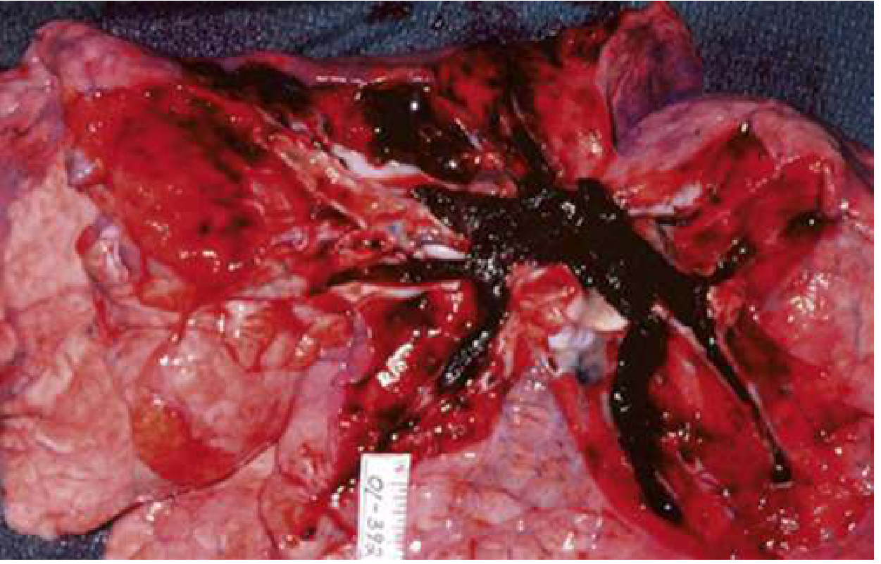
Cardiac arrest due to PE: Pulseless electrical activity (PEA) is the most common arrest rhythm. Survival even with witnessed arrest and bolus fibrinolysis is ~20%.
Diagnosis
Step 1 - Pretest Probability (Clinical Decision Rules)
Wells Score for PE:
| Criterion | Points |
|---|---|
| Clinical signs/symptoms of DVT | 3 |
| PE more likely than alternative diagnosis | 3 |
| Heart rate > 100 | 1.5 |
| Immobilization or surgery in previous 4 weeks | 1.5 |
| Previous DVT or PE | 1.5 |
| Hemoptysis | 1 |
| Malignancy (on treatment, treated in last 6 months, or palliative) | 1 |
- Score > 4 = PE likely
- Score ≤ 4 = PE unlikely (proceed to D-dimer)
Step 2 - D-Dimer
- High sensitivity (~95%), low specificity
- A negative D-dimer in a low-pretest-probability patient effectively excludes PE
- Age-adjusted D-dimer threshold: age × 10 ng/mL (in patients > 50 years) - increases specificity without significantly reducing sensitivity
- YEARS algorithm: combines 3 criteria (DVT signs, hemoptysis, PE as most likely diagnosis) with D-dimer thresholds (< 500 ng/mL if any YEARS item present; < 1000 ng/mL if none present) to rule out PE safely in many patients
Step 3 - Imaging
CT Pulmonary Angiography (CTPA) - first-line imaging
- High sensitivity and specificity for PE down to the segmental level
- Preferred in most non-pregnant patients
- Can identify alternative diagnoses
- Maternal breast radiation exposure is significant (7-70 mSv); consider V/Q in young women
Ventilation-Perfusion (V/Q) Scan
- Preferred in pregnancy (lower maternal breast dose) and when contrast is contraindicated
- Results: normal (excludes PE), high probability (diagnoses PE), or indeterminate
- In pregnancy: perfusion-only scan with half-dose is standard practice
Echocardiography
- Bedside TTE/TEE: RV dilation, septal bowing, reduced RV function, McConnell sign (RV free wall hypokinesis with preserved apex)
- Not sensitive for PE itself but useful for risk stratification and resuscitation guidance
- May directly visualize saddle embolus in massive PE
Chest X-ray
- Usually normal or non-specific
- Classic signs: Hampton's hump (wedge-shaped opacity - infarction), Westermark sign (oligemia)
Leg compression ultrasound (CUS)
- If positive for DVT in a patient with clinical suspicion for PE, may be sufficient to start treatment without CTPA
Biomarkers
- Troponin (I or T): elevated = RV myocardial injury; strongly predicts short-term adverse outcomes and mortality - Murray & Nadel's, p. 893
- BNP/NT-proBNP: elevated with RV strain; used for risk stratification
- D-dimer: see above
Risk Stratification
| Category | Definition | Mortality |
|---|---|---|
| Massive (High-risk) | Sustained hypotension, cardiac arrest, or shock | >15% |
| Submassive (Intermediate-risk) | Hemodynamically stable but RV dysfunction or troponin elevation | 3-15% |
| Low-risk | No RV dysfunction, no biomarker elevation (PESI score low) | <1% |
PESI (Pulmonary Embolism Severity Index) - incorporates age, cancer, chronic cardiopulmonary disease, HR, BP, SpO2, and mental status to stratify risk and guide disposition.
Management
1. Immediate Resuscitation
- Supplemental O2; intubation if respiratory failure (caution: intubation drops preload and can precipitate RV collapse)
- IV fluids (cautiously - RV is preload dependent but volume overload worsens septal shift)
- Vasopressors for refractory hypotension: norepinephrine preferred
2. Anticoagulation (All confirmed PE without contraindication)
Start as soon as diagnosis is confirmed (or suspected while awaiting imaging in high-probability cases).
| Agent | Notes |
|---|---|
| Unfractionated heparin (UFH) | Preferred in massive PE, renal failure, patients likely to need procedures |
| LMWH (enoxaparin, dalteparin) | Preferred in most intermediate/low-risk; weight-based dosing |
| DOACs (rivaroxaban, apixaban) | First-line for most low/intermediate-risk PE; apixaban/rivaroxaban approved for monotherapy without parenteral bridging |
| Warfarin | Requires bridging; largely superseded by DOACs for non-cancer PE |
Recent 2025 meta-analysis (PMID 40578592) confirms DOACs are preferred over LMWH even in cancer-associated thrombosis in most settings, with similar efficacy and lower bleeding risk.
3. Reperfusion Therapy
Systemic fibrinolysis - for massive PE with cardiovascular collapse:
- Alteplase (tPA) 100 mg IV over 2 hours (or 0.6 mg/kg over 15 min in arrest)
- Reduces clot burden, RV dilation, and hemodynamic instability
- Major bleeding (including intracranial hemorrhage ~2-3%) is the principal risk
- Contraindicated with recent surgery, active bleeding, recent stroke
Meta-analysis evidence (PMID 39267429): Thrombolysis reduces short-term mortality vs anticoagulation alone in high-risk PE, but carries significantly higher bleeding rates.
Catheter-directed therapy (CDT): Lower-dose fibrinolytic delivered directly via catheter into pulmonary arteries - used for submassive PE when systemic lysis is relatively contraindicated.
Surgical embolectomy: Reserved for massive PE when fibrinolysis is contraindicated or failed.
IVC filter: Used when anticoagulation is absolutely contraindicated; not a substitute for anticoagulation when it can be given.
4. Duration of Anticoagulation
| Situation | Duration |
|---|---|
| First provoked PE (transient risk factor) | 3 months |
| First unprovoked PE | ≥ 3 months; consider indefinite based on bleeding risk |
| Recurrent unprovoked PE | Indefinite |
| Active cancer | Indefinite (LMWH or DOAC while cancer active) |
5. Outpatient vs. Inpatient Treatment
Low-risk PE (PESI Class I-II, no RV dysfunction, good social support) can be safely treated as outpatients with DOAC therapy. This is supported by validated protocols reducing hospitalization without increasing adverse outcomes. - Rosen's Emergency Medicine, p. 1207
Pregnancy-Specific Considerations
- D-dimer is physiologically elevated in pregnancy; standard thresholds unreliable
- Pregnancy-adapted YEARS algorithm may reduce unnecessary imaging in some patients, but limited data means caution is warranted
- LMWH is the anticoagulant of choice throughout pregnancy (DOACs are contraindicated; warfarin crosses placenta)
- Imaging: Start with lower-limb CUS; if negative, proceed to CTPA or V/Q (V/Q preferred by some due to lower maternal breast dose)
- Fetal radiation from CTPA is low (0.01-0.66 mSv) vs. natural background (~0.5-1.0 mSv/gestation) - Rosen's Emergency Medicine, Table 74-6
Key ECG Findings
- Sinus tachycardia - most common
- S1Q3T3 pattern - classic but present in minority
- New right bundle branch block (RBBB)
- T-wave inversions in V1-V4 (RV strain pattern)
- PEA in cardiac arrest
Summary Algorithm
Suspected PE
    │
    ├─ Hemodynamically unstable?
    │       YES → Immediate resuscitation + CTPA/bedside echo
    │               Massive PE confirmed → Systemic thrombolysis (if no CI)
    │
    └─ Hemodynamically stable?
            │
            ├─ Wells Score
            │       Low/Moderate (PE unlikely, score ≤4) → D-dimer
            │           Negative → PE excluded
            │           Positive → CTPA
            │
            └─ High (PE likely, score >4) → CTPA directly
                    PE confirmed → Anticoagulation ± risk stratify
                    Low risk → Consider outpatient DOAC
                    Submassive → Anticoagulation ± CDT
Key Sources:
- Rosen's Emergency Medicine: Concepts and Clinical Practice, Chapter 74 (Pulmonary Embolism)
- Murray & Nadel's Textbook of Respiratory Medicine (Chapters 81-82 on PE, pathophysiology of hypoxemia)
- Murray & Nadel's Textbook of Respiratory Medicine (Chapter on PE in pregnancy)
- Recent evidence: anticoagulation vs. thrombolysis meta-analysis (PMID 39267429); DOACs in cancer-associated VTE (PMID 40578592)
This is a shared conversation. Sign in to Orris to start your own chat.