
Velopharyngeal insufficiency in detail and rhinolalia aperta management in detail according to standard ENT textbooks and in simplified manner along
velopharyngeal insufficiency anatomy closure pattern endoscopy
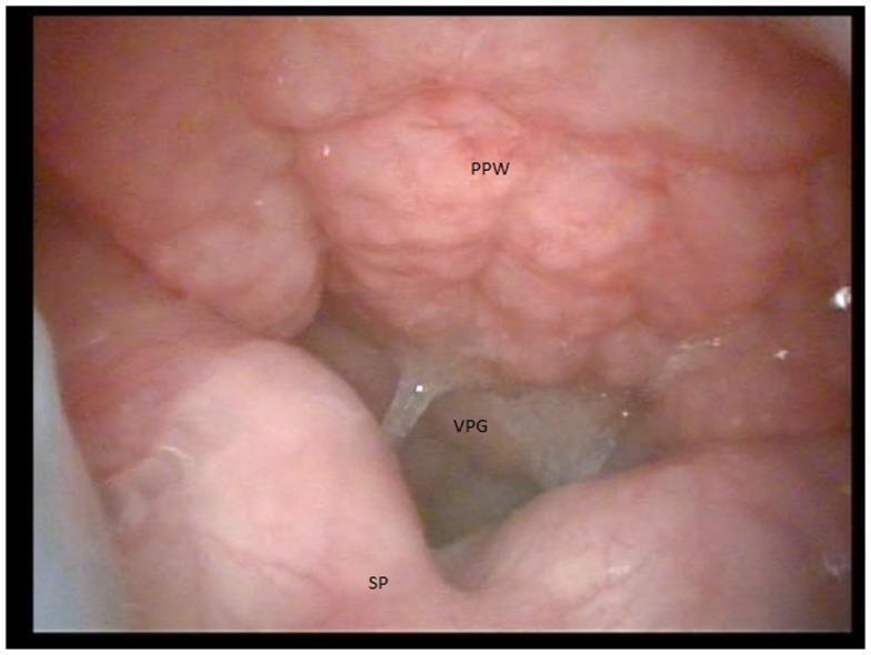
This diagnostic clinical image shows a view from a video nasopharyngeal endoscopy during a velopharyngeal function assessment. The superior aspect of the frame displays the posterior pharyngeal wall (PPW), while the inferior portion shows the superior surface of the soft palate (SP). Centrally, a persistent and significant velopharyngeal gap (VPG) is visible, indicating a failure of the velopharyngeal valve to achieve complete closure during speech or swallow. This condition, known as velopharyngeal insufficiency (VPI), is illustrated here in the clinical context of a submucous cleft palate. The mucosal surfaces appear pink and somewhat irregular, with the gap appearing as a darkened, central opening between the dynamic structures of the palate and the stationary pharyngeal wall. The image is used in otolaryngology and speech-language pathology to determine surgical interventions, such as pharyngeal flap or sphincter pharyngoplasty, based on the size and shape of the observed deficiency.
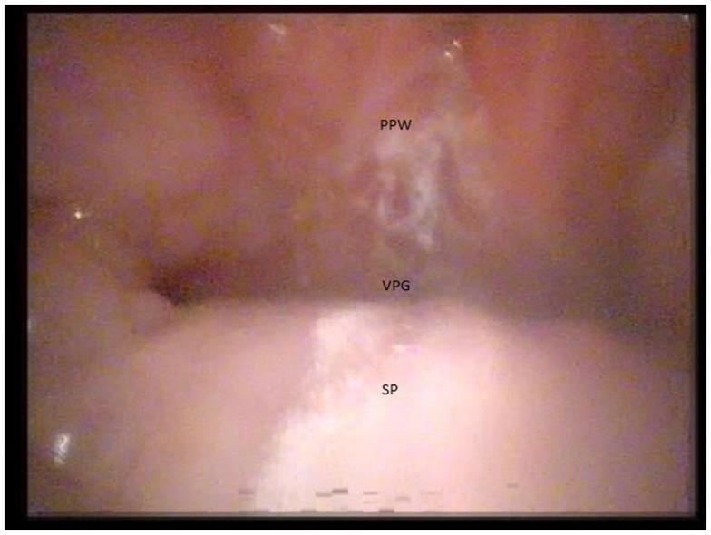
This diagnostic image is a frame from a video nasopharyngeal endoscopy (laryngoscopy) demonstrating velopharyngeal insufficiency (VPI). The view identifies three primary landmarks: the posterior pharyngeal wall (PPW) at the top, the soft palate (SP) at the bottom, and a persistent large velopharyngeal gap (VPG) between them. The PPW and SP exhibit typical mucosal characteristics, appearing moist and pinkish. The VPG represents a failure of the velum to achieve full contact with the pharyngeal walls during speech or swallowing, a finding clinically significant in conditions like 22q11.2 deletion syndrome. The gap is large and irregularly shaped, indicating a significant adynamic or structural defect in the velopharyngeal mechanism. This type of endoscopic evaluation is critical for surgical planning, such as determining the need for a pharyngeal flap versus sphincter pharyngoplasty based on the size and shape of the closure failure.
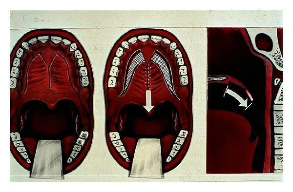
A medical illustration depicting the surgical push-back procedure for treating velopharyngeal insufficiency (VPI). The image is divided into three panels showing the anatomical approach and mechanism. The leftmost panel illustrates the baseline oral anatomy, focusing on the hard and soft palate within the dental arch. The center panel shows the surgical intervention where mucoperiosteal flaps (indicated by a mesh pattern) are mobilized and sutured along the midline, with a white arrow indicating the posterior movement of the secondary palate toward the uvula. The rightmost panel provides a sagittal view of the head and neck, clearly showing the nasal cavity, oral cavity, tongue, and pharyngeal wall. A white arrow in this view demonstrates the 'push-back' movement of the soft palate posteriorly to reduce the velopharyngeal gap. This procedure aims to improve speech by lengthening the palate to facilitate better closure against the posterior pharyngeal wall, specifically targeting hypernasality (hyperrhinophony).
Velopharyngeal Insufficiency (VPI) and Rhinolalia Aperta
PART 1: VELOPHARYNGEAL INSUFFICIENCY (VPI)
🔹 What is VPI? (Simplified)
Terminology (Cummings Classification)
| Term | Cause | Example |
|---|---|---|
| VPI (Insufficiency) | Not enough tissue to close the valve | Cleft palate, short palate |
| Velopharyngeal Incompetence | Motor/neurological - tissue is adequate but doesn't move right | Cerebral palsy, myasthenia gravis, stroke |
| Velopharyngeal Mislearning | Learned habit - no structural or neurological cause | Phoneme-specific VPD after cleft repair |
Cummings Otolaryngology, p. 3620
Anatomy of the Velopharyngeal Mechanism
- Velum (soft palate) - moves superoposteriorly during closure; most important contributor
- Lateral pharyngeal walls - move medially to meet the velum
- Posterior pharyngeal wall - may develop a prominence called the Passavant ridge during closure
- Levator veli palatini - the primary muscle that lifts the soft palate. In cleft palate, this muscle is abnormally oriented longitudinally (runs forward to insert into the hard palate) instead of transversely (across the midline as a sling). Surgical repair reorients this sling.
- Musculus uvulae - adds bulk to the dorsal velum; its absence is a marker of occult submucous cleft
- Superior pharyngeal constrictor - contributes to lateral wall movement
- Palatopharyngeus and palatoglossus - provide minor contributions
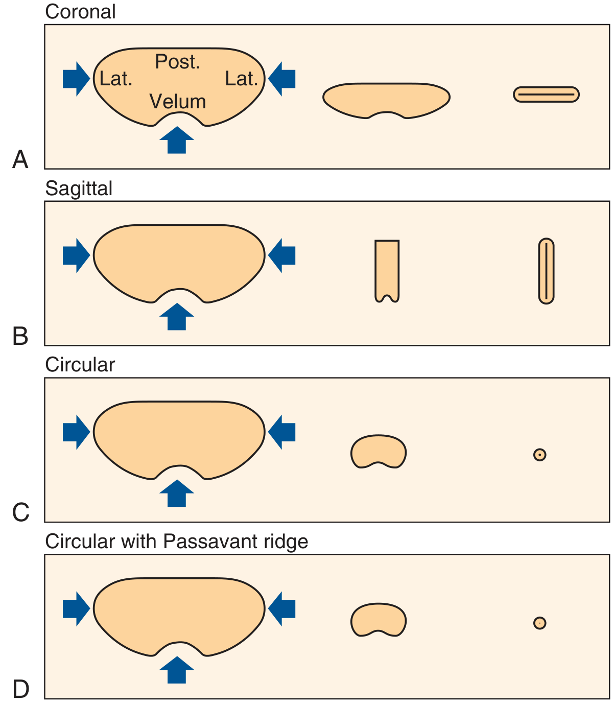
- Coronal - velum fails to reach posterior wall; gap runs side to side (most common, ~55%)
- Sagittal - lateral walls fail to meet; vertical gap in midline
- Circular - all walls contract but don't close; central round gap
- Circular with Passavant ridge - circular but posterior wall also contributes a ridge
Knowing the closure pattern guides which surgery is best.
Causes of VPI
- Cleft palate - most common; incompletely or incorrectly repaired levator sling
- Submucous cleft palate - triad: bifid uvula + zona pellucidum (translucent zone in midline soft palate) + palpable bony notch at posterior hard palate
- Occult submucous cleft - absent musculus uvulae only (detected endoscopically)
- Congenitally short palate / palatopharyngeal disproportion - palate too short relative to deep nasopharynx
- After adenoidectomy - large adenoids may compensate for a borderline palate; removing them exposes the dysfunction. Occurs in ~1/1500 adenoidectomies. Pre-existing submucous cleft must be excluded before adenoidectomy.
- After pharyngeal/skull base surgery - disruption of palatal muscle supply
- Cerebral palsy, muscular dystrophies, myasthenia gravis
- Traumatic brain injury, brainstem stroke
- 22q11.2 deletion syndrome (velocardiofacial/DiGeorge) - most common genetic cause; often combined incompetence + insufficiency
Cummings Otolaryngology, p. 3620-3621
Clinical Features
- Hypernasality - the hallmark; too much nasal resonance on oral sounds
- Nasal air emission - audible air escape through nose during pressure consonants (p, b, t, d, k, g, s, z)
- Weak pressure consonants - plosives and fricatives are muffled
- Compensatory articulation errors - child learns abnormal patterns to overcome VPD:
- Glottal stops (closing larynx instead of lips for /p/, /b/)
- Pharyngeal fricatives (tongue base substituted for tongue tip)
- Backing (posterior placement of tongue)
- These become habituated and persist even after surgery
- Nasal grimacing - child scrunches nose to reduce air escape
- Reduced speech intelligibility
Assessment
- Standardized rating of hypernasality, nasal emission, compensatory errors
- Use of phrases with no nasal sounds (e.g., "Katie's sister was six yesterday") vs. nasal phrases
- Hold cold steel spatula under the nostrils during speech
- Misting only on nasal sounds (m, n, ng) = normal
- Misting on purely oral phrases = VPD
- Measures ratio of nasal-to-oral sound intensity via a headset
- Provides a "nasalance score" with normative data
- Useful for documenting severity and tracking therapy/surgical progress
- A discrepancy may exist between nasalance score and perceived hypernasality
- Measures nasal airflow and oral pressure during speech
- Can calculate cross-sectional nasopharyngeal area and timing of air escape
- Flexible endoscope passed through nose into nasopharynx
- Patient speaks standardized words/sentences with correct articulation
- Identifies: closure pattern, size and location of gap, lateral wall motion, presence of adenoid, musculus uvulae status (detects occult submucous cleft)
- Scope positioned in middle meatus to reduce parallax
- Voice + video recorded simultaneously
- Lateral and Towne's (frontal) views during speech
- Complements endoscopy - shows sagittal velum movement
- Less preferred due to radiation exposure in children
Cummings Otolaryngology, p. 3622
PART 2: RHINOLALIA APERTA - MANAGEMENT IN DETAIL
🔹 What is Rhinolalia Aperta? (Simplified)
Scott-Brown's Otorhinolaryngology Vol 2, p. (block 2, line 6328)
Management Framework
Step 1: Speech Therapy (First-Line for Most Cases)
- Mild or inconsistent hypernasality
- VPD due to mislearning (phoneme-specific)
- Post-operative rehabilitation after surgical VPD correction
- Children too young for surgery
- Studies confirm that sucking, blowing, gagging, swallowing exercises and electrical stimulation have no direct impact on velopharyngeal closure during speech.
- Oral motor exercises are not appropriate unless there is specific oral motor weakness.
-
Articulation therapy - correct glottal stops, pharyngeal fricatives, backing patterns
- Whispering technique for glottal stops: child whispers "pa-pa" then gradually adds voicing
- Tactile, visual, and auditory feedback during correct/incorrect phoneme production
- Key: do not start until dentition is adequate (not missing teeth causing articulatory problems)
-
Resonance therapy - for mild/inconsistent hypernasality:
- Auditory feedback to help child hear nasal vs. oral resonance
- Microphones, accelerometers, nasal tubes
- Nasal occlusion technique: temporarily occlude nares during speech to demonstrate oral resonance, then remove occlusion and attempt to replicate
- This works only if VP muscles are capable but inconsistently used
-
Biofeedback-based therapy - real-time nasometry display to help child self-monitor
Cummings Otolaryngology, p. 3625-3626
Step 2: Prosthetic Management
- Poor surgical candidates (comorbidities, anesthesia risk)
- Neurological VPD with expected recovery (stroke, trauma) - temporary use
- Patients with obstructive sleep apnea (prosthesis removed at night)
- Adolescents/adults who prefer non-surgical management
- Children during active dental treatment (when surgery must wait)
-
Palatal lift prosthesis - physically lifts the soft palate to the level of the posterior pharyngeal wall. Useful in neurological incompetence (palate is mobile but weak).
-
Palatal bulb obturator - fills the nasopharyngeal gap with an acrylic bulb. Lateral pharyngeal walls contact the bulb and may actually improve over time ("weaning" or bulb reduction therapy).
Cummings Otolaryngology, p. 3624
Step 3: Surgical Management (Definitive Treatment)
- Structural VPI (cleft palate or submucous cleft) with inadequate tissue
- VPD persisting after adequate speech therapy trial
- Imaging/endoscopy confirms anatomical/structural gap
A. Intravelar Veloplasty (IVV / Levator Repositioning)
- Principle: The levator muscle in cleft/submucous cleft runs abnormally forward; this surgery detaches it from the hard palate and reorients it transversely as a proper muscular sling.
- Best for: Primary or re-do palatoplasty in patients with longitudinally oriented levator muscles; submucous cleft
- Technique: Hard palatal flaps raised, levator dissected free from hard palate and nasal mucosa, sutured across midline with mattress sutures
- Note: Results are variable when used as a secondary procedure
B. Furlow Double-Opposing Z-Plasty (DOZ Palatoplasty)
- Principle: The classic Z-plasty technique applied to the palate. Reorients the levator AND lengthens + thickens the soft palate. The Z-flap transposition provides bulk in the posterior palate.
- Best for: Small midline gaps; submucous cleft; cases where velum is short; can be used as primary palatoplasty or secondary repair
- Technique: 4 flaps created - two myomucosal flaps (posteriorly based) and two mucosal flaps (anteriorly based); transposed to lengthen the palate and recreate the levator sling
- Advantage: Addresses both the VPI and palatal length simultaneously
C. Superiorly Based Pharyngeal Flap
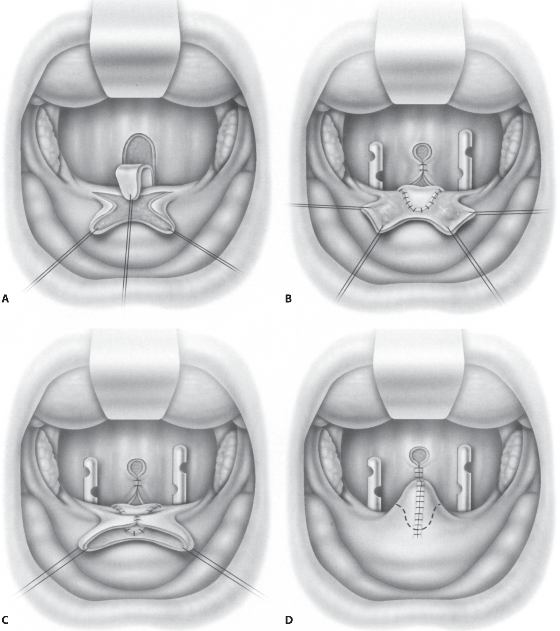
- Principle: A midline flap of mucosa + muscle raised from the posterior pharyngeal wall, based superiorly, and inset into the soft palate. This creates a central bridge that physically obstructs the midline of the velopharynx, leaving two lateral ports on either side.
- Best for:
- Large velopharyngeal gaps
- Sagittal or circular closure pattern with good lateral wall motion (lateral walls must close over the lateral ports)
- Adynamic velopharyngeal mechanisms (poor or absent velar movement)
- All cases of VPI as a versatile option
- Port size control: Catheters placed during surgery to calibrate lateral port size (adequate for breathing but small enough to reduce hypernasality)
- Complication: Risk of obstructive sleep apnea (OSA) - all patients require postoperative airway monitoring; CPAP may help while edema resolves
- Can be revised if ports are too wide (persistent hypernasality) or too narrow (hyponasality/OSA)
D. Sphincter Pharyngoplasty
- Principle: Bilateral superiorly based myomucosal flaps raised from the posterior tonsillar pillars (palatopharyngeus muscle) on both sides, transposed medially, and sutured to each other or to the posterior pharyngeal wall at the level of VP closure. This creates a central opening (like a Passavant ridge effect) and narrows the lateral ports.
- Best for:
- VPI with lateral velopharyngeal gaps or "bowtie" closure pattern (the two sides can't meet laterally)
- Good velar elevation but poor lateral wall motion
- Can be combined with Furlow Z-plasty for added benefit
- Advantage over pharyngeal flap: Maintains central opening (better nasal breathing); good for patients at higher OSA risk
E. Posterior Pharyngeal Wall Augmentation
- Principle: Injecting or implanting material into the posterior pharyngeal wall to reduce the gap between the wall and velum.
- Materials used: Autologous fat, calcium hydroxyapatite, Teflon (historical), Proplast (historical)
- Best for: Small, central velopharyngeal gaps only
- Limitation: Tendency for migration, extrusion, foreign body reactions over time; not suitable for large gaps
F. V-to-Y Pushback Palatoplasty
- Lengthens the palate by advancing soft palate tissue posteriorly
- An option when primary repair left the palate short
- Less commonly used in modern practice
Choosing the Right Surgery (Summary Table)
| Closure Pattern / Gap Type | Preferred Surgery |
|---|---|
| Short velum, submucous cleft | Furlow Z-plasty or IVV |
| Large midline gap, good lateral wall motion | Superiorly based pharyngeal flap |
| Lateral gaps / bowtie pattern | Sphincter pharyngoplasty |
| Small central gap | Posterior pharyngeal wall augmentation |
| Longitudinally oriented levator | IVV ± Furlow |
| 22q11.2 deletion / complex VPD | Usually pharyngeal flap; manage expectations |
Cummings Otolaryngology, p. 3626-3628; KJ Lee's Essential Otolaryngology, p. 983-984
Post-Operative Care
- All patients need monitoring for airway obstruction after VPD surgery (pharyngeal flap and sphincter pharyngoplasty both narrow the nasopharynx)
- CPAP can bridge the early post-operative period
- Persistent OSA may require revision or widening of ports
- Post-operative speech therapy is essential to eliminate compensatory articulation errors that persist even after anatomical correction
Quality of Life Considerations
Cummings Otolaryngology, p. 3622
Special Situation: Adenoidectomy and VPI
- Always screen for submucous cleft palate before adenoidectomy
- Signs of submucous cleft: bifid uvula, zona pellucidum, palpable notch at posterior hard palate
- If these signs are present, partial adenoidectomy (preserving a lateral adenoid buttress) may reduce VPI risk
- VPD after adenoidectomy occurs in ~1/1500 cases; rhinolalia aperta is usually transient (resolves in weeks-months)
- If persistent (0.01%), surgical intervention is needed
Scott-Brown's Otorhinolaryngology Vol 2; Cummings p. 3621
🔹 Quick Summary for Exam (Simplified Cheat Sheet)
- Causes: Cleft palate (most common), submucous cleft, short palate, adenoidectomy, neurological conditions, 22q11.2 deletion
- Symptom: Hypernasality = rhinolalia aperta + nasal air emission + glottal stops
- Diagnose with: Endoscopy (gold standard) + nasometry + perceptual speech evaluation
- Treat:
- Speech therapy first (for mild/mislearning)
- Surgery for structural causes:
- Furlow Z-plasty = reorient levator + lengthen palate
- Pharyngeal flap = block midline (good lateral walls needed)
- Sphincter pharyngoplasty = narrow lateral gaps
- Augmentation = small gaps only
- Prosthesis = for poor surgical candidates
- Always check for 22q11.2 deletion in unexplained VPD
- Never adenoidectomy without checking for submucous cleft first