
Vibrio chilera tests
Vibrio cholerae TCBS agar culture yellow colonies laboratory
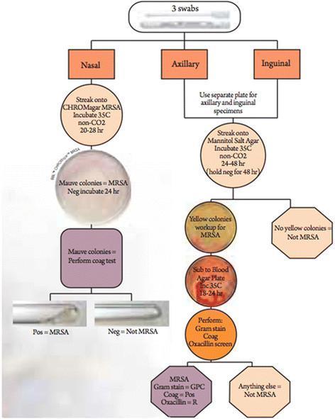
A clinical microbiology flowchart detailing the systematic isolation and identification of Methicillin-resistant Staphylococcus aureus (MRSA) from three anatomical sites: nasal, axillary, and inguinal. The nasal pathway utilizes CHROMagar MRSA, where mauve colonies indicate presumptive MRSA, followed by a confirmatory coagulase plasma test shown in test tubes. The axillary and inguinal pathways use Mannitol Salt Agar (MSA) for initial screening. Yellow colonies on MSA (indicating mannitol fermentation) are subcultured onto Blood Agar Plates for 18-24 hours. Final identification of MRSA requires a battery of tests confirming Gram-positive cocci (GPC) morphology, positive coagulase production, and oxacillin resistance via screening agar. The diagram incorporates photographs of culture plates and biochemical test results to illustrate the diagnostic criteria. This algorithm is designed for laboratory medicine and infectious disease training to demonstrate standard microbial culture protocols and differential media selection based on sample source.
A diagnostic microbiology image showing a fungal culture on an agar slant, specifically Sabouraud Dextrose Agar (SDA). The image illustrates the macroscopic morphology of Purpureocillium lilacinum, characterized by its distinctive lilac-to-pinkish coloration. The colonies are flat and exhibit a densely floccose, velvety texture across the surface of the medium. There is evidence of confluent growth spreading along the slant, with the reverse side (visible at the periphery) appearing off-white to pale yellow. This visual demonstration is critical for identifying hyphomycetes in clinical specimens, particularly in immunocompromised patients presenting with deep-seated mycoses or skin and soft tissue infections. The image highlights the typical maturation and pigmentation used by microbiologists to differentiate this opportunistic pathogen from other filamentous fungi.
This clinical diagnostic image displays three cylindrical glass culture bottles containing Sabouraud's dextrose agar (SDA) slants, used for the isolation and identification of fungal pathogens. The center and right bottles contain patient specimens identifying Madurella mycetomatis, while the left bottle serves as a control. The active fungal cultures exhibit characteristic leathery, light gray to white surface colonies. A hallmark diagnostic feature is the production of a dark brown diffusible pigment that stains the normally translucent yellow agar to a deep red-brown or brownish-black hue. This visual presentation is indicative of Eumycetoma, a chronic subcutaneous fungal infection. The bottles are sealed with metallic screw-top lids (gold and silver-toned), and the third bottle contains a blue marking for identification. Such cultures are essential in differentiating between actinomycotic and eumycotic mycetoma based on colonial morphology and pigment production.
**Imaging Modality:** Laboratory photograph of a microbiological culture on an agar plate. **Anatomical/Biological Entity:** Bacterial culture of *Cedecea neteri* (strain SSMD04). **Observed Pathology/Phenotype:** The image displays bacterial colonies streaked onto a specialized agar medium supplemented with 0.5% corn oil. The primary diagnostic feature is the presence of distinct "halo" zones (zones of clearance or precipitation) surrounding the bacterial growth. **Characteristic Visual Features:** * **Colony Morphology:** Small, circular, opaque white to light-gray colonies distributed along streak lines. * **Enzymatic Activity:** Clear translucency or a discernible halo effect in the agar adjacent to the colonies, indicative of extracellular lipase activity and lipid hydrolysis. * **Growth Pattern:** Standard quadrant streaking technique used to isolate individual colonies. **Diagnostic Significance:** This biochemical assay demonstrates the metabolic capability of *C. neteri* to utilize lipids as a carbon source. The visible halo serves as a positive indicator for lipase production, a key phenotypic trait used in microbial classification and the study of bacterial virulence or industrial potential.
Laboratory Diagnosis of Vibrio cholerae (Cholera)
1. Specimen Collection
- Rubber catheter (No. 26-28) introduced 4-5 cm into rectum - the gold standard under clinical conditions
- Rectal swab - dipped in holding medium before rectal insertion
- In the absence of transport medium, a cotton-tipped swab soaked in liquid stool placed in a sterile plastic bag is acceptable
| Medium | Notes |
|---|---|
| Alkaline peptone water (APW) | pH 8.5-9.0; most commonly used |
| Venkatraman-Ramakrishnan (VR) medium | Classic cholera transport medium |
| Cary-Blair medium | Suitable for rectal swabs; can be refrigerated |
- Park's Textbook of Preventive and Social Medicine, p. 1240
2. Direct Examination
Dark-Field Microscopy
- Vibrios appear as "shooting stars in a dark sky" due to their darting, rapid polar flagellar motility
- Motility stops (immobilization) when a drop of polyvalent anti-cholera diagnostic serum is added - this constitutes a presumptive diagnosis
- Sensitivity improves after 5-6 hours of pre-incubation in alkaline peptone water
Gram Stain
- Gram-negative, comma-shaped curved rods, 1 × 2-4 μm (arrows below)
- On prolonged culture, may straighten and resemble other enteric Gram-negative bacteria
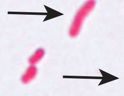
- Jawetz Melnick & Adelbergs Medical Microbiology 28E, p. 268
3. Culture Methods
Enrichment (Pre-plating)
- Inoculate specimen into Peptone Water Tellurite (PWT) or alkaline peptone water at 37°C for 4-6 hours
- A loopful from the surface is then subcultured onto selective plating media
Selective Plating Media
| Medium | V. cholerae Result | Notes |
|---|---|---|
| TCBS agar (Thiosulfate-Citrate-Bile Salts-Sucrose) | Yellow colonies (sucrose fermenter) | Against dark-green background; 2-3 mm glistening colonies |
| Bile Salt Agar (BSA) pH 8.6 | Translucent, moist, raised, smooth colonies ~1 mm | Screened under oblique (Tyndall) illumination |
| MacConkey agar / Blood agar | Grows but non-selective | Used with oxidase test screening |
| TTG agar (Taurocholate-Tellurite-Gelatin) | Grey colonies with hazy/cloudy zones (gelatinase) | Used in Harrison's protocol |
Key point on TCBS: V. cholerae produces yellow colonies (sucrose positive). V. parahaemolyticus and V. vulnificus produce green colonies (sucrose negative) on the same medium. This is a critical differential point.
- Jawetz Melnick & Adelbergs Medical Microbiology 28E, p. 267; Harrison's Principles of Internal Medicine 22E, p. (Diagnosis section)
4. Biochemical Tests
| Test | Result for V. cholerae | Significance |
|---|---|---|
| Oxidase test | Positive | Key preliminary ID step; all vibrios are oxidase-positive |
| String test | Positive (viscous string forms when colony mixed with 0.5% sodium deoxycholate) | Distinguishes vibrios from other Gram-negatives |
| Sucrose fermentation | Positive (acid, no gas) | Yellow on TCBS |
| Mannose fermentation | Positive | |
| Arabinose fermentation | Negative | |
| Voges-Proskauer (VP) test | Positive (El Tor biotype only) | Differentiates El Tor from Classical biotype |
| Polymyxin B sensitivity | Resistant (El Tor) | Classical biotype is sensitive |
| Hemolysin production | Positive (El Tor) | Classical biotype does not produce hemolysin |
| MALDI-TOF MS | Can discriminate V. cholerae from aeromonads | Useful when culture mimics other organisms |
- Park's Textbook, p. 1244; Jawetz, p. 268; Tietz Textbook of Laboratory Medicine 7th Ed.
5. Serological / Immunological Tests
Slide Agglutination (Serogroup Identification)
- Colonies suspended in 0.85% sterile saline + one drop of polyvalent anti-cholera diagnostic serum
- Agglutination = presumptive V. cholerae
- Further sub-typing with O1-specific and O139-specific antisera
- V. cholerae O1 is further serotyped into: Ogawa, Inaba, and Hikojima (rare)
- Non-agglutinating suspicious colonies are tested by oxidase and string tests
Other Rapid Immunological Tests
| Test | Notes |
|---|---|
| Immunofluorescence | Rapid detection |
| Latex agglutination | Rapid, field-adaptable |
| Immunochromatographic dipstick (POC) | Commercially available; antigen detection; useful in field or resource-limited settings where lab facilities are lacking |
- Park's Textbook, p. 1244; Jawetz, p. 268; Harrison's 22E, Diagnosis section
6. Molecular Tests
-
Multiplex PCR targeting:
- ctxA gene (cholera toxin gene)
- O1-specific rfb gene
- O139-specific rfb gene
-
Used in reference/specialized lab settings
-
Useful when culture is not possible or for rapid outbreak confirmation
-
Yamada's Textbook of Gastroenterology 7th Ed., Differential diagnosis section
Summary Table: Tests at a Glance
| Category | Test | Result |
|---|---|---|
| Direct | Dark-field microscopy (wet mount) | Darting "shooting star" motility |
| Direct | Immobilization with anti-cholera serum | Motility stops |
| Direct | Gram stain | Gram-negative curved rods |
| Culture | TCBS agar | Yellow colonies |
| Culture | BSA / TTG agar | Translucent grey colonies |
| Culture | Enrichment in APW/PWT | Pre-plating step |
| Biochemical | Oxidase test | Positive |
| Biochemical | String test | Positive (viscous thread) |
| Biochemical | VP test | Positive (El Tor only) |
| Serological | Slide agglutination (O1/O139 antisera) | Agglutination confirms serogroup |
| Rapid/POC | Dipstick antigen detection | Field-applicable |
| Molecular | Multiplex PCR (ctxA, rfb O1/O139) | Genetic confirmation |
- Positive VP test (El Tor positive, Classical negative)
- Polymyxin B resistance (El Tor resistant, Classical sensitive)
- Hemolysin production (El Tor positive)