
Breast
What aspect of the breast would you like to learn about?
breast cancer staging TNM mammography
ductal carcinoma in situ DCIS breast invasive lobular
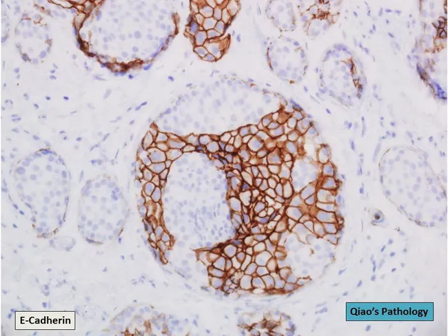
This immunohistochemistry slide of breast tissue was prepared for E-cadherin staining and captured by brightfield microscopy at high magnification. The specimen shows a ductal compartment containing ducts with cohesive epithelial cells consistent with ductal carcinoma in situ (DCIS) that demonstrate strong membranous brown staining for E-cadherin. Adjacent lobular-type cellular nests show absent or markedly reduced E-cadherin staining, compatible with lobular carcinoma in situ (LCIS). Notably, one duct exhibits a hybrid region in which both DCIS and LCIS phenotypes are present within the same duct, illustrating intraductal heterogeneity. The DCIS component forms well-defined, angulated outlines; the LCIS component is less cohesive, with smaller, uniform cells and loose cell packing. The staining pattern confirms the differential adhesion molecule expression between ductal and lobular neoplasia. Clinically, E-cadherin immunostaining is critical for accurate classification of in situ lesions, guiding surveillance, surgical margins, and adjuvant therapy decisions. This image is valuable for educational comparisons of DCIS versus LCIS morphology and for research on combined in situ phenotypes, clonal relationships, and diagnostic workflows in breast pathology. It may aid differential diagnosis against invasive carcinoma and aid optimization of pathology reporting and multidisciplinary management. Clinical correlation with imaging findings and margin status should be integrated into reporting.
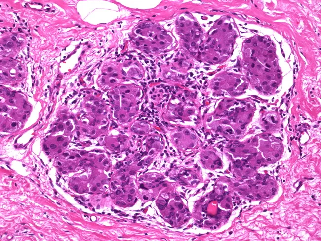
This is a histopathology micrograph of breast tissue stained with Hematoxylin and Eosin (H&E) at high magnification, illustrating apocrine-type ductal carcinoma in situ (DCIS) with extension into lobular structures. The image highlights solid and cribiform ductal proliferations composed of large polygonal cells with abundant eosinophilic, granular cytoplasm, prominent nuclei, and conspicuous nucleoli, consistent with apocrine differentiation. These neoplastic apocrine cells are arranged within ductular and lobulocentric spaces, and are embedded in fibrous stroma with focal sclerosing adenosis, raising potential mimics of invasive carcinoma if the myoepithelial boundary is disrupted. The apocrine DCIS demonstrates an in-situ pattern without overt stromal invasion. In practice, E-cadherin immunostaining may be used to differentiate this entity from lobular neoplasia: apocrine DCIS typically preserves membranous E-cadherin expression, whereas lobular carcinoma in situ shows absent or fragmented staining. Myoepithelial markers (p63, smooth muscle actin, calponin) can aid in confirming an in-situ process by highlighting an intact/basal myoepithelial layer surrounding ducts and lobules. This image underscores the diagnostic challenge when apocrine DCIS extends into sclerosing adenosis and may simulate invasive carcinoma; careful histomorphology combined with ancillary immunohistochemistry is essential for accurate classification and subsequent clinical management. Ancillary testing and multidisciplinary review guide appropriate surgical and adjuvant therapy decisions.
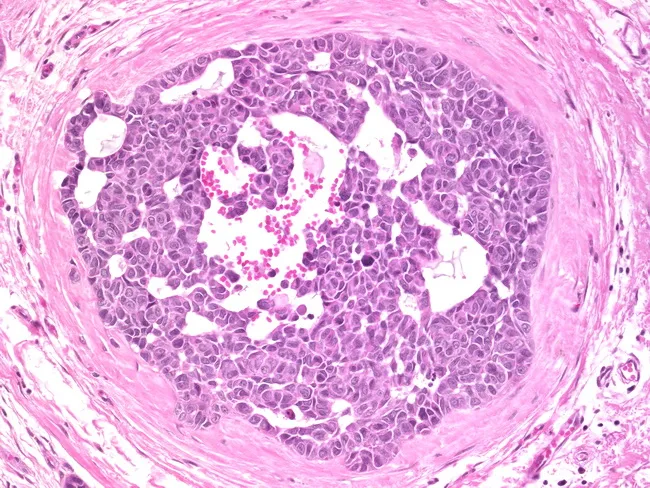
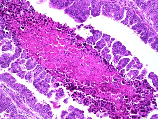
Histopathology slide of breast tissue; transverse cross-section through a breast duct; densely packed neoplastic ductal epithelial cells fill the lumen; cells exhibit high nuclear grade with marked pleomorphism, hyperchromasia, conspicuous nucleoli; the cytoplasm is relatively abundant and borders are sharply demarcated. The epithelial proliferation forms solid/cribriform arrangements; the surrounding basement membrane appears intact; there is no obvious stromal invasion. Central luminal material may show eosinophilic debris consistent with necrosis in some ducts. The tissue architecture is characteristic of ductal carcinoma in situ, high-grade subtype. Diagnostic significance: Non-invasive precursor lesion with potential progression to invasive cancer; high-grade DCIS carries higher risk and often requires surgical excision with clear margins and consideration of radiotherapy; may be associated with microcalcifications on imaging. Differential considerations: low-grade DCIS, cribriform or comedo patterns, invasive ductal carcinoma without invasion; lobular neoplasia unlikely due to ductal architecture; clinical correlation with mammography and breast imaging is essential. Clinical use cases: pathology teaching slides, diagnostic review, AI training for histopathology DCIS recognition, radiologic-pathologic correlation, surveillance planning. Expressed terms include intraductal, ductal carcinoma in situ, high-grade, solid, cribriform, pleomorphic; emphasizes need for margin assessment and multidisciplinary review.
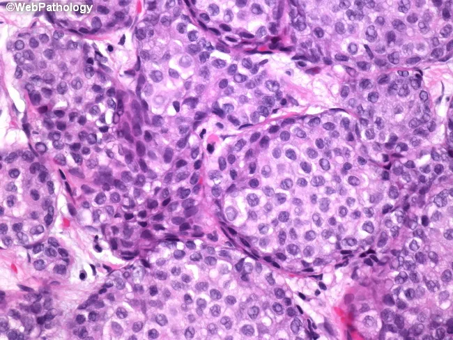
Brightfield histopathology image of breast tissue stained with hematoxylin and eosin (H&E). The specimen shows lobular units filled by small, uniform cells with round to oval nuclei, inconspicuous nucleoli, and scant cytoplasm. Cells are discohesive and expand the terminal duct-lobular units without forming ductal structures, a characteristic pattern of lobular carcinoma in situ (LCIS). In this case, classic LCIS is observed without cytologic atypia, pleomorphism, increased mitotic activity, or necrosis; features that would otherwise designate pleomorphic LCIS when present alone or in combination. The architecture remains confined to lobules with preservation of basement membrane and no evidence of stromal invasion. Background breast parenchyma shows mild fibrous stroma and occasional benign ductal hyperplasia. The image emphasizes in-situ proliferation of mammary epithelial cells that express loss of cohesion and a monomorphic cytologic appearance. Clinically, classic LCIS indicates a marker of increased risk for concurrent or subsequent breast carcinoma in either breast and may influence surgical and surveillance management. Diagnostic significance lies in distinguishing classic LCIS from pleomorphic LCIS and from other in-situ or invasive lesions such as ductal carcinoma in situ (DCIS). This histologic pattern underscores the importance of careful histopathologic assessment and correlates with imaging and clinical risk assessment.
Breast Cancer: Types, Staging & Treatment
Epidemiology
Robbins, Cotran & Kumar Pathologic Basis of Disease, p. 975-976
Risk Factors
| Category | Factors |
|---|---|
| Hormonal | Early menarche, late menopause, nulliparity, delayed childbearing, exogenous estrogen use |
| Genetic | BRCA1, BRCA2, PALB2, TP53 mutations (~12% of all breast cancers) |
| Age | Risk rises rapidly after 30 years |
| Imaging history | Prior atypical hyperplasia or LCIS on biopsy |
| Lifestyle | Obesity, alcohol use, reduced breastfeeding |
Robbins Basic Pathology, p. 719
In Situ Carcinomas (Precursors)
Ductal Carcinoma In Situ (DCIS)
- Non-invasive proliferation of malignant cells within ducts, basement membrane intact
- Most often detected on mammography as microcalcifications
- A true precursor - untreated DCIS progresses to invasive ductal carcinoma in the same breast
- High-grade DCIS (comedo type) shows central necrosis
Lobular Carcinoma In Situ (LCIS)
- Proliferation of discohesive cells filling the terminal duct-lobular units
- Acts as a bilateral risk marker - when carcinoma develops, 2/3 are ipsilateral, 1/3 contralateral
- Cells characteristically lose E-cadherin expression (used in IHC to differentiate from DCIS)
Robbins Basic Pathology, p. 719
Invasive Carcinoma Types
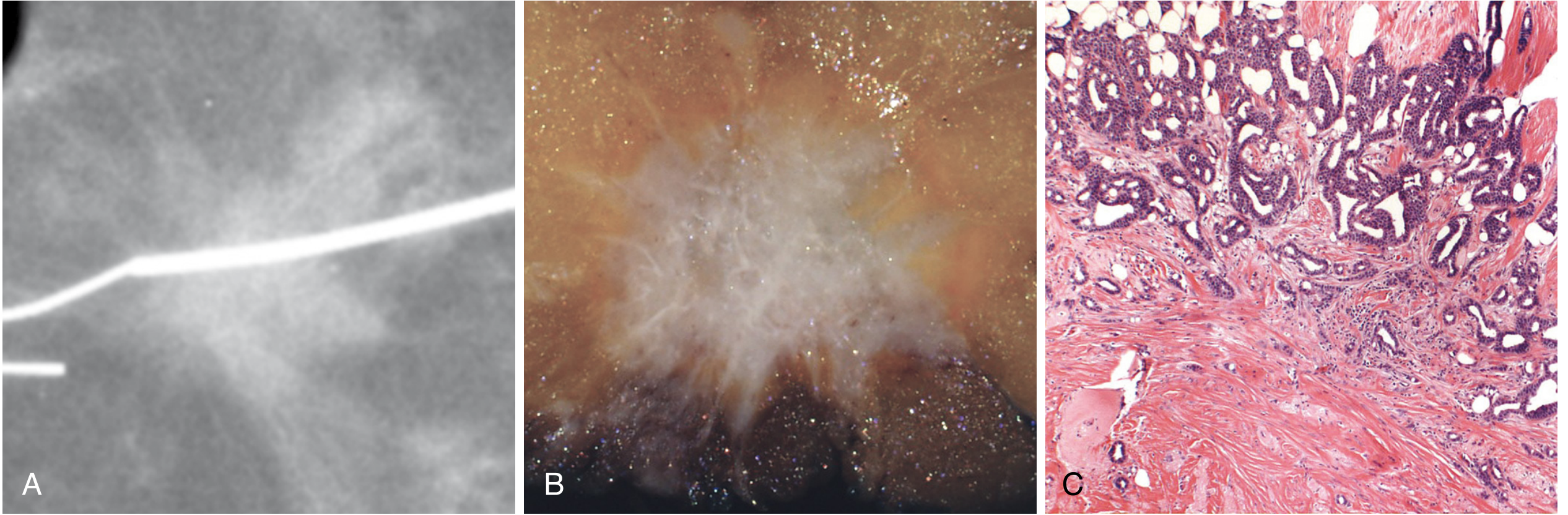
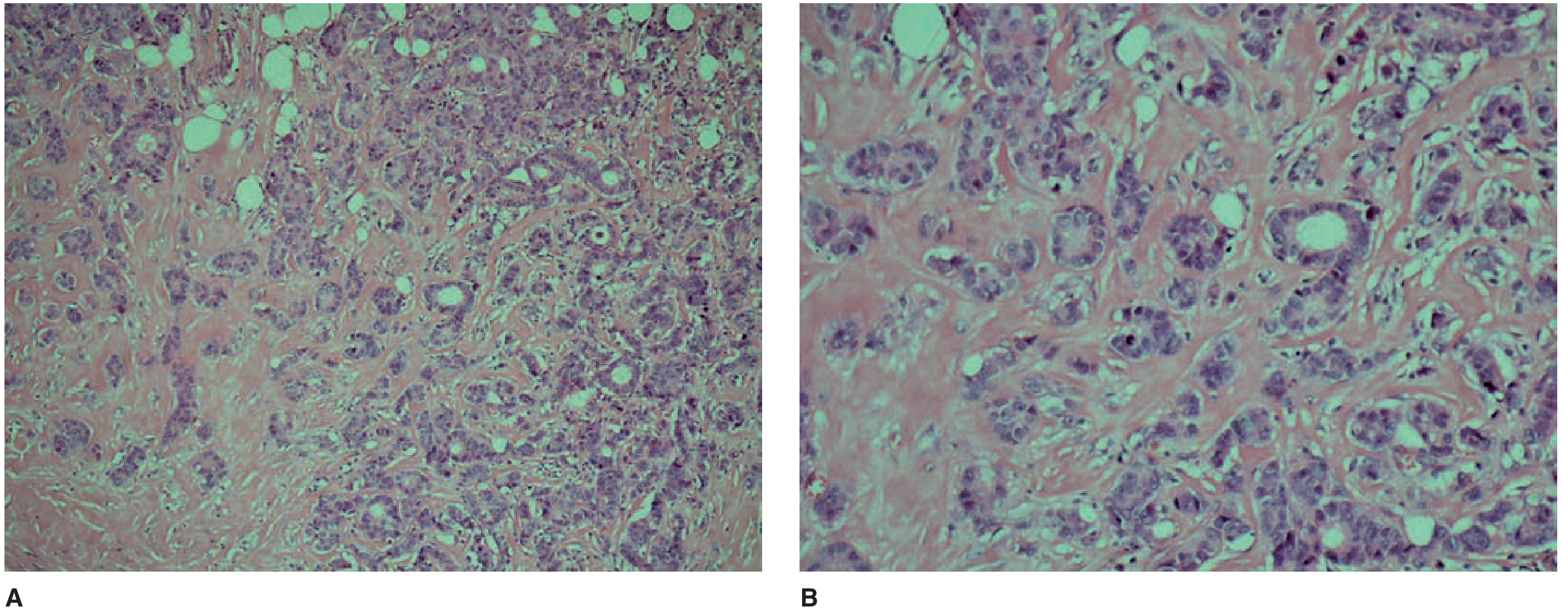
1. Invasive Ductal Carcinoma - No Special Type (NST) - ~75-80%
- Most common subtype
- Presents as a hard, irregular mass with spiculated margins on imaging
- Shows desmoplastic stromal reaction; produces a grating sound on cut surface
- Worse prognosis than special-type cancers
2. Invasive Lobular Carcinoma - ~10%
- Second most common type
- Characterized by single-file (Indian file) infiltration pattern
- Often bilateral and multifocal
- Loss of E-cadherin; tends to metastasize to unusual sites (peritoneum, GI tract, meninges)
3. Special-Type Carcinomas (~10% total)
| Type | % | Key Features | Prognosis |
|---|---|---|---|
| Medullary carcinoma | 4% | Dense lymphocytic infiltrate, pleomorphic nuclei, sheet-like growth; frequent in BRCA1 carriers | Better than NST |
| Mucinous (colloid) | 2% | Extracellular mucin pools surrounding low-grade cells; >90% ER+ | Favorable |
| Tubular carcinoma | 2% | Haphazard tubular elements; ~94% ER+; often found on screening | Excellent |
| Papillary carcinoma | 2% | Papillae with fibrovascular stalks; ~87% ER+; elderly women | Favorable |
| Paget's disease of nipple | Rare | Eczematous nipple lesion; underlying DCIS or invasive cancer; large pale Paget cells in epithelium | Variable |
| Inflammatory breast carcinoma | ~1-3% | Dermal lymphatic invasion; peau d'orange; no discrete mass; worst prognosis | Poor |
Schwartz's Principles of Surgery, pp. 592-595
Molecular/Biological Classification
| Group | Receptor Profile | Key Biology | Treatment Implications |
|---|---|---|---|
| Luminal A | ER+/PR+, HER2-, low grade | Most common; driven by estrogen signaling | Endocrine therapy; low chemo benefit |
| Luminal B | ER+/PR+, HER2- or HER2+, high grade | Higher proliferation (Ki-67) | Endocrine + consider chemo |
| HER2-enriched | ER-/PR-, HER2+ | HER2 amplification/overexpression | HER2-targeted therapy (trastuzumab, pertuzumab) |
| Triple-Negative (TNBC) | ER-/PR-, HER2- | Genomically unstable; BRCA1/TP53 mutations common | Chemotherapy ± immunotherapy; no endocrine or HER2 target |
Robbins, Cotran & Kumar Pathologic Basis of Disease, p. 975
Histologic Grading (Nottingham Score)
| Component | Score 1 | Score 2 | Score 3 |
|---|---|---|---|
| Tubule formation | >75% tubules | 10-75% | <10% |
| Nuclear pleomorphism | Uniform small nuclei | Moderate variation | Marked variation |
| Mitotic rate | Low | Moderate | High |
- Grade 1 (3-5 points): Well differentiated - tubular/cribriform growth, uniform nuclei
- Grade 2 (6-7 points): Moderately differentiated
- Grade 3 (8-9 points): Poorly differentiated - ragged sheets, irregular nuclei, necrosis
Staging (AJCC 8th Edition TNM)
Anatomic Staging
| Stage | T | N | M | Description |
|---|---|---|---|---|
| 0 | Tis | N0 | M0 | In situ only (DCIS/LCIS) |
| I | T1 (≤2 cm) | N0 | M0 | Node-negative, small tumor |
| IIA | T0-T1 | N1 | M0 | Small tumor + 1-3 LN; or T2 N0 |
| IIB | T2 | N1 | M0 | 2-5 cm + 1-3 LN; or T3 N0 |
| IIIA | T0-T3 | N2 | M0 | 4-9 axillary LN; or T3 N1 |
| IIIB | T4 | N0-N2 | M0 | Chest wall/skin invasion |
| IIIC | Any T | N3 | M0 | ≥10 axillary LN or IMA nodes |
| IV | Any T | Any N | M1 | Distant metastasis |
Sabiston Textbook of Surgery, p. 1392
Diagnosis
Triple Assessment
- Clinical examination - palpation, inspection for skin changes (peau d'orange, retraction)
- Imaging - mammography (first line); ultrasound (young women, dense breasts); MRI (high-risk patients, extent of disease)
- Biopsy - core needle biopsy preferred for histology + receptor testing
Key Biomarkers
- ER/PR (by IHC): Guide endocrine therapy decisions
- HER2 (IHC + FISH): Guide anti-HER2 therapy
- Ki-67: Proliferation index; helps distinguish Luminal A vs B
- Oncotype DX (21-gene): Predicts chemotherapy benefit in ER+/HER2-/N0 patients
- CA 15-3, CA 27.29, CEA: Used for monitoring metastatic disease, not screening
Treatment Overview
Surgery
| Procedure | Indication |
|---|---|
| Breast-conserving surgery (BCS/lumpectomy) + radiotherapy | Early-stage (Stage I-II); equivalent survival to mastectomy |
| Simple mastectomy | DCIS, prophylactic, patient preference |
| Modified radical mastectomy | Larger/locally advanced tumors |
| Sentinel lymph node biopsy (SLNB) | Clinically node-negative; avoids full axillary dissection |
| Axillary lymph node dissection (ALND) | Clinically positive nodes, failed SLNB |
Systemic Therapy
- Pre-menopausal: Tamoxifen (SERM) ± ovarian suppression (GnRH agonist)
- Post-menopausal: Aromatase inhibitors (anastrozole, letrozole, exemestane) - superior to tamoxifen
- Duration: 5-10 years adjuvant therapy
- Indicated for triple-negative, HER2+, high-risk ER+, and node-positive disease
- Common regimens: AC (doxorubicin + cyclophosphamide), CMF, AC-T (with taxane), TC
- Neoadjuvant chemotherapy (pre-surgical): Allows breast conservation, assesses response; pathologic complete response (pCR) is a major prognostic milestone
- Trastuzumab (Herceptin) + pertuzumab - first-line for HER2+ disease
- Neratinib, lapatinib (TKIs) for later lines
- T-DM1 (ado-trastuzumab emtansine) - for residual disease after neoadjuvant therapy
Radiation Therapy
- Standard after BCS (whole-breast RT reduces local recurrence)
- Post-mastectomy RT for T3/T4 tumors or ≥4 positive nodes
- Partial breast irradiation for selected early-stage cases
Special Entities
Inflammatory Breast Cancer (IBC)
- Rapid-onset breast erythema, warmth, peau d'orange without a discrete mass
- Due to dermal lymphatic invasion by tumor emboli
- Classified as Stage IIIB at minimum (T4d)
- Treatment: Neoadjuvant chemotherapy first, then surgery + radiation
Male Breast Cancer
- ~1% of all breast cancers
- Almost always invasive ductal carcinoma; usually ER+
- BRCA2 mutation is the most significant genetic risk factor in males
- Treatment mirrors female breast cancer; tamoxifen preferred over AIs (due to testosterone conversion)
BRCA-Associated Cancers
- BRCA1: Triple-negative phenotype, Grade 3, younger onset
- BRCA2: ER+, later onset, similar to sporadic breast cancer
- Prophylactic bilateral mastectomy reduces risk by ~90-95%
Prognosis
- Molecular subtype (TNBC worst; Luminal A best)
- Anatomic stage (nodal status most critical)
- Response to neoadjuvant therapy (pCR strongly favorable)
Sources: Robbins, Cotran & Kumar Pathologic Basis of Disease | Schwartz's Principles of Surgery, 11th Ed. | Sabiston Textbook of Surgery | Robbins Basic Pathology