
nerve monitoring in ent
intraoperative nerve monitoring ENT surgery facial nerve recurrent laryngeal nerve 2024
facial nerve monitoring EMG intraoperative ENT surgery
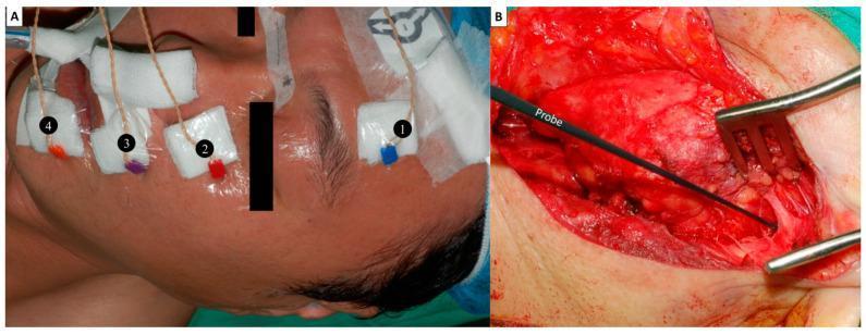
Two-panel clinical photograph illustrating an intraoperative facial nerve (FN) monitoring setup. (A) Lateral view of a patient's face showing the placement of four paired subdermal needle electrodes for electromyography (EMG) recording. The electrodes are secured with white gauze and transparent tape at four anatomical sites corresponding to FN branches: 1) lower forehead (temporal branch), 2) infraorbital area (zygomatic branch), 3) superolateral upper lip (buccal branch), and 4) inferolateral lower lip (marginal mandibular branch). Connectors are color-coded (blue, red, purple, orange) for channel identification. (B) Intraoperative view of a parotid gland dissection. A black 1.0 mm ball-tip monopolar stimulating probe is positioned within the surgical field to identify the FN trunk via electrical localization. Surgical retractors are visible, exposing the deep tissue layers. This setup demonstrates the methodology for continuous neural monitoring to prevent iatrogenic nerve injury during head and neck surgery.
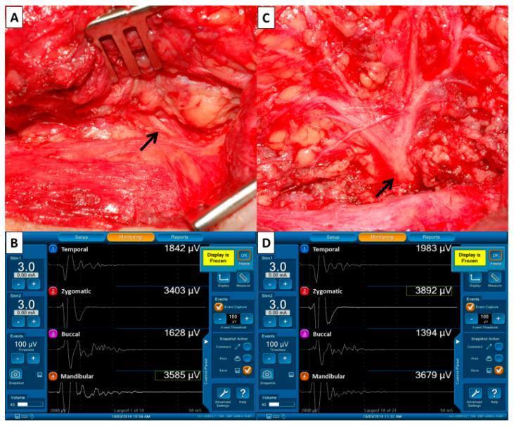
A multi-panel medical image illustrating intraoperative facial nerve monitoring (FNM) during parotid surgery. Panel A is a clinical photograph of the surgical field showing the identification of the facial nerve trunk (arrow) amidst soft tissue and temporo-parotid fascia. Panel C shows the same surgical field following dissection and parotid tumor resection, where the facial nerve trunk and its distal branches are more clearly visualized. Panels B and D display corresponding four-channel electromyography (EMG) monitoring screens. The channels record electrical activity from the temporal, zygomatic, buccal, and marginal mandibular branches. Waveforms and peak amplitudes (measured in µV) are shown for each branch: Panel B (F1) represents baseline signals pre-dissection, and Panel D (F2) represents signals post-dissection. This comparison is used to evaluate nerve integrity and function throughout the procedure. The interface highlights stimulus intensity (3.0 mA), event thresholds (100 µV), and real-time amplitude variations indicative of possible neuropraxia or successful nerve preservation.
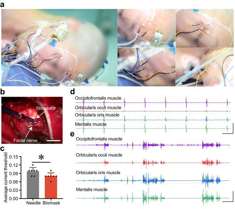
This multi-panel figure demonstrates intraoperative facial nerve monitoring during vestibular schwannoma surgery. Panel A shows clinical photographs of a patient's face in the surgical position, illustrating the concurrent placement of traditional needle electrodes and a novel, flexible 'Biomask' sensor array over key facial muscles, including the occipitofrontalis, orbicularis oculi, orbicularis oris, and mentalis. Panel B is a close-up intraoperative photograph showing an electrical stimulator probe applied to the exposed facial nerve (indicated by a dashed white line). Panel C provides a quantitative comparison of stimulation current thresholds between the two monitoring systems, indicating lower thresholds for the Biomask. Panels D and E display electromyography (EMG) waveforms recorded by the Biomask. Panel D shows rhythmic, stimulation-induced EMG signals, while Panel E illustrates erratic, high-frequency waveforms typical of mechanical traction or surgical manipulation of the nerve. The figure evaluates the clinical efficacy of flexible microneedle arrays in providing real-time neurophysiological feedback during neurosurgery to prevent iatrogenic nerve injury.
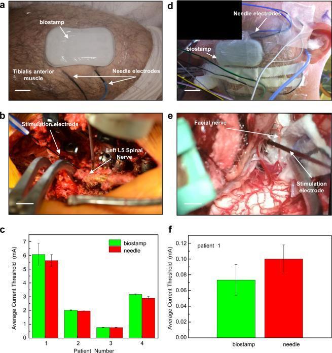
A multi-panel clinical and diagnostic composite demonstrating intraoperative electromyography (EMG) monitoring during spinal and cranial nerve surgeries. (a) Clinical photograph of the lower leg showing the placement of a flexible 'biostamp' sensor on the upper left tibialis anterior muscle and conventional needle electrodes positioned distally. (b) Intraoperative view of a spinal surgical field exposing the left L5 spinal nerve, with a stimulation electrode in direct contact with the neural tissue. (c) Bar graph comparing average current thresholds (mA) between biostamp and needle electrodes for four spinal surgery patients, showing comparable performance. (d) Clinical photograph of the lateral face showing biostamp and needle electrode placement over the facial muscle for cranial nerve monitoring. (e) Intraoperative view of a cranial surgical site showing the exposed facial nerve and an associated stimulation electrode. (f) Bar graph of average current thresholds for a cranial surgery patient, illustrating statistically similar results between the wearable biostamp and traditional needle-based monitoring systems. The composite illustrates the clinical utility of thin, stretchable biosensors in specialized neurosurgical environments.
Intraoperative Nerve Monitoring in ENT Surgery
1. Nerves Monitored in ENT
| Nerve | Surgery | Monitoring Method |
|---|---|---|
| Facial nerve (CN VII) | Parotidectomy, mastoidectomy, CPA tumor, cochlear implant | Free-run EMG + triggered EMG |
| Cochlear nerve (CN VIII) | Vestibular schwannoma, posterior fossa | ABR, DENM, electrocochleography |
| Recurrent laryngeal nerve (RLN) | Thyroid, parathyroid surgery | Endotracheal tube EMG |
| Superior laryngeal nerve (SLN) | Thyroid surgery | ETT EMG |
| Trigeminal nerve (CN V) | CPA/Meckel's cave tumors | Masseter/temporalis EMG |
| Glossopharyngeal (CN IX), Vagus (CN X) | Jugular foramen tumors | Cricothyroid EMG, CoMEP |
| Spinal accessory (CN XI) | Neck dissection, CPA | Trapezius needle EMG |
| Hypoglossal (CN XII) | Skull base surgery | Tongue needle EMG |
2. Facial Nerve Monitoring (CN VII)
Why It Matters
EMG Modalities
- Free-run EMG (FEMG) - Continuous real-time recording with sweep durations of 200 ms to 5 seconds. Captures spontaneous activity triggered by mechanical, thermal, or electrical irritation.
- Triggered EMG (TEMG) - Records and saves spontaneous responses that exceed a preset voltage (typically 100 µV), with an audible alarm alerting the surgeon.
- Stimulated EMG (SEMG) - Records evoked responses to a hand-held monopolar or bipolar stimulating probe. Used for nerve identification and integrity testing.
Electrode Configuration
- Monopolar: Active needle electrodes placed in target facial myotomes (orbicularis oculi for temporal/zygomatic branch; orbicularis oris/mentalis for buccal/marginal mandibular branch) with a neutral reference electrode on the contralateral forehead.
- Bipolar: Both active and reference electrodes placed within the same muscle - less noise, more specificity.
- Frontalis (temporal branch)
- Orbicularis oculi (zygomatic branch)
- Orbicularis oris (buccal branch)
- Mentalis/depressor anguli oris (marginal mandibular branch)
Commercial Systems
Nerve Stimulation for Mapping
- Smaller tumors (CPA component ≤1 cm): Start at brainstem entry zone and stimulate at 3× threshold across tumor surface before dissection.
- Larger tumors: Sweep at 0.3 mA, then 0.5 mA, then 1.0 mA. No response at 1.0 mA = nerve not on exposed surface; safe to dissect.
- Once response obtained: Reduce to 0.1-0.2 mA to narrow the nerve location precisely.
Corticobulbar Motor-Evoked Potentials (CoMEP)

Ephaptic Response (for Hemifacial Spasm)
Anesthesia Considerations
3. Indications for Facial Nerve Monitoring
| Setting | Evidence/Recommendation |
|---|---|
| CPA tumor (acoustic neuroma) surgery | Standard of care; reduces complete permanent paralysis; CMAP amplitude after resection correlates with immediate postoperative function |
| Revision mastoidectomy | Established; distorted anatomy increases risk |
| Parotidectomy (especially malignancy, revision) | Useful for nerve identification; may reduce temporary palsy even if permanent palsy rates unchanged |
| Cochlear implantation | Widely practiced |
| Congenital ear abnormalities | Established |
| Middle ear/primary mastoidectomy | Recommended; cost-effective; monitoring both primary and revision surgeries has greatest effectiveness and lowest cost (Wilson et al.) |
4. Recurrent Laryngeal Nerve (RLN) Monitoring
Technique
Clinical Value
- Provides real-time assessment of nerve function during thyroid/parathyroid surgery.
- Facilitates intraoperative nerve identification - reduces inadvertent injury risk.
- Guides decision-making for contralateral exploration when ipsilateral RLN signal is lost.
- Loss of IONM signal has variable positive predictive value (10-90%) for postoperative vocal cord palsy, and signal loss does not always correlate to permanent dysfunction.
- Continuous (C-IONM) vs. intermittent (I-IONM): Continuous monitoring allows real-time detection of traction injury as it develops, not just after the fact.
Limitations
- Requires general anesthesia and specialized equipment.
- NMBA must be avoided (same rule as for facial nerve monitoring).
- Endotracheal tube must be positioned precisely - rotation can cause false-negative results.
- Evidence unequivocally demonstrating reduction in permanent RLN palsy rate is still limited by study heterogeneity.
5. Cochlear / Auditory Nerve (CN VIII) Monitoring
Methods (in order of increasing proximity to nerve):
- Auditory Brainstem Response (ABR) - Most commonly used. Click or tone stimulus via insert earphone; Wave V latency and amplitude tracked. Described since 1971; easy to apply, low cost, good localization. Wave V prolongation or loss warns of cochlear nerve compromise.
- Direct Eighth Nerve Monitoring (DENM) / Direct nerve action potential - Electrode placed on the cochlear nerve or within the internal auditory canal. More sensitive than ABR; identifies neural continuity even when ABR is absent.
- Cochlear Nucleus Surface Recordings - Electrode on cochlear nucleus in brainstem; useful in rare circumstances.
- Electrocochleography (ECoG) - Electrode as close to cochlea as possible; records compound action potential from most distal cochlear nerve activity.
- Evoked Otoacoustic Emissions (OAEs) - Proposed as an adjunct; less commonly used intraoperatively.
6. Other Cranial Nerve Monitoring (Jugular Foramen / Skull Base)
- CN V (trigeminal - motor): Needle electrodes in masseter or temporalis.
- CN X (vagus): Direct laryngoscopy for vocalis muscle needle placement, or ETT surface electrodes. CoMEP from cricothyroid muscle.
- CN XI (spinal accessory): Needle electrodes in trapezius.
- CN XII (hypoglossal): Needle electrodes in tongue.
- Direct nerve monitoring: A C-shaped electrode secured around vagus, spinal accessory, or hypoglossal nerve in the neck provides direct nerve action potential recording - used for skull base tumors.
7. Key Technical Pitfalls and Caveats
| Pitfall | Solution |
|---|---|
| Electrocautery artifact | Cease monitoring briefly during cautery near nerve; use bipolar cautery |
| NMB not reversed | Confirm train-of-four recovery before beginning nerve dissection |
| False-negative stimulation | Always confirm system is working at start (stimulate a known structure) |
| ETT rotation (RLN monitoring) | Reposition and confirm electrode contact before relying on signals |
| Reduced anesthesia depth | Diffuse multi-channel EMG increase = anesthesia issue, not nerve stimulation |
| Repetitive vs. non-repetitive responses | Repetitive = nerve irritability/thermal injury; non-repetitive = direct mechanical stimulation |
8. Evidence Summary and Current Status
- IONM is standard for CPA tumor surgery and increasingly routine for thyroid, parotid, and otologic procedures.
- The 2021 Laryngoscope Best Practices guideline recommends routine FNM for otologic surgery as cost-effective and safety-improving.
- A 2024 review in J Clin Med (PMID 38673494) by Brunet et al. confirms IONM is well-established for thyroid/parathyroid surgery and expanding evidence supports its use in esophageal and tracheal surgery for RLN preservation.
- No RCT has definitively proven reduction in permanent facial nerve or RLN palsy rates, but retrospective and cohort data consistently support the technique for nerve identification, trainee safety, and medico-legal protection.