
Complications of chronic otitis media (squamousal type) in detail with their respective management in detail and in simplified manner from standard ent textbooks
cholesteatoma chronic otitis media squamous type complications
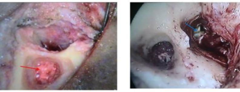
Two intraoperative otologic endoscopic photographs illustrating pathological findings during a tympanomastoidectomy for chronic otitis media. The first image (left) shows a red arrow pointing to a localized area of beefy red, friable-appearing granulation tissue within the mastoid cavity or middle ear space. The second image (right) displays a blue arrow indicating a light-colored, whitish, keratin-filled cholesteatoma sac situated within a retraction pocket, surrounded by inflamed mucosa and surgical debris. These images demonstrate the surgical landmarks and pathological manifestations of squamous disease, emphasizing the difference between vascular granulation tissue and the characteristic opaque appearance of a cholesteatoma. This clinical imaging serves as an educational resource for identifying middle ear pathologies during mastoid surgery and understanding the surgical management of chronic middle ear infections.
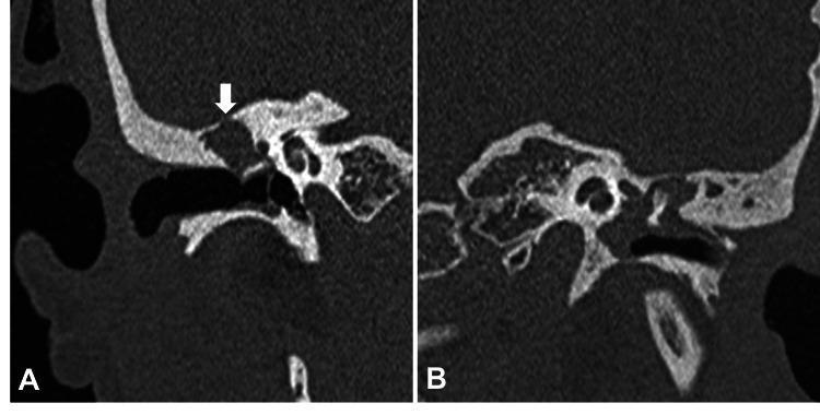
Diagnostic imaging: Two reformatted coronal CT scans of the temporal bone demonstrating complications of chronic suppurative otitis media (CSOM) with cholesteatoma. Panel A shows the right middle ear where a non-dependent, expansive soft tissue density occupies the attic and middle ear space. A white arrow highlights focal bony erosion of the tegmen tympani, indicating potential intracranial extension. Panel B displays the left middle ear, where a similar soft tissue density is present, resulting in the erosion and deossification of the ossicular chain, specifically involving the head of the malleus. These images illustrate the hallmark radiologic features of cholesteatoma, including non-dependent soft tissue masses and characteristic destructive patterns of adjacent bony structures in the middle ear and mastoid. Clinical relevance includes the preoperative assessment of ossicular integrity and the evaluation of potential tegmen defects in patients with middle ear disease.
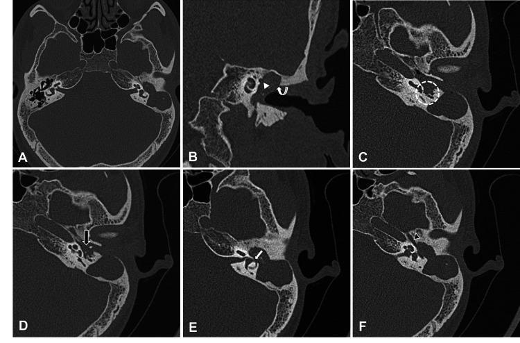
A multi-panel series of High-Resolution Computed Tomography (HRCT) images of the temporal bone in axial (A, C, D, E, F) and coronal (B) planes, demonstrating complications of Chronic Suppurative Otitis Media (CSOM) with cholesteatoma. Panel A shows a large, well-defined soft tissue density in the left middle ear extending into the aditus and mastoid antrum, causing ossicular destruction (malleus and incus) and loss of the 'ice cream cone' configuration. Panel B (coronal) highlights erosion of the scutum (curved arrow) and the inferior wall of the facial canal (arrowhead). Further panels illustrate specific structural involvement: extension into the sinus tympani and facial recesses (C, dashed circle), stapes destruction and mastoid cortex dehiscence with dural sinus plate thinning (D, black arrow), a focal defect in the lateral semicircular canal indicating a perilymphatic fistula (E, white arrow), and soft tissue extension into the eustachian tube (F, black arrowhead). The series serves as a diagnostic guide for identifying bony erosion patterns and soft tissue distribution characteristic of middle ear cholesteatoma.
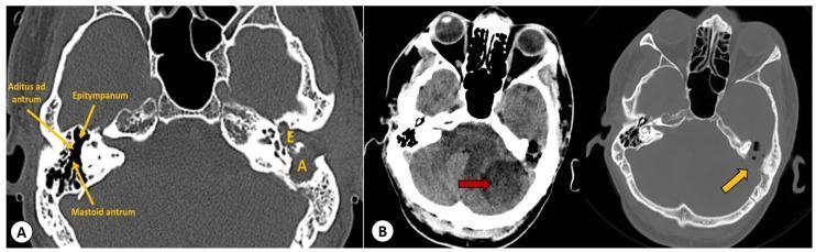
This medical image consists of axial CT scans of the head and temporal bones illustrating complications of chronic suppurative otitis media with cholesteatoma. Panel A provides a bone-window view of the skull base, comparing the normal right middle ear anatomy (epitympanum, aditus ad antrum, and mastoid antrum) with the pathological left side. The left ear shows characteristic findings of cholesteatomatous otomastoiditis, including widening of the aditus and the formation of a confluent, soft-tissue-filled cavity between the epitympanum and aditus. Panel B presents two axial slices at the level of the posterior fossa. The soft-tissue window (left) highlights a well-defined hypodense lesion (red arrow) in the left cerebellar hemisphere, consistent with an intracranial abscess. The corresponding bone-window slice (right) demonstrates erosion of the posterior petrous wall of the temporal bone (yellow arrow), indicating a direct communication pathway between the mastoid infection and the posterior cranial fossa. These images illustrate the progression from middle ear disease to life-threatening intracranial complications.
Complications of Chronic Otitis Media - Squamous Type (Unsafe/Dangerous Type / COM with Cholesteatoma)
Sources: Cummings Otolaryngology Head & Neck Surgery, Shambaugh Surgery of the Ear, KJ Lee's Essential Otolaryngology
Background
Classification of Complications
| Category | Complications |
|---|---|
| Extracranial (Intratemporal) | Conductive/SNHL, Coalescent Mastoiditis, Subperiosteal Abscess, Bezold's Abscess, Labyrinthine Fistula, Labyrinthitis, Facial Nerve Paralysis, Petrous Apicitis |
| Intracranial | Meningitis, Extradural (Epidural) Abscess, Subdural Empyema, Brain Abscess (Temporal Lobe / Cerebellar), Lateral (Sigmoid) Sinus Thrombophlebitis, Otic Hydrocephalus |
EXTRACRANIAL COMPLICATIONS
1. Conductive Hearing Loss (CHL) and Sensorineural Hearing Loss (SNHL)
- CHL: Ossicular erosion by cholesteatoma - the incus (long process) is most commonly destroyed. Severity depends on the status of the ossicular chain and the position of the cholesteatoma sac.
- SNHL: Results from secondary suppurative labyrinthitis, or direct loss of cochlear hair cells adjacent to the cholesteatoma. Cochlear fistula is the most common cause of SNHL in this setting.
- Mixed hearing loss is possible when both mechanisms are operative.
- Surgical: Tympanomastoidectomy (canal wall-up or canal wall-down) to eradicate disease. Ossicular chain reconstruction (ossiculoplasty) is performed either at the primary surgery or as a staged second procedure.
- Canal wall-up (intact canal wall) allows better hearing reconstruction but carries a higher risk of residual/recurrent disease (residual 11-27%, recurrent 5-13%); requires second-look surgery or DWI-MRI follow-up.
- Canal wall-down (modified radical mastoidectomy) has lower recurrence (2-10%) but hearing reconstruction is more challenging and the mastoid cavity requires lifelong periodic cleaning.
2. Coalescent Mastoiditis
- Persistent otalgia and purulent otorrhea for >2 weeks
- Fever, toxicity, post-auricular tenderness, erythema
- Anterior-inferior displacement of the pinna and obliteration of the post-auricular crease (pointing towards subperiosteal abscess)
- Sagging of the posterosuperior EAC wall
- CT temporal bone: Opacification of mastoid with loss of bony septa (coalescence). Bone window shows destruction of mastoid cortex.
- Enhanced CT or MRI if intracranial extension is suspected (nearly 25% of children with coalescent mastoiditis have concomitant intracranial complications).
- Cortical mastoidectomy with ventilating tube placement + IV broad-spectrum antibiotics.
- Medical management alone (IV antibiotics + tympanostomy tube) may be attempted in early-stage disease without osteitis, but osteitis with bone destruction mandates surgery.
- The underlying cholesteatoma is addressed once the acute infection is controlled.
3. Subperiosteal Abscess
- Fluctuant, tender post-auricular swelling with lateral displacement of the pinna
- Post-auricular skin erythema
- Incision and drainage of the abscess + cortical mastoidectomy + IV antibiotics.
- Culture-directed antibiotic therapy (common organisms: S. pneumoniae, S. pyogenes, S. aureus, Pseudomonas in COM).
4. Bezold's Abscess
- Deep neck swelling below the mastoid tip
- Neck stiffness and torticollis
- Can track into the posterior triangle or parapharyngeal space
- Surgical drainage of the neck abscess + mastoidectomy + prolonged IV antibiotics.
- CT neck with contrast to delineate the extent of the abscess before surgery.
5. Labyrinthine Fistula
- Symptoms of the underlying cholesteatoma (foul-smelling otorrhea)
- Vertigo (only in ~60% of patients with a fistula)
- Positive fistula test (Hennebert's sign) - only 30-50% sensitivity
- Tullio phenomenon (vertigo induced by loud sounds)
- SNHL in 60-70% (especially cochlear fistula)
- Often asymptomatic - so the surgeon must always assume possible fistula in cholesteatoma surgery
- Fistula test: high specificity, low sensitivity.
- CT temporal bone: Only ~60% of confirmed fistulas are visible preoperatively.
- Definitive diagnosis is only made intraoperatively.
-
Option A (Conservative - Leave matrix in place): Canal wall-down procedure; leave the cholesteatoma matrix intact over the labyrinth. Removing the bulk of the cholesteatoma sac eliminates pressure and reduces further erosion. This is the preferred approach for large fistulae and cochlear fistulae. Complete removal increases risk of SNHL significantly.
-
Option B (Complete removal + fistula repair): Remove cholesteatoma entirely and repair the fistula with bone wax, fascia/bone paste (pâté). Eliminates infection and continued erosion. Preferred for small fistulae without perilymph exposure. Do NOT use for cochlear fistulae.
-
If the endosteum is torn during surgery → immediately convert to canal wall-down and leave matrix in place.
-
High incidence of concurrent facial nerve dehiscence and tegmen dehiscence in cases with labyrinthine fistula - requires high surgical alertness.
6. Labyrinthitis
- Serous labyrinthitis: Toxins diffuse through intact membranes - reversible.
- Suppurative labyrinthitis: Direct bacterial invasion - leads to complete loss of cochlear and vestibular function (dead ear + profound vertigo).
- Sudden severe vertigo, nausea, vomiting
- Rapid and profound SNHL (dead ear in suppurative type)
- Loss of vestibular function
- Medical: IV antibiotics (broad-spectrum); steroids for serous type.
- Surgical: Urgent tympanomastoidectomy to eradicate the source. Labyrinthectomy may be needed for suppurative labyrinthitis with dead ear.
- Vestibular rehabilitation after resolution of acute phase.
7. Facial Nerve Paralysis
- Erosion of the bony fallopian canal by cholesteatoma, particularly the tympanic segment (which has congenital dehiscences in up to 30% of people, making it vulnerable).
- Can also occur acutely from infection (bacterial exotoxins, inflammatory edema → avascular necrosis of axons).
- In COM with cholesteatoma: usually a slow, progressive course (paresis → paralysis).
- Ipsilateral lower motor neuron facial palsy (House-Brackmann grading)
- COM findings in the ear (with or without active discharge)
- Pain and facial spasm are typically absent (distinguishes from Bell's palsy)
- Slow progression is characteristic of cholesteatoma-induced paralysis vs. sudden onset in AOM
- Clinical: House-Brackmann scale
- CT temporal bone: Evaluates extent of canal erosion, labyrinthine involvement, tegmen dehiscence.
- IV antibiotics (broad-spectrum, transitioning to culture-directed oral therapy)
- Urgent tympanomastoidectomy:
- Remove matrix and granulation tissue from epineurium
- Decompress facial nerve proximal and distal to the dehiscent segment
- Opening the epineurium is not required unless cholesteatoma has actually invaded the nerve sheath
- Outcome: Recovery to House-Brackmann grade I-II in 52-83% of patients. Prognosis depends heavily on whether axonal degeneration has occurred prior to surgery.
8. Petrous Apicitis (Gradenigo's Syndrome)
- Retro-orbital (deep ear/facial) pain (CN V irritation)
- Sixth cranial nerve (abducens) palsy - ipsilateral diplopia
- Otorrhea
- IV antibiotics for prolonged duration (essential)
- Surgical drainage when necrotic bone is apparent - routes depend on available air cell pathways:
- Posterior to posterior semicircular canal
- Subcochlear/infralabyrinthine approach
- Middle cranial fossa approach (if other routes inadequate)
- The petrous apex cannot be fully excised due to proximity of the labyrinth and carotid artery; partial apicectomy reduces bacterial load enough for antibiotics to control disease.
INTRACRANIAL COMPLICATIONS
- Direct extension through eroded bone (most common with cholesteatoma)
- Hematogenous spread (retrograde thrombophlebitis)
- Through preformed pathways (Hyrtl's fissure, perivascular spaces)
- Persistent/severe headache, high fever, meningism (neck stiffness, Kernig's, Brudzinski's signs)
- Altered consciousness (lethargy to coma)
- Focal neurological deficits (seizures, hemiplegia, aphasia, cerebellar signs)
- Papilledema, diplopia, cranial nerve deficits
9. Meningitis
- High fever, severe headache, neck stiffness
- Positive Kernig's and Brudzinski's signs
- Photophobia, phonophobia
- CSF: raised protein, low glucose, neutrophilic pleocytosis
- High-dose broad-spectrum IV antibiotics (adjusted per CSF culture results)
- Urgent tympanostomy tube/tympanocentesis to obtain material for culture
- Dexamethasone (adjunct to reduce inflammation)
- Neurosurgical consultation
- Cortical mastoidectomy or tympanoplasty with mastoidectomy once the patient is stabilized - to eradicate the source
10. Extradural (Epidural) Abscess
- Often subtle/occult, discovered incidentally at mastoid surgery
- Headache, fever, persistent otorrhea
- Usually no focal neurological signs unless the dura is breached
- IV broad-spectrum antibiotics
- Surgical drainage: Mastoidectomy with careful debridement of granulation tissue and pus from the epidural space
- Neurosurgical involvement if the dura is involved
- Follow-up enhanced MRI post-treatment
11. Subdural Empyema
- Rapidly progressive fever, headache, meningism
- Focal neurological signs: hemiplegia, seizures
- Deteriorating consciousness
- Urgent neurosurgical drainage (craniotomy or burr hole) - primary priority
- IV antibiotics (prolonged course)
- Mastoidectomy to eradicate the primary source (staged or simultaneous depending on clinical condition)
- Follow-up MRI to confirm resolution
12. Otogenic Brain Abscess
- Direct spread from the cholesteatoma/mastoid into the adjacent temporal lobe (most common)
- Posteriorly into the cerebellum via perisinus routes
- Via adjacent complications (lateral sinus thrombophlebitis, petrositis, meningitis)
- Temporal lobe abscess: Most common
- Cerebellar abscess: Via posterior fossa extension
- Initially insidious (malaise, low-grade fever) → gradual deterioration
- Headache, papilledema, raised ICP
- Temporal lobe signs: receptive aphasia (dominant side), homonymous hemianopia, personality changes
- Ipsilateral cerebellar signs: dysdiadochokinesia, dysmetria, intention tremor, ataxia, nystagmus
- Nuchal rigidity
- CT/MRI brain with contrast: ring-enhancing lesion with surrounding edema
- CSF culture may be negative if abscess is deep
- IV broad-spectrum antibiotics (prolonged course - 4-6 weeks)
- Surgical drainage of the abscess: Aspiration (stereotactic or open) vs. excision - depending on size, location, and stage
- In certain cases, long-term parenteral antibiotics alone may suffice
- Mastoidectomy to eradicate the primary source - staged: mastoidectomy first → allow inflammation to resolve → definitive cholesteatoma removal
- Staged third surgery often required to ensure no residual disease
13. Lateral (Sigmoid) Sinus Thrombophlebitis
- "Picket fence" (spiking/hectic) fever - pathognomonic
- Septicemia, rigors, chills
- Nausea and vomiting
- Headache (retro-orbital or occipital)
- Tenderness over the jugular vein in the neck (Griesinger's sign - edema over mastoid emissary vein)
- Papilledema if propagation causes raised ICP
- MRI with gadolinium (MRV): Shows absence of flow signal/filling defect in sigmoid/lateral sinus ("empty delta sign" on contrast CT)
- Blood cultures: May grow causative organism
- IV antibiotics (broad-spectrum; prolonged 6-week course)
- Mastoidectomy - to drain the perisinus abscess and remove the cholesteatoma
- Anticoagulation (heparin then warfarin): controversial but generally recommended for propagating or septic thrombus
- Surgical ligation or excision of the internal jugular vein is rarely needed today
- Follow-up MRV to confirm sinus recanalization
14. Otic Hydrocephalus (Otitic Hydrocephalus)
- Headache, papilledema
- Blurred vision, diplopia (CN VI palsy from raised ICP)
- No meningism, no fever
- Normal or near-normal CSF findings
- Treat the underlying ear infection (mastoidectomy)
- Lumbar puncture - therapeutic and diagnostic
- Acetazolamide or repeated LPs to control ICP
- In severe cases: optic nerve sheath fenestration to protect vision
Surgical Approach Summary for Squamosal COM with Complications
| Situation | Preferred Surgery |
|---|---|
| Cholesteatoma, no complications | Canal wall-up (CWU) or canal wall-down (CWD) based on extent |
| Labyrinthine fistula (small) | CWU/CWD + complete removal + fistula repair |
| Labyrinthine fistula (large/cochlear) | CWD + leave matrix over labyrinth |
| Facial nerve paralysis | Urgent tympanomastoidectomy + nerve decompression |
| Intracranial complication | Mastoidectomy (radical/modified radical) to eradicate source; neurosurgical co-management |
| Active infection + cholesteatoma | Stage 1: mastoidectomy; Stage 2 (several months later): definitive cholesteatoma clearance |
| Subperiosteal abscess | I&D + cortical mastoidectomy |
| Bezold's abscess | Neck drainage + mastoidectomy |
Key Bacteriology
- Aerobic: Pseudomonas aeruginosa (31%), S. aureus (19%), Proteus mirabilis
- Anaerobic: Peptococcus (12%), Bacteroides spp. (12%), Peptostreptococcus, Clostridium
- Cummings Otolaryngology Head & Neck Surgery, Chapters 140-141 (Chronic Otitis Media with Cholesteatoma; Complications of Otitis Media)
- Shambaugh Surgery of the Ear, Chapter 26 (Complications of Chronic Otitis Media)
- KJ Lee's Essential Otolaryngology, Chapter on Facial Nerve Paralysis & Labyrinthine Fistula in COM