
Urolithiasis - Pathology, Medicine, Surgery
Reading File
Urolithiasis (kidney stones) involves stone formation in the urinary tract from supersaturation of urine with lithogenic substances. The four main stone types are calcium oxalate/phosphate (~80%), uric acid (~10%), struvite (~10%), and cystine (~1%). Clinically it presents with colicky flank pain, hematuria, and nausea. Management depends on stone size and location: conservative (hydration, analgesics, alpha-blockers) for stones <10mm, or intervention (ESWL, ureteroscopy, PCNL) for larger or obstructing stones.
Searching sources...
Finding Sources
Searching PubMed
"urolithiasis"[MeSH Terms] AND "management"[MeSH Terms]
Excellent results. Now I'll read the key textbook blocks in parallel - Robbins for pathology, Bailey & Love for surgery, and Brenner & Rector / Campbell Walsh for medicine/nephrology/urology.
Reading File
Reading File
Reading File
Good start. Now I need the Robbins pathology section on urolithiasis specifically, plus deeper content from Brenner & Rector and Bailey & Love's surgery section on adult urolithiasis.
Finding Sources
Reading File
Finding Sources
Now I'll read the Robbins Basic Pathology section (most focused pathology content) and Bailey & Love adult urolithiasis section in parallel.
Reading File
Reading File
Good content flowing in. Now I need to read more of Bailey & Love for surgical management details, and get the Robbins full pathology block with Table 12.5 and morphology.
Reading File
Reading File
I got the Bailey & Love surgical content. Now let me read the full Robbins Basic Pathology urolithiasis section (the correct block) and more of Bailey & Love for treatment details.
Reading File
Reading File
Excellent - now I have rich content. Let me read the last part of Bailey & Love for PCNL/ureteroscopy details and get the clinical features from Robbins.
Reading File
Reading File
I now have comprehensive content from three major textbooks. Let me also quickly fetch the key stone image from Robbins and get the Bailey & Love stone type details.
Loading Image
Reading File
I now have comprehensive content from all three disciplines. Let me update todos and compile the full response.---
Urolithiasis - Comprehensive Review
1. PATHOLOGY
Definition
Urolithiasis refers to calculus formation at any level of the urinary collecting system; most calculi arise in the kidney (nephrolithiasis). It is the common final manifestation of a variety of underlying pathophysiologic processes - not a single diagnosis. - Brenner & Rector's The Kidney, p. 1277
Epidemiology
-
Lifetime prevalence varies from 1-20%; rising steadily over four decades in Western populations.
-
By age 70, approximately 11% of men and 5.6% of women in the United States will have developed a symptomatic kidney stone.
-
Recurrence is high: ~50% within the first decade of diagnosis.
-
Men are twice as likely to form stones; peak incidence in the 4th-6th decades. Women show a bimodal peak (3rd decade and postmenopause).
-
White individuals are at higher risk than other ethnic groups.
-
Strong family history: patients with a positive family history are 2.5x more likely to develop stone disease.
-
Robbins & Kumar Basic Pathology, p. 527; Bailey & Love's Surgery 28th ed., p. 1496
Pathogenesis
Stone formation results from a cascade of events:
- Supersaturation - When urinary concentration of lithogenic solutes (calcium, oxalate, uric acid, cystine) exceeds their solubility, crystallization is favored.
- Inhibitor failure - Normal urine contains inhibitors of crystal growth: citrate, potassium, magnesium, Tamm-Horsfall mucoprotein. When these are depleted or overwhelmed, nucleation proceeds.
- Crystal nucleation and growth - Crystals either pass with urine flow or anchor onto renal papillae forming Randall's plaques - subepithelial calcium phosphate deposits at the papillary tips that serve as a nidus.
- pH effects - Acidic urine (pH <5.5) favors uric acid and cystine stone precipitation. Alkaline urine favors calcium phosphate and struvite stone formation.
- Stasis - Urinary stasis promotes crystal aggregation, producing multiple round "milk of calcium" stones.
- Organic matrix - All stone types contain ~2.5% organic mucoprotein matrix by weight.
- Bailey & Love's Surgery 28th ed., p. 1496; Robbins & Kumar Basic Pathology, p. 527
Stone Types
| Stone Type | Prevalence | Key Metabolic Factors | Urine pH | Appearance/Notes |
|---|---|---|---|---|
| Calcium oxalate ± phosphate | ~80% | Hypercalciuria (idiopathic 50%), hypercalcemia, hyperoxaluria, hyperuricosuria, hypocitraturia | Any (CaOx); alkaline (CaPO4) | Hard, spiculated; radiopaque |
| Struvite (Mg-NH3-PO4) | ~10% | Urease-producing UTI (Proteus, Klebsiella, Serratia) | Alkaline | Staghorn calculi; radiopaque |
| Uric acid | 6-9% | Gout, myeloproliferative disorders, persistently acidic urine (pH <5.5) | Acidic | Radiolucent on plain X-ray |
| Cystine | 1-2% | Autosomal recessive cystinuria (defective proximal tubular reabsorption) | Acidic | Very hard (disulfide bonds); fail SWL |
| Other | ~1-2% | Drug-induced (e.g. indinavir, corticosteroids) | Variable | Indinavir stones are radiolucent even on CT |
- Robbins & Kumar Basic Pathology, Table 12.5, p. 527-528; Bailey & Love's Surgery, p. 1496-1497
Calcium Stones - Causes in Detail
- Absorptive hypercalciuria: excessive intestinal calcium absorption - most common mechanism
- Renal hypercalciuria: primary tubular reabsorption defect
- Primary hyperparathyroidism: most common hypercalcemic cause - PTH increases bone resorption and activates 1,25-dihydroxyvitamin D3, driving intestinal calcium absorption
- Hyperoxaluria: enteric (after small bowel disease/resection - increased colonic oxalate absorption) or primary (rare enzyme defect)
- Hypocitraturia: citrate normally chelates calcium in urine, inhibiting crystallization; distal RTA is a key cause
- Hyperuricosuria: uric acid crystals act as nidi for calcium oxalate aggregation
Struvite Stones
- Exclusively occur with urease-producing bacteria (Proteus, Klebsiella, Serratia, Enterobacter)
- Urease hydrolyzes urea to CO2 and ammonium, alkalinizing urine
- Staghorn calculi fill and branch through the entire pelvicalyceal system
- Can grow to massive size before detection; cause chronic infection, obstructive uropathy, and renal failure
- Complete clearance is mandatory - residual fragments cause rapid recurrence
Uric Acid Stones
- 50% of uric acid stone formers have neither hyperuricemia nor increased urine urate - they simply excrete persistently acidic urine (pH <5.5)
- Radiolucent on plain radiograph; visible on CT
- Regional variation: highest prevalence in Middle East and some European countries
Cystine Stones
- Autosomal recessive defect in renal and intestinal amino acid transport (cystine, ornithine, lysine, arginine - COLA)
- Insoluble at physiologic pH; worsens with acidic urine
- Extremely hard due to disulfide bonds; resistant to ESWL
Morphology
-
Unilateral in ~80% of patients
-
Common sites: renal pelvis, calyces, bladder
-
Average size: 2-3 mm; may be smooth or jagged
-
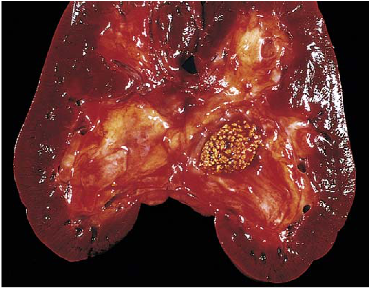
Staghorn calculi: progressive accretion forms branching cast of the pelvicalyceal system; almost always struvite
-
Stones cause mucosal ulceration and hemorrhage; obstruction leads to hydronephrosis
-
Robbins & Kumar Basic Pathology, p. 528
2. MEDICINE (Clinical Assessment & Medical Management)
Clinical Presentation
-
Asymptomatic: increasingly diagnosed incidentally on imaging (ultrasound, CT) - particularly large pelvic stones
-
Ureteric colic (most common acute presentation): sudden-onset, excruciating, colicky flank pain radiating to the groin, scrotum/labia or inner thigh; caused by hyperperistalsis of ureteric musculature against the obstructing stone
- Small 3-5 mm calculi lodge most commonly at the ureterovesical junction (UVJ)
- Lower ureteric/UVJ stones cause urgency, frequency, dysuria
-
Hematuria: gross or microscopic, present in most cases during colic
-
Calculuria: passing of sand or gravel
-
Nausea/vomiting: due to shared celiac innervation
-
Fever + rigors: suggests concurrent UTI/urosepsis - a surgical emergency
-
Malaise, weight loss: in longstanding infection stones or renal failure
-
Bailey & Love's Surgery 28th ed., p. 1497; Robbins & Kumar Basic Pathology, p. 528
Differential Diagnosis of Ureteric Colic
- Urinary: clot colic (anticoagulation, haemophilia), papillary necrosis (diabetes, NSAIDs, sickle cell disease)
- Surgical: acute appendicitis, bowel obstruction, AAA
- Gynaecologic: ectopic pregnancy, ovarian torsion
- Malingering (opioid-seeking behaviour)
Investigations
Emergency setting:
- Urinalysis: hematuria (present in majority); pyuria (sterile or infected); urine pH
- Urine culture: if infection suspected or planned intervention
- Bloods: FBC (leukocytosis suggests infection), serum creatinine, electrolytes, serum calcium, uric acid, phosphorus; pregnancy test in women
- Plain KUB radiograph: detects radiopaque stones (calcium, struvite); misses uric acid and cystine stones
- Ultrasound: detects stones in kidney and UVJ; preferred first-line in children and pregnant women (avoids radiation)
- Non-contrast CT (NCCT): gold standard - detects virtually all stone types except indinavir; determines stone size, location, and degree of obstruction
Non-emergency/metabolic evaluation:
- 24-hour urine: calcium, oxalate, uric acid, citrate, sodium, creatinine, phosphate, volume
- Serum: PTH (if hypercalcemic), uric acid
- Stone analysis: ideally analyze all passed or retrieved stones
- Risk stratification into low-risk (single stone, no metabolic abnormality) vs. high-risk formers
Medical Management
Acute episode (ureteric colic):
- Analgesia: NSAIDs (first-line; e.g., diclofenac) + paracetamol; antispasmodics are not necessary
- Opioids: for refractory pain
- IV hydration: if unable to tolerate orally or signs of sepsis
- Antibiotics: broad-spectrum empirical coverage if sepsis suspected; cultures before starting
Medical expulsive therapy (MET):
- Alpha-blockers (tamsulosin): causes smooth muscle relaxation of distal ureteric wall; used for distal ureteric stones >5 mm and post-ESWL fragment passage
- Calcium channel blockers: also reduce dysmotive ureteric contractions
- Spontaneous passage rates: nearly all stones <4 mm pass; ~50% of 5-10 mm stones pass; >10 mm stones rarely pass spontaneously
Stone-specific pharmacological prevention:
| Stone Type | Pharmacological Prevention |
|---|---|
| Calcium oxalate (hypercalciuria) | Thiazide diuretics (hydrochlorothiazide) to reduce urinary calcium |
| Calcium oxalate (hypocitraturia) | Potassium citrate (alkalinizes urine, increases citrate) |
| Uric acid | Potassium citrate/sodium bicarbonate (alkalinize urine to pH 6.5-7.0); allopurinol if hyperuricemic |
| Struvite | Urease inhibitors (acetohydroxamic acid); antibiotic chemoprophylaxis post-clearance |
| Cystine | High fluid intake; D-penicillamine or tiopronin; urine alkalinization |
General preventive measures (all patients):
-
Fluid intake >2.5 litres/day (target urine output >2 L/day)
-
Dietary calcium should NOT be restricted (paradoxically increases oxalate absorption); supplemental calcium at mealtimes
-
Reduce animal protein and salt intake
-
Reduce oxalate-rich foods (spinach, nuts, chocolate) in hyperoxaluric patients
-
Bailey & Love's Surgery 28th ed., p. 1498-1499; Brenner & Rector's The Kidney, p. 1319
3. SURGERY
Indications for Intervention
-
Failure of conservative/medical management
-
Impaired or deteriorating renal function
-
Complete ureteric obstruction
-
Chronic/recurrent infection (staghorn calculi, matrix calculi)
-
Urosepsis with obstruction (emergency decompression)
-
High-risk occupation (pilots, sailors, long-distance drivers) or remote geographical location
-
Patient preference
-
Bailey & Love's Surgery 28th ed., p. 1498
Emergency Urinary Decompression
When urosepsis or complete obstruction is present, immediate decompression takes priority over stone removal:
- Ureteric stenting (JJ/double-J stent): retrograde approach; provides internal drainage
- Percutaneous nephrostomy (PCN): antegrade approach; preferred when retrograde approach fails or is not feasible
- Stone removal (e.g., ureteroscopy) can be attempted in select stable patients without infection
Extracorporeal Shock Wave Lithotripsy (ESWL)
- Non-invasive; introduced by Christian Chaussy in 1980
- Mechanism: acoustic pulse waves focused on the stone (localized by fluoroscopy and/or ultrasound); stone fragments by direct mechanical stress from the shock wave and indirect cavitation (bubble collapse)
- Indications: renal and ureteric stones <2 cm; first-line for most renal stones <2 cm (unless lower-pole anatomy unfavorable)
- Contraindications: pregnancy, uncorrected bleeding diathesis, aortic/renal artery aneurysm, morbid obesity (depth to stone >10 cm), pacemaker (relative), distal obstruction, acute UTI
- Complication - Steinstrasse ("street of stones"): row of stone fragments lining the distal ureter after ESWL, particularly with high stone burden; usually pass spontaneously but may obstruct
- "Clinically insignificant residual fragments" (CIRFs) = ≤4 mm post-treatment; still 20-40% fail to clear and form a nidus
Ureteroscopy (URS) / Retrograde Intrarenal Surgery (RIRS)
- Rigid URS: for ureteric stones (proximal ureter to UVJ)
- Flexible URS / RIRS: for intrarenal stones; allows access to all calyces
- Energy sources: holmium:YAG laser (gold standard - most efficient; works on all stone types); also pneumatic, electrohydraulic, ultrasonic
- Indications: ureteric stones at any level; renal stones <2 cm; failed ESWL
- Avoided in children <5 years due to ureteric caliber
- Can be done as day-case procedure; does not require a skin incision
Percutaneous Nephrolithotomy (PCNL)
- Stone removal via a dilated percutaneous track (standard: >28 Fr) between skin and pelvicalyceal system
- Typically performed in prone position; posterolateral calyx is the usual entry point
- Localization: fluoroscopy or ultrasound guidance
- Energy sources: ultrasound in combination with pneumatic and laser lithotripsy
Indications for PCNL:
- Renal stones >2 cm
- Lower-pole renal stones with unfavorable anatomy for ESWL
- Failed ESWL or RIRS for renal calculi
- Staghorn calculi (complete clearance is mandatory)
Contraindications:
- Pregnancy, untreated UTI, bleeding diathesis, current anticoagulation
Complications:
- Bleeding (may require selective angioembolization for severe hemorrhage)
- Infection/sepsis
- Pleural violation (supracostal puncture - pneumothorax, hydrothorax)
- Perforation of adjacent organs
Mini-PCNL: uses smaller access tracks (<22 Fr); preferred for smaller stone burdens and children; less bleeding
Endoscopic combined intrarenal surgery (ECIRS): simultaneous flexible ureteroscopy + PCNL; for complex multi-stone disease or stones in challenging locations
Open/Laparoscopic Surgery
- Reserved for complex, infected stones with anatomical abnormalities (e.g. horseshoe kidney, pelvic kidney, kyphoscoliosis) where endourological access is impossible
- Procedures: pyelolithotomy (stones in renal pelvis), anatrophic nephrolithotomy (complex intrarenal staghorn)
- Rarely performed in modern practice
Special Situation: Pregnancy
-
Renal colic is the most common cause of non-obstetric hospital admission in pregnancy
-
Physiological changes: increased GFR, ureteric dilation from progesterone and uterine compression
-
Imaging: ultrasound first-line; MRI (no radiation); avoid CT if possible
-
Management: conservative (most stones pass spontaneously); ureteric stent or PCN if obstruction; ESWL is contraindicated; URS can be performed if necessary
-
Bailey & Love's Surgery 28th ed., p. 1498-1500
Summary Table
| Feature | Calcium Oxalate | Struvite | Uric Acid | Cystine |
|---|---|---|---|---|
| Prevalence | 60-80% | ~10% | 6-9% | 1-2% |
| pH | Any (CaOx); alkaline (CaPO4) | Alkaline | Acidic <5.5 | Acidic |
| X-ray | Radiopaque | Radiopaque (staghorn) | Radiolucent | Faintly opaque |
| CT | Visible | Visible | Visible | Visible |
| Key cause | Hypercalciuria, hyperoxaluria | UTI (Proteus, Klebsiella) | Gout, myeloproliferative | Cystinuria (AR) |
| ESWL | Yes | Difficult (hard) | Yes | No (very hard) |
| Medical Rx | Thiazides, K-citrate | Urease inhibitors | K-citrate, allopurinol | D-penicillamine, tiopronin |
Sources:
- Robbins & Kumar Basic Pathology, 10th ed. (Table 12.5, p. 527-528)
- Robbins, Cotran & Kumar Pathologic Basis of Disease, 10th ed.
- Bailey & Love's Short Practice of Surgery, 28th ed. (Chapter 82, p. 1496-1500)
- Brenner & Rector's The Kidney, 2-Vol Set (Chapter 38, p. 1277-1319)
- Campbell-Walsh-Wein Urology---
Urolithiasis - Comprehensive Review
1. PATHOLOGY
Definition
Urolithiasis refers to calculus formation at any level of the urinary collecting system; most calculi arise in the kidney (nephrolithiasis). It is the common final manifestation of a variety of underlying pathophysiologic processes - not a single diagnosis per se - and although the stones are localized to the urinary tract, it is fundamentally a systemic disorder. - Brenner & Rector's The Kidney, p. 1277
Epidemiology
-
Lifetime prevalence: 1-20%; rising steadily over four decades in Western populations
-
By age 70: ~11% of men and 5.6% of women in the US will develop a symptomatic kidney stone
-
Recurrence is high: ~50% within the first decade
-
Men are 2x more likely to form stones; peak incidence in the 4th-6th decades; women have a bimodal peak (3rd decade and postmenopause)
-
White individuals have higher risk; family history confers 2.5x risk
-
Estimated economic burden in the United States approaching $4.7 billion by 2030
-
Robbins & Kumar Basic Pathology, p. 527; Bailey & Love's Surgery 28th ed., p. 1496; Brenner & Rector's The Kidney, p. 1277
Pathogenesis
Stone formation follows a cascade:
- Supersaturation - When urinary concentrations of lithogenic solutes exceed their solubility product, crystallization begins
- Inhibitor failure - Normal urine contains crystal-growth inhibitors: citrate, potassium, magnesium, Tamm-Horsfall mucoprotein. When these are depleted or overwhelmed, nucleation proceeds
- Crystal nucleation and anchoring - Crystals either pass with urine or anchor onto renal papillae as Randall's plaques (subepithelial calcium phosphate deposits), which serve as a fixed nidus
- pH effects - Acidic urine (pH <5.5) favors uric acid and cystine precipitation; alkaline urine favors calcium phosphate and struvite formation
- Urinary stasis - Promotes crystal aggregation; stasis stones are typically multiple, round, smooth ("milk of calcium stones")
- Organic matrix - All stone types contain ~2.5% mucoprotein matrix by weight
- Bailey & Love's Surgery 28th ed., p. 1496; Robbins & Kumar Basic Pathology, p. 527
Stone Types
Gross pathology: small calculi impacted in the renal pelvis (Robbins & Kumar Basic Pathology)
| Stone Type | Prevalence | Urine pH | Radiology | Key Cause |
|---|---|---|---|---|
| Calcium oxalate ± phosphate | ~80% | Any (CaOx); Alkaline (CaPO4) | Radiopaque | Hypercalciuria, hyperoxaluria, hypocitraturia |
| Struvite (Mg-NH3-PO4) | ~10% | Alkaline | Radiopaque (staghorn) | UTI - urease producers (Proteus, Klebsiella) |
| Uric acid | 6-9% | Acidic <5.5 | Radiolucent (X-ray); visible on CT | Gout, myeloproliferative disorders, persistently acidic urine |
| Cystine | 1-2% | Acidic | Faintly opaque | Autosomal recessive cystinuria |
- Robbins & Kumar Basic Pathology, Table 12.5, p. 527-528
Calcium Stones - Causes
- Idiopathic hypercalciuria (50% of calcium stone formers): subdivided into absorptive (excessive gut calcium absorption) and renal (tubular reabsorption defect)
- Primary hyperparathyroidism: most common hypercalcemic cause; PTH drives bone resorption and 1,25-dihydroxyvitamin D3 synthesis, increasing intestinal calcium absorption
- Hyperoxaluria: enteric (ileal disease/resection increases colonic oxalate absorption) or primary (rare enzymatic defect)
- Hypocitraturia: distal RTA is the classic cause; citrate normally chelates urinary calcium
- Hyperuricosuria: uric acid crystals act as heterogeneous nidi for calcium oxalate crystallization
- No identified metabolic abnormality in 15-20% of calcium stone formers
Struvite Stones
- Form only in the presence of urease-producing bacteria: Proteus, Klebsiella, Serratia, Enterobacter
- Urease hydrolyzes urea to CO2 and ammonium, raising urine pH and precipitating magnesium-ammonium-phosphate
- Staghorn calculi - branch through the entire pelvicalyceal system; cause progressive renal destruction through chronic infection and obstructive uropathy; complete clearance is mandatory since residual fragments cause rapid recurrence and persistent bacteriuria
Uric Acid Stones
- Radiolucent on plain radiograph (but visible on CT - important diagnostic point)
- 50% of uric acid stone formers have neither hyperuricemia nor increased urinary urate; they simply excrete persistently acidic urine (pH <5.5) - unexplained
- Conditions causing hyperuricosuria: gout, myeloproliferative disorders post-cytotoxic therapy
- Highest geographic prevalence in the Middle East
Cystine Stones
- Autosomal recessive defect in renal proximal tubular and intestinal transport of cystine, ornithine, lysine, arginine (COLA)
- Cystine insoluble even at physiologic pH; worsens with acidic urine
- Very hard due to disulfide bonds - essentially resistant to ESWL
Morphology
-
Unilateral in ~80% of patients
-
Most common sites: renal pelvis, calyces, bladder
-
Typically 2-3 mm; may be smooth or jagged
-
Staghorn calculi: massive branching structures creating a cast of the pelvicalyceal system - almost always struvite
-
Stones cause mucosal ulceration, hemorrhage, and obstruction; the latter leads to hydronephrosis and, if chronic, loss of renal function
-
Robbins & Kumar Basic Pathology, p. 528
2. MEDICINE
Clinical Presentation
- Asymptomatic: increasingly diagnosed incidentally on imaging, especially large pelvic stones
- Ureteric colic: sudden-onset excruciating flank pain radiating to groin, scrotum/labia; caused by hyperperistalsis of ureteric musculature against an obstructing stone
- Small (3-5 mm) calculi most commonly lodge at the ureterovesical junction (UVJ)
- Lower ureteric/UVJ stones cause additional urgency, frequency, dysuria
- Hematuria: gross or microscopic in most cases
- Calculuria: passage of sand or gravel
- Nausea and vomiting: from shared celiac innervation
- Fever and rigors: indicates concurrent UTI/urosepsis - a urological emergency
- Malaise and weight loss: with longstanding infection stones or renal failure
Differential Diagnosis of Ureteric Colic
- Clot colic (anticoagulation, haemophilia), papillary necrosis
- Acute appendicitis, bowel obstruction, abdominal aortic aneurysm
- Ectopic pregnancy, ovarian torsion
- Malingering
Investigations
Emergency:
- Urinalysis: hematuria, pyuria (sterile or infected), pH
- Urine culture: if infection suspected or intervention planned
- Bloods: FBC, serum creatinine, electrolytes, calcium, uric acid; pregnancy test (women)
- Plain KUB: detects radiopaque stones; misses uric acid and most cystine stones
- Ultrasound: first-line in children, pregnant women; detects hydronephrosis and stones at kidney/UVJ
- Non-contrast CT (NCCT): gold standard - detects all stone types except indinavir; best for size, location, and degree of obstruction
Non-emergency/Metabolic Evaluation:
- 24-hour urine: calcium, oxalate, uric acid, citrate, sodium, creatinine, volume
- Serum: PTH (if hypercalcemic), uric acid
- Stone analysis: analyze all passed or retrieved stones
- Stratify into low-risk (single stone, no metabolic abnormality) vs. high-risk formers
Medical Management
Acute (ureteric colic):
- NSAIDs (diclofenac, ketorolac) - first-line analgesic; antispasmodics are not necessary
- Opioids for refractory pain
- IV hydration if unable to tolerate orally or sepsis is present
Medical Expulsive Therapy (MET):
- Alpha-1 blockers (tamsulosin): relaxes smooth muscle of the distal ureter; used for distal ureteric stones >5 mm and post-ESWL fragment passage
- Calcium channel blockers: also reduce dysmotive ureteric contractions while preserving expulsive peristalsis
- Spontaneous passage rates: ~95-100% for stones <4 mm; ~50% for 5-10 mm; rarely for >10 mm
Prevention - Stone-Specific Pharmacotherapy:
| Stone Type | Medication | Mechanism |
|---|---|---|
| Calcium oxalate (hypercalciuria) | Hydrochlorothiazide | Reduces urinary calcium excretion |
| Calcium oxalate (hypocitraturia) | Potassium citrate | Alkalinizes urine; increases citrate; chelates calcium |
| Uric acid | Potassium citrate / NaHCO3 | Alkalinizes urine to pH 6.5-7.0 → uric acid dissolves |
| Uric acid (hyperuricemic) | Allopurinol | Reduces uric acid production |
| Struvite | Acetohydroxamic acid (urease inhibitor); post-clearance antibiotics | Inhibits urease; prevents recurrence |
| Cystine | High fluid intake; D-penicillamine or tiopronin; urine alkalinization | Chelation; reduces cystine crystallization |
General measures (all patients):
-
Fluid intake >2.5 L/day (target urine output >2 L/day)
-
Do NOT restrict dietary calcium (paradoxically increases oxalate absorption - intestinal calcium binds oxalate in the gut and prevents its absorption)
-
Supplemental calcium should be taken at mealtimes
-
Reduce animal protein and sodium intake
-
Bailey & Love's Surgery 28th ed., p. 1498-1499; Brenner & Rector's The Kidney, p. 1319
3. SURGERY
Indications for Surgical Intervention
- Failure of medical/conservative management
- Impaired or deteriorating renal function
- Complete ureteric obstruction
- Chronic or recurrent infection (staghorn calculi, matrix calculi)
- Urosepsis with obstruction (emergency)
- High-risk occupation (pilots, sailors, long-distance locomotive drivers) or remote geographical location
- Patient preference
Emergency Urinary Decompression
When urosepsis or complete obstruction is present, decompression takes priority over stone removal:
- JJ ureteric stent (retrograde; cystoscopy-guided): provides internal drainage
- Percutaneous nephrostomy (PCN) (antegrade): for failed retrograde approach or anatomical difficulty
- In selected stable patients without sepsis, ureteroscopy with stone removal can be performed primarily
Extracorporeal Shock Wave Lithotripsy (ESWL)
- Non-invasive, outpatient procedure; introduced by Christian Chaussy (1980)
- Mechanism: focused acoustic pulse waves (localized by fluoroscopy ± ultrasound) cause stone fragmentation through direct mechanical stress and cavitation (bubble collapse)
- Indications: renal and ureteric stones <2 cm; first-line for most renal stones <2 cm with favorable anatomy
- Absolute contraindications: pregnancy, uncorrected coagulopathy, active UTI, distal obstruction, aortic/renal artery aneurysm
- Relative contraindications: pacemaker, morbid obesity, musculoskeletal deformities, renal anomalies (horseshoe kidney, pelvic kidney)
- Complications:
- Steinstrasse ("street of stones"): row of stone fragments lining the distal ureter; usually pass spontaneously but may obstruct
- "Clinically insignificant residual fragments" (CIRFs, ≤4 mm): 20-40% fail to clear and form a nidus for recurrence
Ureteroscopy (URS) / Retrograde Intrarenal Surgery (RIRS)
- Rigid URS: ureteric stones from proximal ureter to UVJ
- Flexible URS / RIRS: intrarenal stones; access to all calyces
- Energy sources: holmium:YAG laser (gold standard - works on all stone compositions); pneumatic, electrohydraulic, ultrasonic also used
- Indications: ureteric stones at any level; renal stones <2 cm; failed ESWL
- Avoided in children <5 years due to ureteric caliber
- Day-case procedure; no skin incision required
Percutaneous Nephrolithotomy (PCNL)
- Removal of renal stones via a dilated percutaneous track (standard >28 Fr) between skin and pelvicalyceal system
- Position: typically prone; posterolateral calyx used as entry point
- Guidance: fluoroscopy or ultrasound
- Energy sources: ultrasound + pneumatic ± laser
Indications:
- Renal stones >2 cm
- Lower-pole renal stones with anatomy unfavorable for ESWL
- Failed ESWL or RIRS
- Staghorn calculi (first-line; complete clearance mandatory)
Contraindications: pregnancy, untreated UTI, bleeding diathesis, active anticoagulation
Complications:
- Hemorrhage (can require selective angioembolization)
- Sepsis/infection
- Pleural violation (supracostal puncture): pneumothorax, hydrothorax
- Injury to adjacent organs (bowel, spleen, liver)
Mini-PCNL (<22 Fr): lower bleeding risk; preferred for smaller stone burdens and children
ECIRS (Endoscopic Combined Intrarenal Surgery): simultaneous flexible URS + PCNL; for complex, multiple, or anatomically challenging stones
Open/Laparoscopic Surgery
- Reserved for complex/infected stones with anatomical abnormalities preventing endourological access
- Procedures: pyelolithotomy (pelvic stones), anatrophic nephrolithotomy (complex staghorn)
- Rarely performed in the modern era
Special Situation: Pregnancy
-
Renal colic = the most common cause of non-obstetric hospital admission in pregnancy
-
Physiological changes: increased GFR, progesterone-mediated ureteric dilation, uterine compression
-
Imaging: ultrasound first-line; MRI if needed; CT avoided where possible
-
Treatment: conservative (most stones pass spontaneously); ureteric stent or PCN for obstruction; ESWL contraindicated; URS can be done if necessary
-
Bailey & Love's Surgery 28th ed., p. 1499-1500
Quick Reference Summary
| Procedure | Stone Size/Location | Key Point |
|---|---|---|
| Conservative (MET + analgesia) | <10 mm (especially <5 mm) | Tamsulosin for distal ureteric stones >5 mm |
| ESWL | <2 cm renal/ureteric | Non-invasive; fails for cystine; Steinstrasse complication |
| Ureteroscopy / RIRS | <2 cm ureteric/renal | Holmium laser; day-case |
| PCNL | >2 cm renal; staghorn | Prone; most invasive; bleeding risk |
| Ureteric stent / PCN | Any size with sepsis or obstruction | Decompression first, stone removal later |
| Open surgery | Complex anatomy; failed endourology | Rare in modern practice |
Sources used:
- Robbins & Kumar Basic Pathology, 10th ed., Table 12.5, p. 527-528
- Robbins, Cotran & Kumar Pathologic Basis of Disease, 10th ed.
- Bailey & Love's Short Practice of Surgery, 28th ed., Chapter 82, p. 1496-1500
- Brenner & Rector's The Kidney, 2-Vol Set, Chapter 38, p. 1277-1319
- Campbell-Walsh-Wein Urology, 12th ed.
This is a shared conversation. Sign in to Orris to start your own chat.