
Types of arthritis
types of arthritis comparison joint changes
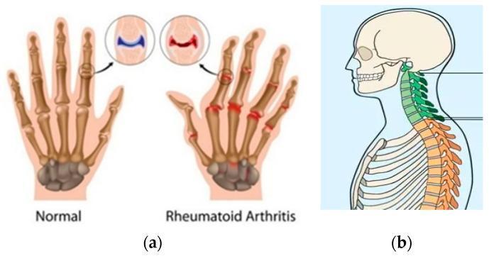
Educational comparison diagrams illustrating musculoskeletal changes in inflammatory arthritis. (a) A comparative anatomical diagram of a normal hand versus a hand with Rheumatoid Arthritis (RA). The normal hand shows clear joint spaces and smooth bone alignment. The RA hand depicts ulnar deviation, swelling of the metacarpophalangeal and interphalangeal joints, and joint space narrowing. Inset circles provide a high-magnification view of a healthy synovial joint versus an RA joint characterized by cartilage erosion and synovial inflammation (pannus). (b) A sagittal anatomical illustration of the head, neck, and upper torso highlighting changes associated with Ankylosing Spondylitis (AS). The cervical spine (green) and thoracic spine (orange) show indicative features of syndesmophyte formation and vertebral fusion, leading to a loss of the normal physiological curvature and the development of a 'bamboo spine' appearance. These visuals are intended to demonstrate the distinctive clinical manifestations and progressive joint degradation found in chronic rheumatological conditions.
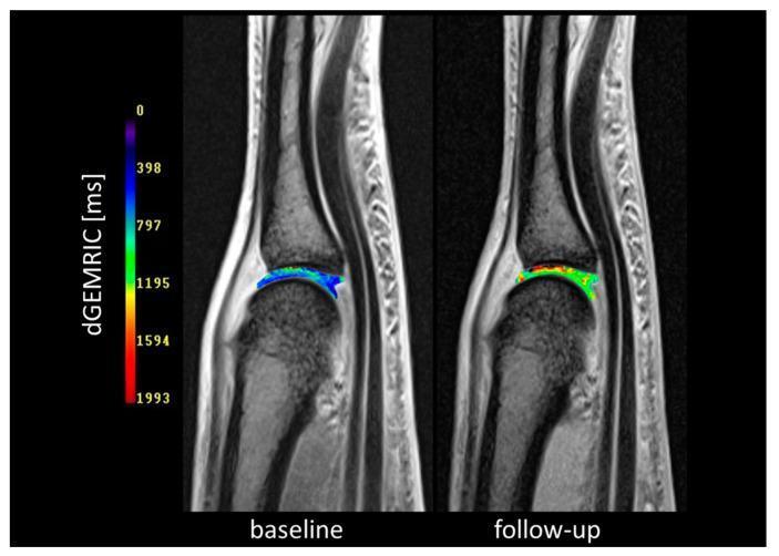
Comparison of delayed Gadolinium-Enhanced Magnetic Resonance Imaging (dGEMRIC) maps of the third metacarpophalangeal (MCP3) joint, illustrating cartilage composition changes in a patient with early rheumatoid arthritis. The image displays a side-by-side comparison of 'baseline' and 'follow-up' (24 weeks post-adalimumab plus methotrexate therapy) states, fused with morphological MRI scans. A color-coded scale on the left represents dGEMRIC values in milliseconds (ms), where blue/purple indicate low glycosaminoglycan (GAG) content (cartilage damage) and red/yellow indicate high GAG content (healthy cartilage). At baseline, the articular cartilage is predominantly blue, signifying significant GAG depletion. At follow-up, there is a visible spatial shift toward green and red hues centrally within the joint space, indicating increased dGEMRIC values and cartilage biochemical regeneration. This imaging modality demonstrates the utility of functional MRI in monitoring pharmacological response to biologic therapy beyond simple structural assessment.
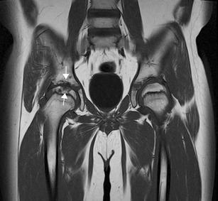
This diagnostic image is a coronal T1 Turbo Spin Echo (TSE) MRI of a pediatric pelvis, illustrating musculoskeletal manifestations of Juvenile Idiopathic Arthritis (JIA). The scan provides a side-by-side comparison of the hip joints. The right hip joint exhibits significant pathological changes, including marked joint space narrowing and prominent bony erosions (indicated by white arrows) on both the superior aspect of the femoral head and the adjacent acetabular margin. These erosions appear as irregular, low-signal intensity focal concavities and lytic lesions that disrupt the smooth cortical contour. In contrast, the left hip joint maintains a relatively preserved joint space and smooth articular surfaces without evidence of bone destruction. The image demonstrates the clinical utility of MRI in detecting late-stage structural damage, such as cartilage loss and erosive disease, which are key indicators of joint destruction in chronic inflammatory arthritis. This content is intended for advanced medical education in pediatric radiology and rheumatology.
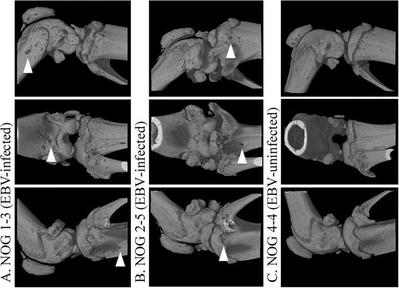
Three-dimensional computed tomography (3D-CT) comparison of mouse knee joints in a model of erosive arthritis related to Epstein-Barr virus (EBV) infection. The figure is divided into three columns representing EBV-infected subjects (A and B) and an EBV-uninfected control (C), with multiple anatomical views for each. Column A and B display significant pathological changes in the bone architecture of humanized NOG mice. White arrowheads highlight focal bone erosions, visible as irregular pitting, cortical discontinuity, and structural breakdown at the distal femur and proximal tibia. These morphological features resemble the erosive changes seen in Rheumatoid Arthritis (RA). In contrast, column C displays the control joint, characterized by smooth, intact cortical surfaces and a preserved joint space without signs of osseous degradation. This diagnostic imaging serves to demonstrate the development of virus-induced erosive joint disease and the role of humanized immune cells in bone resorption.
Types of Arthritis
1. Degenerative Arthritis
Osteoarthritis (OA)
- Most common form of joint disease worldwide.
- Caused by biomechanical stress exceeding cartilage synthesis capacity - matrix breakdown is the primary driver; inflammation is minimal and secondary.
- Local inflammatory cytokines (e.g., IL-1, TNF) contribute to progression.
- Affects weight-bearing joints (knees, hips), spine, and hands (DIP joints, CMC joints).
- Key features: joint space narrowing, osteophytes, subchondral sclerosis, bony crepitus.
- No systemic features; ESR/CRP usually normal.
2. Inflammatory / Autoimmune Arthritis
Rheumatoid Arthritis (RA)
- Chronic autoimmune disease driven by cellular and humoral immune response against citrullinated proteins (anti-CCP antibodies positive in 60-80%; specificity 85-99%).
- TNF plays a central pathogenic role; anti-TNF biologics are effective.
- Symmetric, small-joint polyarthritis (MCPs, PIPs, wrists); spares DIP joints.
- Hallmarks: pannus formation, synovitis, articular erosions, morning stiffness >1 hour.
- Systemic manifestations: rheumatoid nodules, interstitial lung disease, vasculitis, pericarditis.
Juvenile Idiopathic Arthritis (JIA)
- Arthritis onset before age 16, lasting >6 weeks, with no other identifiable cause.
- Several subtypes: oligoarticular (most common), polyarticular (RF+ or RF-), systemic (Still disease), enthesitis-related, psoriatic.
- Complication: uveitis (especially in ANA-positive oligoarticular JIA).
Systemic Lupus Erythematosus (SLE) - Lupus Arthritis
- Arthritis/arthralgias occur in up to 90% of SLE patients.
- Symmetric inflammatory arthritis, predominantly knees, wrists, and small hand joints.
- Key feature: Jaccoud's arthropathy - reducible deformities (ulnar deviation, swan-neck) due to ligamentous laxity without typical radiographic erosions.
- Synovial effusions are less inflammatory than in RA.
3. Seronegative Spondyloarthropathies (SpA)
| Subtype | Key Features |
|---|---|
| Ankylosing Spondylitis (AS) | Young men; chronic low back pain/stiffness; sacroiliitis; "bamboo spine"; uveitis |
| Psoriatic Arthritis (PsA) | Associated with psoriasis; DIP involvement; dactylitis ("sausage digit"); asymmetric |
| Reactive Arthritis | Follows GI (Salmonella, Campylobacter) or genitourinary (Chlamydia) infection; asymmetric oligoarthritis; urethritis/conjunctivitis |
| IBD-associated Arthritis | Peripheral (parallels bowel activity) or axial (independent of bowel disease) |
| Undifferentiated SpA | Features of SpA without fitting a defined subtype |
4. Crystal Arthropathy
Gout
- Caused by monosodium urate (MSU) crystal deposition in joints due to hyperuricemia.
- Needle-shaped crystals with strong negative birefringence on polarized microscopy.
- Classically affects the first MTP joint (podagra), ankles, and knees.
- Acute flares + chronic tophaceous gout; urate-lowering therapy (allopurinol, febuxostat) is the long-term treatment.
Pseudogout (CPPD - Calcium Pyrophosphate Deposition Disease)
- Caused by calcium pyrophosphate dihydrate (CPPD) crystals.
- Rhomboid-shaped crystals with weakly positive birefringence.
- Most common in knees and wrists.
- Chondrocalcinosis on X-ray is characteristic.
5. Infectious Arthritis
Septic (Suppurative) Arthritis
- Bacterial infection of a joint space, usually acquired hematogenously.
- Most common organism: Staphylococcus aureus (adults); Neisseria gonorrhoeae (sexually active young adults).
- Medical emergency - requires joint aspiration and IV antibiotics.
- Hot, swollen, single joint (monoarthritis); WBC in synovial fluid typically >50,000/µL.
Lyme Arthritis
- Caused by Borrelia burgdorferi (tick-borne).
- Oligoarthritis, especially knees, weeks to months after infection.
- May have autoimmune component in chronic stages.
6. Other Notable Types
| Type | Key Points |
|---|---|
| Psoriatic Arthritis | Listed separately above but also classified as SpA; nail changes + arthritis |
| Enteropathic Arthritis | Seen in Crohn's and UC; peripheral or axial pattern |
| Hemophilic Arthropathy | Recurrent hemarthrosis causing chronic joint destruction |
| Neuropathic (Charcot) Joint | Progressive joint destruction due to loss of pain sensation (diabetes, syphilis, syringomyelia) |
| Viral Arthritis | Parvovirus B19, Hepatitis B/C, HIV, Chikungunya; usually self-limiting |
Quick Comparison: The Big Three
| Feature | Osteoarthritis | Rheumatoid Arthritis | Gout |
|---|---|---|---|
| Mechanism | Mechanical wear | Autoimmune | Crystal deposition |
| Age | >50 (older adults) | Any age (peak 40-60) | Typically men >40 |
| Joint pattern | Asymmetric; DIP, knees, hips | Symmetric; MCP, PIP, wrists | Asymmetric; MTP, knees |
| Morning stiffness | <30 minutes | >1 hour | Not a feature |
| Systemic features | No | Yes | No (except tophi) |
| Labs | Normal ESR/CRP | Elevated CRP/ESR, RF+, anti-CCP+ | Elevated uric acid |
| X-ray | Osteophytes, sclerosis | Erosions, periarticular osteopenia | Punched-out erosions, tophi |
Rheumatic arthritis
rheumatoid arthritis joint pathology hand deformity
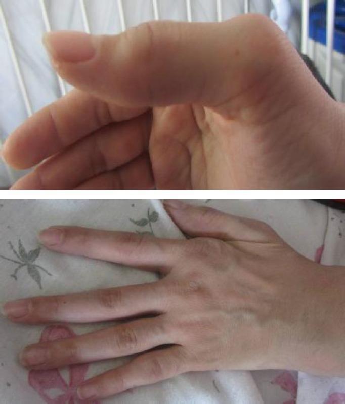
Clinical photographs of a hand demonstrating chronic structural deformities characteristic of rheumatoid arthritis. The upper image provides a lateral view focusing on a digit showing a prominent deformity at the proximal interphalangeal (PIP) joint and enlargement of the metacarpophalangeal (MCP) joint. The lower image provides a dorsal view of the hand, illustrating significant ulnar deviation of the fingers at the MCP joints, which is a classic hallmark of progressive inflammatory arthritis. There is visible soft tissue swelling and joint enlargement at the 2nd metacarpal and multiple PIP joints. The images exemplify the long-term musculoskeletal manifestations of active rheumatoid arthritis, showcasing joint misalignment and chronic inflammatory changes in the small joints of the hand. This content is suitable for teaching clinical signs of autoimmune rheumatological diseases and physical examination findings in hand pathology.
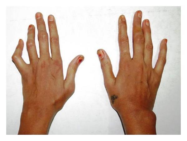
Clinical photograph of the dorsal aspect of bilateral hands and wrists in a patient with Rheumatoid Arthritis (RA). The image demonstrates characteristic chronic inflammatory joint deformities. On the left hand, there is a prominent swan-neck deformity of the fifth digit, characterized by hyperextension of the proximal interphalangeal (PIP) joint and flexion of the distal interphalangeal (DIP) joint. Swelling is visible across the interphalangeal joints of the third and fourth digits. The right hand shows a significant, localized soft tissue swelling on the lateral/ulnar aspect of the wrist joint, consistent with synovial hypertrophy or a rheumatoid nodule. General architectural changes include joint stiffness and minor deviations of the fingers. This visual illustrates the progressive peripheral joint involvement and resulting anatomical deformities typical of systemic inflammatory polyarthritis, emphasizing the clinical signs used in diagnosing the severity of RA.
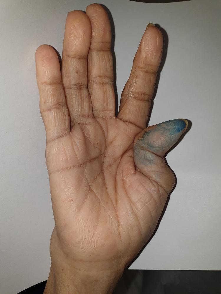
This clinical photograph displays the palmar surface of a right hand, primarily illustrating a classic 'Z-thumb' deformity (Hitchhiker thumb), a hallmark sign frequently associated with advanced rheumatoid arthritis. The deformity is characterized by severe flexion at the metacarpophalangeal (MCP) joint and compensatory hyperextension at the interphalangeal (IP) joint. There is also evident bony swelling and ulnar deviation at the wrist. While the distal interphalangeal (DIP) joints of the digits appear relatively spared, there is subtle swelling suggestive of polyarthropathy in the proximal regions. The thumb's skin shows a blue discoloration, likely from diagnostic marking or external staining. This image serves as an educational example of chronic inflammatory joint destruction and secondary ligamentous laxity leading to fixed musculoskeletal deformities in systemic autoimmune conditions.
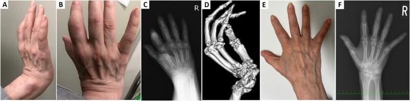
This composite figure displays clinical and multi-modality imaging findings of a patient with Rheumatoid Arthritis (RA) and striatal hand deformity. Panels A and B are clinical photographs showing significant swelling of the proximal interphalangeal (PIP) and metacarpophalangeal (MCP) joints, with a rigid, flexed deformity of the right wrist resembling bony ankylosis. Panel C is a posteroanterior hand radiograph demonstrating periarticular osteopenia, radiocarpal joint space narrowing, and flexion of the index finger distal interphalangeal (DIP) joint. Panel D provides a 3D CT reconstruction that clearly visualizes a dorsal dislocation of the distal ulnar joint and spatial misalignment of the carpal bones without true osseous bridging. Panels E and F show post-treatment clinical and radiographic follow-up, respectively, highlighting a marked reduction in soft tissue swelling of the hand and improved joint alignment and bone density. The collection illustrates the differentiation between fixed dystonic deformities and active inflammatory arthropathy in a rheumatological context, emphasizing the importance of cross-sectional imaging (CT) over 2D radiography for assessing complex joint architecture.
"rheumatoid arthritis"[MeSH Terms] AND "treatment"[MeSH Terms]
Rheumatoid Arthritis (RA)
Pathogenesis
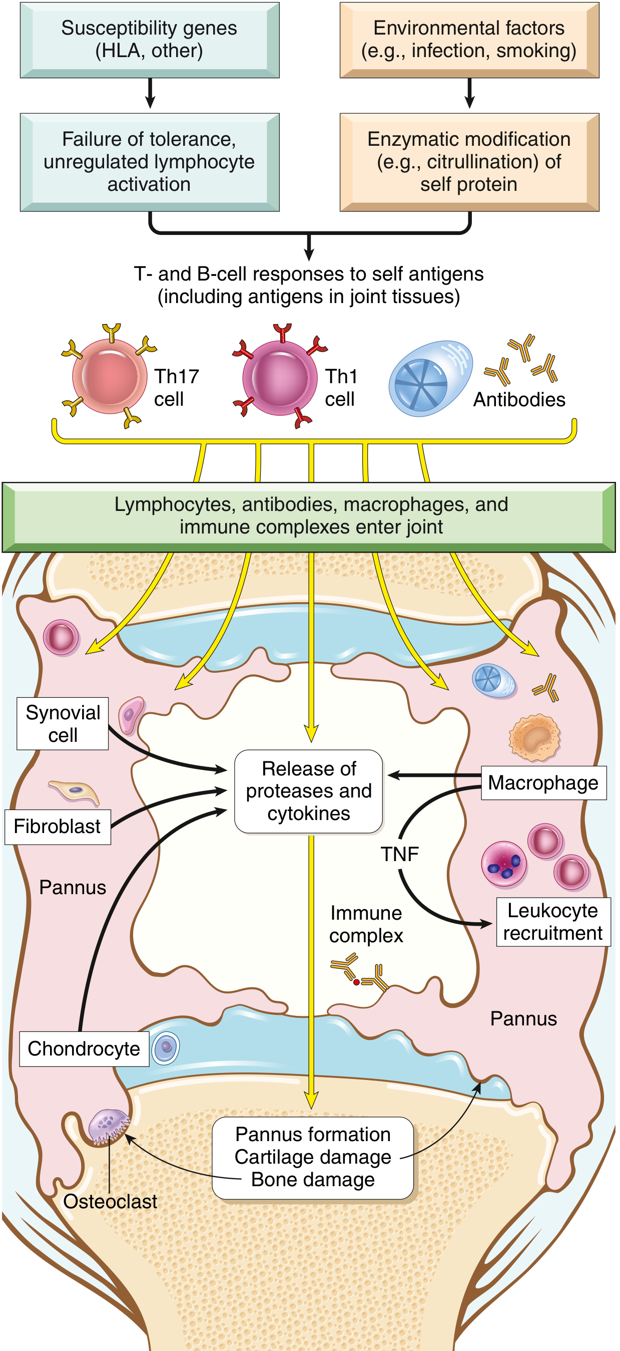
Genetic Factors
- ~50% of RA risk is inherited
- HLA-DR4 is strongly associated with ACPA-positive RA
- The shared epitope hypothesis: a citrullinated protein (vinculin) mimics microbial epitopes that are presented by HLA-DR4 (molecular mimicry)
Environmental Triggers
- Smoking and periodontitis are the best-documented environmental risk factors
- These promote enzymatic citrullination of self-proteins (arginine → citrulline), creating neo-epitopes that break immune tolerance
Immune Cascade
- CD4+ Th1 and Th17 cells react against citrullinated joint antigens
- Cytokine storm: TNF-α, IL-1, IL-6 from macrophages recruit leukocytes and stimulate proteases that destroy cartilage
- IL-17 from Th17 cells recruits neutrophils and monocytes
- RANKL expressed on T cells activates osteoclasts → bone erosion
- B cells and plasma cells produce:
- Rheumatoid factor (RF): IgM/IgA autoantibody against Fc portion of IgG
- Anti-citrullinated protein antibodies (ACPAs): more specific for RA
The Role of Fibroblast-like Synoviocytes (FLS)
Morphology (What Happens in the Joint)
| Feature | Detail |
|---|---|
| Synovial changes | Edematous, thickened, hyperplastic synovium with delicate villi |
| Histology | Synoviocyte hyperplasia; dense infiltrates of CD4+ T cells, B cells, plasma cells, macrophages; angiogenesis |
| Pannus | Mass of edematous synovium + inflammatory cells + granulation tissue + fibroblasts that grows over and erodes articular cartilage |
| Bone erosion | Osteoclast activation at pannus-bone interface; periarticular erosions and subchondral cysts |
| Ankylosis | Fibrous ankylosis → bony ankylosis in advanced disease |
| Rheumatoid nodules | Necrotizing granulomas in subcutaneous tissue (forearm, elbows, occiput) |
Clinical Features
Joint Involvement
- Onset: Gradual in ~50% of cases - weeks to months of malaise, fatigue, and generalized musculoskeletal pain before joint involvement
- Pattern: Symmetric polyarthritis
- Joints affected: MCPs, PIPs, wrists, ankles, elbows, knees - DIP joints are classically spared (distinguishes from OA and psoriatic arthritis)
- Morning stiffness: >1 hour (hallmark; correlates with disease activity)
- Joints are swollen, warm, painful, tender
Classic Hand Deformities (from tendon/ligament inflammation)
- Ulnar deviation at MCPs
- Swan-neck deformity: PIP hyperextension + DIP flexion
- Boutonniere deformity: PIP flexion + DIP hyperextension
- Z-thumb (Hitchhiker's thumb): MCP flexion + IP hyperextension
Radiographic Hallmarks
- Joint effusions
- Juxta-articular osteopenia (periarticular bone loss)
- Marginal erosions
- Narrowing of joint space
- Loss of articular cartilage
Extra-articular Manifestations
| System | Manifestation |
|---|---|
| Skin | Rheumatoid nodules (20-30%), vasculitis |
| Lungs | Interstitial lung disease, pleural effusions, rheumatoid nodules |
| Heart | Pericarditis, accelerated atherosclerosis (major CV risk) |
| Eyes | Sicca syndrome (secondary Sjögren's), episcleritis, scleritis |
| Blood vessels | Vasculitis |
| Systemic | Fatigue, anemia of chronic disease, cognitive impairment, osteoporosis |
| Cervical spine | Atlantoaxial subluxation (C1-C2) - risk of cord compression |
Diagnosis
ACR/EULAR 2010 Classification Criteria
- Joint involvement (0-5 points): number and size of affected joints
- Serology (0-3 points): RF and/or ACPA positivity
- Acute-phase reactants (0-1 point): elevated CRP or ESR
- Duration of symptoms (0-1 point): ≥6 weeks
Key Lab Tests
| Test | Significance |
|---|---|
| Anti-CCP (ACPA) | Positive in 60-70% of RA; specificity 85-99%; may precede symptoms by years |
| Rheumatoid Factor (RF) | Positive in ~80%; less specific (also positive in other conditions) |
| ESR / CRP | Elevated - markers of systemic inflammation |
| CBC | Normocytic anemia of chronic disease; thrombocytosis |
| Synovial fluid | Inflammatory (WBC 5,000-50,000/µL, predominantly neutrophils) |
Treatment
Step 1 - Conventional Synthetic DMARDs (csDMARDs)
- Methotrexate (MTX) is the anchor drug and first-line DMARD for most patients
- Titrated up to 25 mg/week; can be given subcutaneously for better bioavailability
- Requires folic acid supplementation; monitor LFTs and CBC
- Other csDMARDs: hydroxychloroquine, sulfasalazine, leflunomide
- Triple therapy (MTX + HCQ + SSZ) is an alternative to biologics
Step 2 - Biologic DMARDs (bDMARDs)
| Class | Drugs | Target |
|---|---|---|
| TNF inhibitors | Etanercept, infliximab, adalimumab, certolizumab, golimumab | TNF-α |
| IL-6 inhibitor | Tocilizumab, sarilumab | IL-6 receptor |
| Costimulation blocker | Abatacept | CD80/CD86 (T cell activation) |
| B cell depletion | Rituximab | CD20 on B cells |
| IL-1 inhibitor | Anakinra | IL-1 receptor |
Step 3 - Targeted Synthetic DMARDs (tsDMARDs)
- JAK inhibitors: Tofacitinib, baricitinib, upadacitinib
- Oral agents; inhibit intracellular JAK-STAT signaling
- Carry FDA warnings for thrombosis risk and cardiovascular events in high-risk patients
Adjunct Therapy
- NSAIDs: Symptomatic relief only; never used without concurrent DMARDs
- Glucocorticoids: Rapidly effective bridge therapy; should be tapered and eventually discontinued ("best dose for chronic use is zero")
- Physical therapy and occupational therapy
- Surgery: Joint replacement (hip, knee) for end-stage disease; synovectomy in selected cases
Prognosis
- With early diagnosis and treat-to-target strategy, most patients can achieve remission or low disease activity
- Poor prognostic factors: high ACPA/RF titers, high CRP/ESR, many swollen joints, early radiographic erosions, smoking
- Long-term anti-TNF therapy increases risk of opportunistic infections (especially reactivation TB - screen before starting)
- Cardiovascular disease is the leading cause of excess mortality in RA - aggressive CV risk factor management is essential
Rheumatic arthritis
Rheumatoid Arthritis (RA) - Summary
What It Is
Pathogenesis (in brief)
- Trigger: Smoking/infection causes citrullination of self-proteins → immune tolerance breaks down
- Genetics: HLA-DR4 association; ~50% genetic risk
- Key cells: CD4+ T cells, B cells, macrophages, fibroblast-like synoviocytes
- Key cytokines: TNF-α, IL-1, IL-6, IL-17
- Result: Pannus formation → cartilage erosion + bone destruction via osteoclasts (RANKL-driven)
Clinical Features
| Deformity | Description |
|---|---|
| Ulnar deviation | Fingers deviate toward ulnar side at MCPs |
| Swan-neck | PIP hyperextension + DIP flexion |
| Boutonniere | PIP flexion + DIP hyperextension |
| Z-thumb | MCP flexion + IP hyperextension |
Diagnosis
| Test | Finding |
|---|---|
| Anti-CCP (ACPA) | Positive in 60-70%; specificity 85-99% |
| Rheumatoid Factor (RF) | Positive in ~80%; less specific |
| ESR/CRP | Elevated |
| X-ray | Marginal erosions, juxta-articular osteopenia, joint space narrowing |
Treatment (Treat-to-Target: aim for remission or low disease activity)
| Step | Drugs |
|---|---|
| 1st line | Methotrexate (MTX) - anchor DMARD, up to 25mg/week ± folic acid |
| Add-on csDMARDs | Hydroxychloroquine, sulfasalazine, leflunomide |
| Biologic DMARDs | TNF inhibitors (adalimumab, etanercept), IL-6 inhibitors (tocilizumab), rituximab, abatacept |
| JAK inhibitors | Tofacitinib, baricitinib, upadacitinib (oral targeted therapy) |
| Bridge therapy | Short-course glucocorticoids (taper ASAP) |
| Symptom relief | NSAIDs (never alone - always with DMARDs) |